WEEK3!
 Call Kai
 Learn
 Practice Test
 Spaced Repetition
 Match
 Flashcards
 Knowt Play
1/59

There's no tags or description
Looks like no tags are added yet.
60 Terms
What are the six extraocular muscles, and what is their general purpose?
The superior rectus, inferior rectus, medial rectus, lateral rectus, superior oblique, and inferior oblique. They control eye movement in different directions.
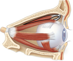
What does the mnemonic LR6 SO4 help you remember?
Lateral Rectus is controlled by cranial nerve VI; Superior Oblique is controlled by cranial nerve IV; all remaining extraocular muscles are controlled by cranial nerve III.

Which cranial nerve controls the superior rectus, inferior rectus, medial rectus, and inferior oblique?
Cranial nerve III (oculomotor nerve).
What movement does the superior rectus produce, and which cranial nerve controls it?
It elevates the eye (moves the eye upward). It is controlled by CN III.

What movement does the inferior rectus produce, and which cranial nerve controls it?
It depresses the eye (moves the eye downward). It is controlled by CN III.

What is the difference between adduction and abduction of the eye?
Adduction moves the eye toward the nose (medially), while abduction moves the eye away from the nose (laterally).

Which muscle is responsible for adduction, and which cranial nerve controls it?
The medial rectus performs adduction and is controlled by CN III.

Which muscle is responsible for abduction, and which cranial nerve controls it?
The lateral rectus performs abduction and is controlled by CN VI.

What are the primary actions of the superior oblique muscle?
It depresses and intorts the eye. It is controlled by CN IV.

What is the trochlea, and what is its relationship to the superior oblique?
The trochlea is a pulley-like structure that the superior oblique tendon passes through, helping redirect the muscle's pull.
What are the primary actions of the inferior oblique muscle?
It elevates and extorts the eye. It is controlled by CN III.

A patient cannot move their right eye laterally. Which muscle and cranial nerve should you suspect are damaged?
The right lateral rectus and right CN VI (abducens nerve), because this nerve controls lateral eye movement.

A patient has difficulty moving one eye downward and inward. Which muscle could be damaged?
The superior oblique, which is controlled by CN IV and contributes to depression and intorsion.

If the oculomotor nerve (CN III) is damaged, which extraocular muscles may lose normal function?
The superior rectus, inferior rectus, medial rectus, and inferior oblique may be affected because CN III controls these four muscles.

Why is the LR6 SO4 mnemonic useful when identifying extraocular muscles on a practical exam?
It quickly identifies the two exceptions to CN III control. Once lateral rectus and superior oblique are identified, the other four muscles are supplied by CN III.
Trace the pathway sound follows from the outside of the ear until vibrations enter the inner ear.
Pinna collects sound → external auditory canal → tympanic membrane → malleus → incus → stapes → oval window → inner ear.

What is the function of the pinna (auricle)?
The visible outer flap of the ear that collects and directs sound waves into the external auditory canal.

What is the external auditory canal, and where does it lead?
It is the tunnel-like passageway that carries sound waves from the pinna to the tympanic membrane.

What is the tympanic membrane, and what happens when sound waves reach it?
The tympanic membrane is the eardrum. Sound waves cause it to vibrate, beginning the mechanical transmission of sound through the middle ear.

What are the three auditory ossicles, and what are their correct order?
Malleus (hammer) → Incus (anvil) → Stapes (stirrup). They transmit and amplify vibrations toward the oval window.

Which ossicle directly contacts the oval window, and what happens when it moves?
The stapes. Its movement pushes against the oval window, creating fluid waves in the inner ear.

What is the function of the oval window?
It transfers vibrations from the stapes into the fluid-filled inner ear, helping initiate waves that stimulate hearing receptors.
What is the function of the round window, and why is it necessary?
It acts as a pressure-relief membrane that allows inner-ear fluid to move when vibrations enter through the oval window.

What is the function of the Eustachian tube?
It equalizes air pressure between the middle ear and the throat (nasopharynx), helping the eardrum vibrate normally.
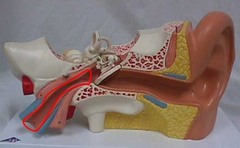
What is the cochlea responsible for, and how does it contribute to hearing?
The cochlea converts mechanical fluid vibrations into electrical nerve signals through sensory hair cells in the organ of Corti.

What is the organ of Corti, and where is it located?
It is the sensory organ for hearing, located inside the cochlea. Its hair cells convert mechanical vibrations into electrical signals.
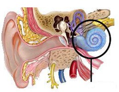
What are the functions of the vestibule, and which two structures are found inside it?
The vestibule detects static equilibrium and linear acceleration. It contains the utricle and saccule.

What is the function of the semicircular canals?
They detect rotational movements of the head and help maintain dynamic equilibrium.
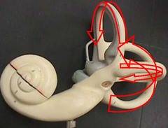
What is the difference between static equilibrium and dynamic equilibrium?
Static equilibrium involves sensing head position relative to gravity and linear acceleration; dynamic equilibrium involves sensing rotational or angular head movements.
Which cranial nerve carries hearing and balance information to the brain?
The vestibulocochlear nerve (CN VIII), which carries both auditory and vestibular information.

A person has difficulty maintaining balance when turning their head. Which inner-ear structures may be involved, and why?
The semicircular canals, because they detect rotational head movements and contribute to dynamic equilibrium.
A person experiences hearing difficulties because sensory hair cells in the organ of Corti are damaged. Which structure is affected, and why does this cause hearing problems?
The cochlea is affected because the organ of Corti is located inside it. Damaged hair cells cannot properly convert mechanical vibrations into electrical signals.
If the Eustachian tube becomes blocked, what problem can occur in the middle ear?
Pressure may become unequal on either side of the tympanic membrane, interfering with normal vibration and potentially causing discomfort or muffled hearing.
Why would damage to the vestibulocochlear nerve potentially affect both hearing and balance?
CN VIII carries sensory information from both the cochlea (hearing) and vestibular apparatus (balance).
What is the corpus callosum, and how does it appear when viewing a midsagittal sheep brain?
It is a thick band of white matter connecting the left and right cerebral hemispheres. It appears as a prominent white band on the cut surface.

What is the arbor vitae, where is it located, and why does it have its name?
It is the branching white matter inside the cerebellum. It resembles a tree with branches when viewed in a cut section.

What is the function of the cerebellum?
It coordinates voluntary movement, balance, posture, and motor learning.

What are the colliculi, where are they located, and how many bumps should you identify?
They are four rounded elevations on the posterior part of the midbrain: two superior colliculi and two inferior colliculi.

What is the function of the superior colliculi compared with the inferior colliculi?
Superior colliculi are primarily involved in visual reflexes and orienting movements; inferior colliculi are primarily involved in auditory reflexes and sound processing.

What does the optic chiasm look like on the underside of a sheep brain, and what is its general function?
It appears as a white X-shaped structure on the ventral surface. It is where some optic nerve fibers cross to the opposite side of the brain.

What are the olfactory bulbs, where are they located, and what sensory information do they process?
They are prominent structures at the anterior (front) end of the brain that receive and process information related to smell.

During a sheep brain practical, you see a white branching, tree-like pattern inside the cerebellum. What structure are you identifying?
The arbor vitae, which is the branching white matter inside the cerebellum.

During a sheep brain practical, you see a thick white band connecting the two cerebral hemispheres on a midsagittal cut. What structure is it?
The corpus callosum.

Why is it important to distinguish the optic chiasm from the olfactory bulbs when viewing the underside of the sheep brain?
The optic chiasm is an X-shaped structure farther back that involves crossing optic nerve fibers, while the olfactory bulbs are located toward the anterior end and process smell.
What is the cornea, where is it located, and what is its primary function?
The transparent curved covering at the front of the eye. It protects the eye and refracts incoming light.

What is the lens, and what is its function?
The clear, firm, flexible structure behind the iris that changes shape to focus light onto the retina.

What is the vitreous humor, and where is it located?
It is the transparent jelly-like substance filling the large posterior cavity behind the lens. It helps maintain the shape of the eyeball.

What is the retina, and what is its function?
The thin, light-sensitive neural layer lining the back of the eye. It contains photoreceptors that convert light into electrical signals.

What is the choroid, and why does it appear dark during a sheep eye dissection?
It is the vascular, pigmented layer between the retina and sclera. Its dark pigment absorbs excess light and helps prevent internal light scattering.

What is the tapetum lucidum, and what is its appearance in a sheep eye?
It is a reflective layer within the choroid that reflects light back through the retina, improving vision in low-light conditions. It often appears shiny blue-green.
What is the optic nerve, where is it located, and what information does it carry?
It is the nerve exiting the back of the eyeball that carries visual information from the retina to the brain.

What is the ciliary body, and what is its role in focusing vision?
It is a muscular structure containing folds around the lens. It helps control lens shape through the suspensory apparatus for accommodation.

During a sheep eye dissection, you remove a firm, clear disc from behind the iris. What structure have you found?
The lens.

You notice a thin, delicate, filmy layer peeling away from the back of the sheep eye. Which structure is this?
The retina, the light-sensitive neural layer.

You observe a shiny blue-green reflective surface inside the sheep eye. What structure are you looking at, and why does it exist?
The tapetum lucidum, which reflects light through the retina to improve low-light vision.
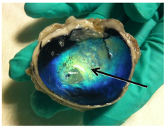
You identify a white stump protruding from the posterior surface of the sheep eyeball. What is it?
The optic nerve, which carries visual information from the retina toward the brain.

Why is it important to distinguish the retina from the choroid during a sheep eye practical?
The retina is a thin, delicate, light-sensitive neural layer that may peel away, while the choroid is a darker, vascular, pigmented layer underneath it.
What is the difference between the vitreous humor and the lens?
The vitreous humor is transparent jelly filling the posterior cavity, while the lens is a firm, clear structure that focuses incoming light.
If the optic nerve is damaged, what sensory information would be affected?
Visual information from the retina would not be transmitted normally to the brain, potentially causing vision loss.
Why does the tapetum lucidum help animals such as sheep see in low-light conditions?
It reflects incoming light back through the photoreceptors, giving them another opportunity to detect the light.