SBMS 1431- Organization of the Nervous System and Autonomic Nervous System
 Call Kai
 Learn
Learn Practice Test
 Spaced Repetition
 Match
Match Flashcards
 Knowt Play
1/68

There's no tags or description
Looks like no tags are added yet.
Name | Mastery | Learn | Test | Matching | Spaced | Call with Kai | Chat |
|---|
No analytics yet
Send a link to your students to track their progress
69 Terms
Neural Tissue
Conducts electrical impulses
Carries information
Overview of the Nervous System
It regulates personal responses to adapt to changes in both the internal and external environment.
It forms the prime communication and coordination network inside the body systems to reflect, respond, and react to the external environment and maintain body’s homeostasis.
How does the nervous system work? (Overview)
Sensory input: The gathered information is called sensory input.
The nervous system uses its millions of sensory receptors to monitor changes occurring both inside and outside the body.
Integration: The nervous system processes and interprets sensory input and decides what should be done at each moment
an integration process.
Motor output: The nervous system sends a motor output by activating effector organs, including muscles and glands.
Central Nervous System (CNS)
Includes brain and spinal cord
Involved in decision-making processes, including a variety of functions:
Interpretation of sensory information
Planning and monitoring movement
Maintenance of homeostasis
Higher mental functions such as language and learning
Peripheral nervous system (PNS)
Consists of spinal nerves, cranial nerves, and sensory receptors.
Motor functions- include stimulation of a muscle cell contraction or a gland secretion; performed by the peripheral nervous system (PNS)
Sensory functions- detection of sensations within and outside the body; also performd by PNS
The Neurons
Basic and Functional Units
Carry electrical signals
Organelles are in the cell body
The dendrites, higher branched extensions that receive signals from other neurons
The axon is typically a much longer extension that transmits signals to other cells at synapses.
The cone-shaped base of an axon is called the axon hillock: it is part of the initial segment of the trigger zone for the membrane potential propagation.
Most axons are covered with myelin; a fatty whitish substance wrapped around axons to facilitate impulse conduction
Types of Neuroms
Sensory Neurons: afferent neurons conduct action potentials toward the CNS
Interneurons: conduct action potentials from one neuron to another within the CNS
Efferent neurons conduct action potentials away from the CNS
Structural Classification of Neurons
Anaxomic Neuron
Bipolar Neuron
Pseudonipolar Neuron
Multipolar Neuron
Processes (neurological term)
Any projection, extension, or “arm” that sticks out from the main body (the soma) of a neuron.
Anaxomic Neuron
Found in brain and sense organs
Have more than two processes, but axons cannot be distinguished from the dendrites
Bipolar Neuron
Found in special sensory organs (sight, smell & hearing)
Bipolar neurons have two processes separated by the cell body.
Pseudounipolar Neuron
Also called a Unipolar Neuron
Pseudounipolar neurons have single elongated processes with the cell body situated to one side
Found in sensory neurons fo PNS, e.g. dorsal root ganglion
Multipolar Neuron
Multipolar neurons have more than two processes; there is a single axon and multiple dendrites
Neuroglial Cells and Tissue
Neuroglial cells + extracellular matrix within the CNS and PNS connective tissue
Neuroglial cells (glial cells) bathe and sustain neurons
Maintain the integrity of the brain and spinal cord by glueing neurons together with blood vessels to form structures
Neuroglial Cells found in the PNS:
Satellite Cells
Schwann Cells
Satellite Cells
Satellite Cells/ Satellite Glial Cells - Surround neuron cell bodies of the dorsal root ganglia (sensory ganglia) in PNS; regulate the supply of O2, CO2, nutrients, and neurotransmitter levels around neurons in the ganglia.
Schwann Cells
Schwann Cells /neurilemma cell - Surround all axons of the nerves in PNS; responsible for myelination of peripheral axons; participate in the repair process after injury; also act as phagocytes to rid a damaged nerve of deteriorating cell debris.
Neuroglial Cells found in the CNS:
Oligodendrocytes
Astrocytes
Microglia
Ependymal cells
Ependymal Cells
Line the lumen of the ventricles of the brain (choroid plexus) and the central canal of the spinal cord
Produce cerebrospinal fluid (CSF)
Oligodendrocytes
Their cytoplasmic extensions wrap tightly around the CNS axons to form the myelin sheaths
Microglia
Roaming scavenger cells (macrophage); engulf; digest, and destroy invading organisms and foreign matter; clean up the debris of dead cells following an injury.
Astrocytes
Most abundant; shaped like stars. From the cell body, they send out projections that grip and stabilize neurons and capillaries
Control the condition of the ECM (ground substances + protein fibers)
Maintain the blood-brain barrier
Frontal Plane of the brain (Pictorial)

The Blood-Brain Barrier (BBB)
Offers molecular protection and protects the brain from harmful substances released from the blood capillaries
Is a protective mechanism that helps to ensure the brain’s environment remains stable
Endothelial Cells of capillaries forming the tight junction with astrocytes and pericytes in the connective tissue of the brain act as a firewall to prevent most substances from directly entering the brain tissue.

Substances that can pass through the BBB
Gases and fat-soluble substances can move across these barriers without assistance
Glucose and amino acids pass through via regular transport processes.
Substances that can pass through the BBB
Gases and fat-soluble substances can move across these barriers without assistance
Glucose and amino acids pass through via regular transport processes.
Tight Junctions
A specialized cell-to-cell adhesion structure that forms a continuous, impermeable seal between adjacent cells (a lot of times the endothelial cells of the capillaries)
Foot processes of astrocytes
Wrap around the outer surfaces of capillaries
Pericytes
(A type of smooth muscle) regulate the cerebral blood flow in capillaries
Contractile cells located on the outer surfaces of capillary endothelial cells, embedded within the capillary basement membrane. In the brain, they regulate cerebral blood flow by adjusting capillary diameter, and they control vascular formation by stabilizing and maturing new vessels. They also support the blood-brain barrier.
Central Nervous System
Consists of:
Brain: Located up the cranial vault of the skull
Spinal cord: Located in the vertebral canal
Brain and spinal cord: Continuous with each other at the foremen magnum
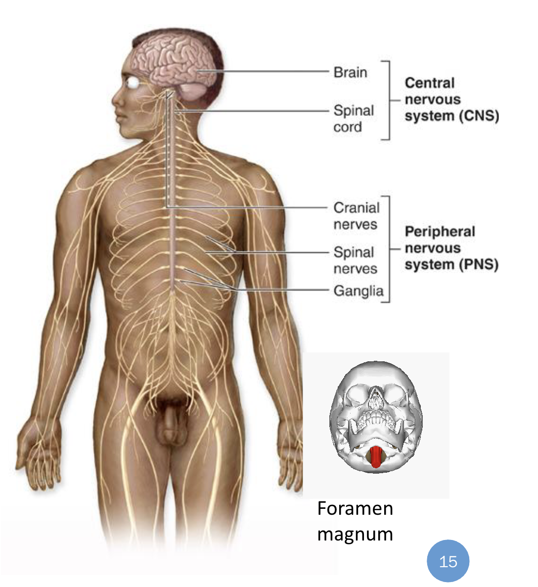
CNS: The Brain
The largest and most complex part of the nervous system. It oversees various aspects of physiology, including sensation and perception, movement, and cognition.
Consists of four divisions, each distinct in the type of input it receives and where it sends its output:
Cerebrum
Diencephalon
Cerebellum
Brainstem
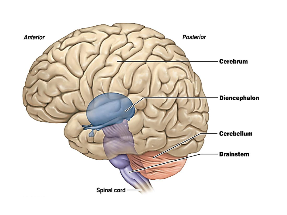
Cerebrum
Conscious thought, voluntary movement, sensation, language, memory and emotion
Diencephalon
Comprises of thalamus and hypothalamus
Relay and regulation- the thalamus relays sensory input to the cortex
The hypothalamus controls homeostasis, hormones, temperature, hunger, thirst and sleep.
Cerebellum
Coordinates movement, balance and posture, making motor activity smooth and accurate
Brainstem
Controls vital automatic functions- breathing, heart rate and blood pressure0 and connects the brain to the spinal cord
CNS: Spinal Cord
Runs through the vertebral canal Brain
Extends from the second lumbar vertebra
Regions/ Segments: Cervicalm Thoracic, Lumbar, Sacrak & Coccygeal
Diameter is not uniform
Cervical enlargement: supplies the upper limbs
Lumbar enlargement: supplies the lower limbs
Conus medullaris- inferior end tip of the spinal cord tapered between L1 and :2
Cauna equina (horse’s tail)- The collection of spinal nerves at the end of conus medullaris below L1 extends to the sacral region.
Film terminale- A fibrous band extend from the conus medullaris to the periosteum of the coccyx. Helps stabilize, fixate, and buffer the distal spinal cord
(Periosteum of the coccyx is the dense connective tissue layer covering the outer surfaces of the tailbone, providing blood supply, nerve innervation, and osteopenia cells for bone growth and repair.

Arrangement of Neurons within the CNS (Gray Matter)
Gray Matter: The dominant cell bodies aggregate (collect together) in locations found in both the brain and spinal cord, with dendrites and unmyelinated axons.
In the brain- gray matter forms the outer few millimeters of the cerebrum (cortex), and is also scattered in deeper regions (e.g., basal nuclei, thalamus)
In the spinal cord- gray matter is located internally (butterfly-shaped, containing nuclei that process information.
Key Point: Gray matter= cell bodies + dendrites + unmyelinated axons.

Arrangement of Neurons within the CNS (White Matter)
White Matter: found in both the brain and spinal cord; the area is dominated by myelinated axons, which give the appearance of white color.
Each lobe of the cerebrum contains bundles of white matter called tracts. This communication connects different parts of the brain and spinal cord.
The spinal cord contains gray matter internally, containing nuclei that process information and tracts of white matter that shuttle this information to and from the brain.
Arrangement of Neurons within the brain
Gross anatomical structures include:
Sulci: shallow grooves on the surface of the cerebrum
Gyri: elevated ridges found between sulci; together increase the surface area of the brain, maximising cerebral space (fits more brain tissue) within the limited space of the skull.
Fissures: Deep grooves found on the surface of the cerebrum
Longitudinal fissure: a long, deep groove that separates the left and right cerebral hemispheres.

Ganglia
Singular: Ganglion= a cluster of neurons cell bodies located outside the CNS (in the PNS)
Structure- mainly cell bodies, with some synapses and support cells (satellite cells)
Function- relay and process signals between the CNS and the body.
Gray matter in the brain
Superficial layer: cortex of the cerebrum contains
Deep layer: basal nuclei/ basal ganglion form the small nodules of the grey matter, which are deep within the cerebrum.
White Matter in the brain
Axons of white matter are organized into bundles, either called:
Association fibers: connect signals between adjacent gyri
Commissural fibers: the corpus callosum connects signals between the left and right cerebral hemispheres
Projection fibers: connect signals between the cerebral cortex and the lower brain or spinal centers

Arrangement of Neurons within the spinal cord
In cross-section, the gray matter of the spinal cord looks like the letter H or a butterfly.
Horns of gray matter:
Dorsal horn- receives sensory impulses from the dorsal root ganglion of the PNS
Ventral and lateral horns- carry motor impulses from the brain/ CNS
Tracts= bundles of axons in the CNS
Gray matter (deep in the spinal cord) = signal integration site
White matter (tracts) encircles the gray matter.
Tracts only transmit signals within the CNS

The Spinal Cord: Dorsal Horn
Location:
The dorsal horn is located on the posterior (dorsal) side of the spinal cord.
Divided into six laters of nuclei, which receive different types of sensory input.
Functions:
The dorsal horn is a primary processing center for somatosensory information, including touch, pressure, temperature, pain, and proprioception, contributing to deep tendon reflex pathways.
It receives inputs from primary afferent fibers via the dorsal roots of the spinal nerves and sends signals to higher brain centers and motor neurons
The spinal cord: Lateral Horn
Location:
The lateral horns is a small projection of grey matter situated between the dorsal and ventral horns, primarily found in the thoracic and upper lumbar regions (T1- L2)
Also known as the intermediolateral nucleus involved in sympathetic functions.
Function:
It contains the cell bodies of preganglionic sympathetic neurons, which are part of the autonomic nervous system. These neurons transmit to the sympathetic ganglia via the ventral roots and white rami communicantes.
( White rami communicantes: connect the ventral roots to the sympathetic chain ganglia )
Spinal cord → ventral roots → white ramus communicans → sympathetic ganglion

The spinal cord: Ventral horn
Location:
located at the anterior aspect of the spinal cord
Prominent in regions that control somatic limb/ skeletal muscle movements, such as the cervical and lumbar enlargements.
Function:
It contains the cell bodies of motor neurons that project their axons through the ventral roots to innervate skeletal muscle, facilitating voluntary and involuntary movements
The Peripheral Nervpus System (PNS)
Composed of mixed nerves and transmits information in two directions:
Afferent Nerve Fibers
Efferent Nerve Fibers
Afferent Nerve Fibers
Carry sensory nerve impulses from sensory receptors, which detect changes in the internal and external environment, to the CNS
Efferent Nerve Fibers
Convey motor nerve signals from the CNS to effectors such as muscles, organs, or glands.
The PNS: Spinal Nerves
There are 31 pairs of mixed spinal nerves.
These are. Emitted from the intervertebral foramens- which contains dendrites of sensory neurons and axons of motor neurons.
Conduct impulses necessary for sensations and voluntary/ involuntary movements
The PNS: The Dorsal Root
Also known as the posterior root, it is one of the two roots that emerge from the spinal cord.
It plays a crucial role in transmitting sensory information from the body to the central
nervous system (CNS).
• The dorsal root emerges from the posterior aspect of the spinal cord and extends to the
dorsal root ganglion (DRG), which contains the cell bodies of sensory neurons.
• The impulse further moves toward the dorsal horn of the spinal cord after synapse and
then moves to the interneuron of the spinal cord within the CNS and further carries the
impulse up to the other neurons inside the brain for integration
The PNS: The Ventral Root
Also known as the anterior root, it is a crucial component of the spinal cord's anatomy. It plays a significant role in the
motor functions of the body by facilitating the transmission of signals from the central nervous system (CNS) to muscles and other peripheral tissues.
• These roots are composed of efferent (motor) nerve fibers. The cell bodies of these motor neurons are located within the spinal cord's central gray matter.
Motor Output
Somatic nervous system
Autonomic nervous system
The somatic nervous system
The somatic nervous system is responsible for voluntary/conscious activity to the effectors, stimulating skeletal muscle.
The upper motor neurons (gray matter of the brain) of the CNS (e.g., cortex) integrate and initiate the impulse to the ventral or lateral horn and synapse with the lower motor neurons (H-shaped gray matter); the signal will be further transmitted to the effectors via the efferent nerves.
Feature | Details |
|---|---|
Sensory input | From somatic senses (skin, skeletal muscles, joints) and special senses |
Control of motor output | Voluntary control from the cerebral cortex, with contributions from basal ganglia, cerebellum, brain stem, and spinal cord |
Motor pathway | A single, myelinated somatic motor neuron — cell body in the CNS, axon extends to skeletal muscle fibers |
Neurotransmitter | Only Acetylcholine (ACh) |
Effector | Skeletal muscle |
Response | Contraction of skeletal muscle |
Autonomic Nervous System
Sympathetic nervous system
Parasympathetic nervous system
Enteric nervous system
Feature | Autonomic Innervation |
|---|---|
General function | Involuntary/subconscious control to regulate the body processes of internal organs |
Sensory input | Mainly from interoceptors; some from somatic senses and special senses |
Control of motor output | Involuntary control from hypothalamus, limbic system, brain stem, and spinal cord; limited control from cerebral cortex. (Action potential can be stimulated by the hypothalamus) |
Motor pathways | Consist of 2 motor neurons (visceral efferent fibers associated with autonomic ganglia) |
1st neuron | Myelinated preganglionic neuron — cell body is in the CNS (brain or spinal cord); axon leaves the CNS to synapse with the 2nd neuron |
2nd neuron | Unmyelinated postganglionic neuron — cell body is in the PNS (specifically in the autonomic ganglion); axon extends to the organ it serves |
Synapse location | The preganglionic axon synapses with the postganglionic neuron at the autonomic ganglion |
Neurotransmitter | Either acetylcholine (ACh) or noradrenaline (NE) |
Effectors | Smooth muscle, cardiac muscle, and glands (e.g., smooth muscle of colon) |
Responses | Contraction or relaxation of smooth muscle; increased or decreased rate and force of contraction of cardiac muscle; increased or decreased secretions of glands |
Autonomic Nervous System — Two Divisions: Compare the effects of the Parasympathetic vs. Sympathetic divisions on each target organ.
Target Organ | Parasympathetic | Sympathetic |
|---|---|---|
Pupils | Constrict | Dilate |
Salivation | Stimulate | Inhibit |
Airways | Constrict | Relax |
Heart | Slow heartbeat | Increase heartbeat |
Stomach | Stimulate activity | Inhibit activity |
Liver / Gallbladder | Inhibit glucose release; Stimulate gallbladder | Stimulate glucose release; Inhibit gallbladder |
Intestines | Stimulate activity | Inhibit activity |
Adrenal Glands | — | Secrete epinephrine & norepinephrine |
Bladder | Contract | Relax |
Reproductive System | Promote erection | Promote ejaculation |
Origin in CNS:
Parasympathetic: Craniosacral (brainstem + S2–S4)
Sympathetic: Thoracolumbar (T1–L2), via the sympathetic chain
General Mnemonic:
Parasympathetic = "Rest and Digest"
Sympathetic = "Fight or Flight"
Autonomic Nervous System — Overview: What is its general function?
General function:
Involuntary / subconscious regulation of internal body processes
Operates automatically
Keeps the body in a state of dynamic balance
Autonomic Nervous System — Overview: how do its sensory and motor components work?
Sensory (Afferent) Nerves:
Receive signals from visceral receptors that monitor internal conditions (e.g., blood pressure)
Send those signals to the CNS
Motor (Efferent) Nerves:
Convey signals from the CNS to the effectors
Effectors = smooth muscle, cardiac muscle, and glands
Effects on target tissue:
Excitation (increased activity), or
Inhibition (decreased activity)
Sensory in → CNS → Motor out → excite or inhibit → balance.
The Autonomic Nervous System:
Parasympathetic Division
The parasympathetic system sends its motor signals out from two places:
The Brain (Cranial Portion) — Signals start in the nuclei of four cranial nerves:
Nerve III
Nerve VII
Nerve IX
Nerve X
The Lower Spine (Sacral Portion) — Signals start in the gray matter of the sacral region and travel out through the spinal nerves there.
How the signal travels:
The first neuron (preganglionic) leaves the brain or spinal cord.
It synapses with a second neuron (postganglionic).
That second neuron sits on or very close to the target organ.
The 12 Cranial Nerves: What are their numbers, names, and functions (Sensory, Motor, Parasympathetic)?
12 pairs associated with the brain.
Most are mixed nerves (sensory + motor).
Two pairs are purely sensory (I and II).
Numbered I–XII from anterior to posterior.
# | Name | Sensory Function | Motor Function | PS Fibers |
|---|---|---|---|---|
I | Olfactory | Yes (smell) | No | No |
II | Optic | Yes (vision) | No | No |
III | Oculomotor | No | Yes | Yes |
IV | Trochlear | No | Yes | No |
V | Trigeminal | Yes (general sensation) | Yes | No |
VI | Abducens | No | Yes | No |
VII | Facial | Yes (taste) | Yes | Yes |
VIII | Vestibulocochlear | Yes (hearing & balance) | Some | No |
IX | Glossopharyngeal | Yes (taste) | Yes | Yes |
X | Vagus | Yes (taste) | Yes | Yes |
XI | Accessory | No | Yes | No |
XII | Hypoglossal | No | Yes | No |
*PS = Parasympathetic
Parasympathetic Responses
Enhances “rest-and-digest” activities
Promotes maintenance activities and conserves body energy
Paradoxical fear
Parasympathetic Responses: Enhances “rest-and-digest” activities
The parasympathetic nervous system (PNS) promotes bodily
functions that occur when the body is at rest, such as digestion and energy conservation.
• Normally dominates over sympathetic impulses: In non-stressful situations, the PANS is more active than the sympathetic nervous system (SNS), which is responsible for the “fight or flight” response.
Parasympathetic Responses: Promotes maintenance activities and conserves body energy
It helps in maintaining homeostasis by
conserving energy and promoting activities like digestion, repair, and growth.
• Illustrated in a person who relaxes and reads after a meal: When a person is relaxed, the PANS is
active, facilitating digestion and relaxation with SLUDD responses:
• Salivation: Increased saliva production to aid in digestion.
• Lacrimation: Production of tears to keep the eyes moist.
• Urination: Promotes the excretion of urine.
• Digestion: Enhances digestive processes.
• Defecation: Facilitates bowel movements.
Parasympathetic Responses: Paradoxical fear
When there is no escape route or no way to win, the body may experience a
massive activation of the PNS.
• This can lead to a sudden and overwhelming response.
• Leads to loss of control over urination and defecation: In extreme cases, this can result in
involuntary urination or defecation due to the intense activation of the PNS.
ANS: Pathway Features Comparisons
Neuron chain: how many neurons are connected in a row to carry the signal from the CNS to the target organ.
ANS: Anatomical Comparison
Division | Origin of Fibers | Length of Fibers | Location of Ganglia |
|---|---|---|---|
Sympathetic | Thoracolumbar region of the spinal cord | Short preganglionic, long postganglionic | Close to spinal cord |
Parasympathetic | Brain and sacral spinal cord (craniosacral) | Long preganglionic, short postganglionic | In visceral effector organs |
The Autonomic Nervous System: Sympathetic Division
Origin:
Thoracic and first two lumbar segments of the spinal cord (T1–L2)
Cell bodies are in the lateral horns of the gray matter
Path:
Preganglionic fiber leaves the spinal cord through the ventral root of the spinal nerve.
Travels down the thoracic or lumbar nerve.
Synapses at the sympathetic trunk / paravertebral ganglia (autonomic ganglia).
After the sympathetic chain:
Once the signal passes through the sympathetic chain, it takes different routes depending on where it's going.
Eventually it synapses with the second neuron (the postganglionic neuron).
From there, the postganglionic fiber carries the signal to the target organ.
These postganglionic fibers are usually much longer than the preganglionic ones.
The Sympathetic Trunk
What it is:
Also called the paravertebral ganglia or sympathetic chain
A series of ganglia running along both sides of the vertebral column
What it does:
Acts as a conduit (pathway) for preganglionic fibers
Allows those fibers to travel up and down to reach different levels of the body
Houses the paravertebral ganglia where many preganglionic fibers synapse with their second neuron
Example mentioned:
Celiac ganglion — one of the ganglia associated with the sympathetic trunk
Sympathetic Responses: What is the "fight or flight" response
What it is:
Alarm reaction → "Fight or Flight"
Prepares the body to confront or escape a threat
Also promotes adjustments during exercise
Body changes:
Change | Purpose |
|---|---|
Blood shunted to skeletal muscles & heart | More O₂ and nutrients for quick response |
Bronchioles dilate; respiratory rate increases | Enhances oxygen intake |
Liver releases glucose | Quick energy source for muscles |
Triggers — "E situations":
Emergency — accidents, natural disasters
Embarrassment — social stressors
Excitement — thrilling activities
Exercise — physical exertion
Why effects last long:
Norepinephrine (noradrenaline) lingers in the synaptic cleft at the effector
This prolongs activation of the sympathetic response