DI exam 2
 Call Kai
 Learn
Learn Practice Test
 Spaced Repetition
 Match
Match Flashcards
 Knowt Play
1/314

There's no tags or description
Looks like no tags are added yet.
Name | Mastery | Learn | Test | Matching | Spaced | Call with Kai | Chat |
|---|
No analytics yet
Send a link to your students to track their progress
315 Terms
What are the standard radiographic views for the abdomen
Left and right lateral and a VD
Should the pelvic limbs be pulled caudally for a VD or lateral of the abdomen
no, the femur should be perpendicular to the lumbar spine
Why should the pelvic limbs not be pulled caudally when radiographing the abdomen
The skin folds can create linear opacities and cause crowding of the caudal abdomen
Describe what a correct abdominal x-ray would look like
The xray would contain from the diaphragm to the urinary bladder, the vertebrae would be lined up with even disc spaces and lined up spinous processes, and the femur would be perpendicular to the lumbar spine
All abdominal xrays should contain what cranial and caudal borders
diaphragm to urinary bladder

Which image is showing correct positioning for a VD abdomen
Left
If an organ is full covered by visceral peritoneum
intraperitoneal
The kidneys are only partially covered by peritoneum making them
retroperitoneal
The abdominal cavity is continuous with what other cavity
pelvic
What species has better serosal detail due to a greater amount of fat in the abdomen
cat

Which image is feline
left
What organs are easily identifiable in an abdominal x-ray
liver, spleen, kidneys, urinary bladder, stomach, small intestine, large intestine

What organ is being outlined in the image
liver
The liver is positioned between the
diaphragm and stomach
The liver should be contained within
the margins of the rib cage
The margins of the liver should be
sharp
The position of the liver in VD depends on
the fill of the stomach

The stomach is filled with soft tissue opacity and gas in the image, how is it displacing the liver
ventrally
What part of the spleen is mobile
tail
The head of the spleen is held in place by the
gastrosplenic ligament
The head of the spleen is close to what other anatomical structure
fundus of the stomach

What is being outlined in this radiograph
tail of the spleen

What is being outlined in this radiograph
head of the spleen
The spleen is located on what side of the abdomen
mid-left
In cats the spleen is only seen in what x-ray positioning
VD
A small radioluscent opacity is seen in the center of a kidney on a feline patient. What is causing this opacity?
fat in the renal hilus
Normal kidney size in a canine
2.5-3.5x L2 body length
Normal kidney size in a feline
2.4-3x L2 body length

What is being outlined in this radiograph
kidney

Which kidney is positioned more cranially
right

What is being outlined in this x-ray
urinary bladder
What might cause gas to be seen within the urinary bladder
U-cath or cystocentesis
What species can you commonly see the urethra on x-ray
feline
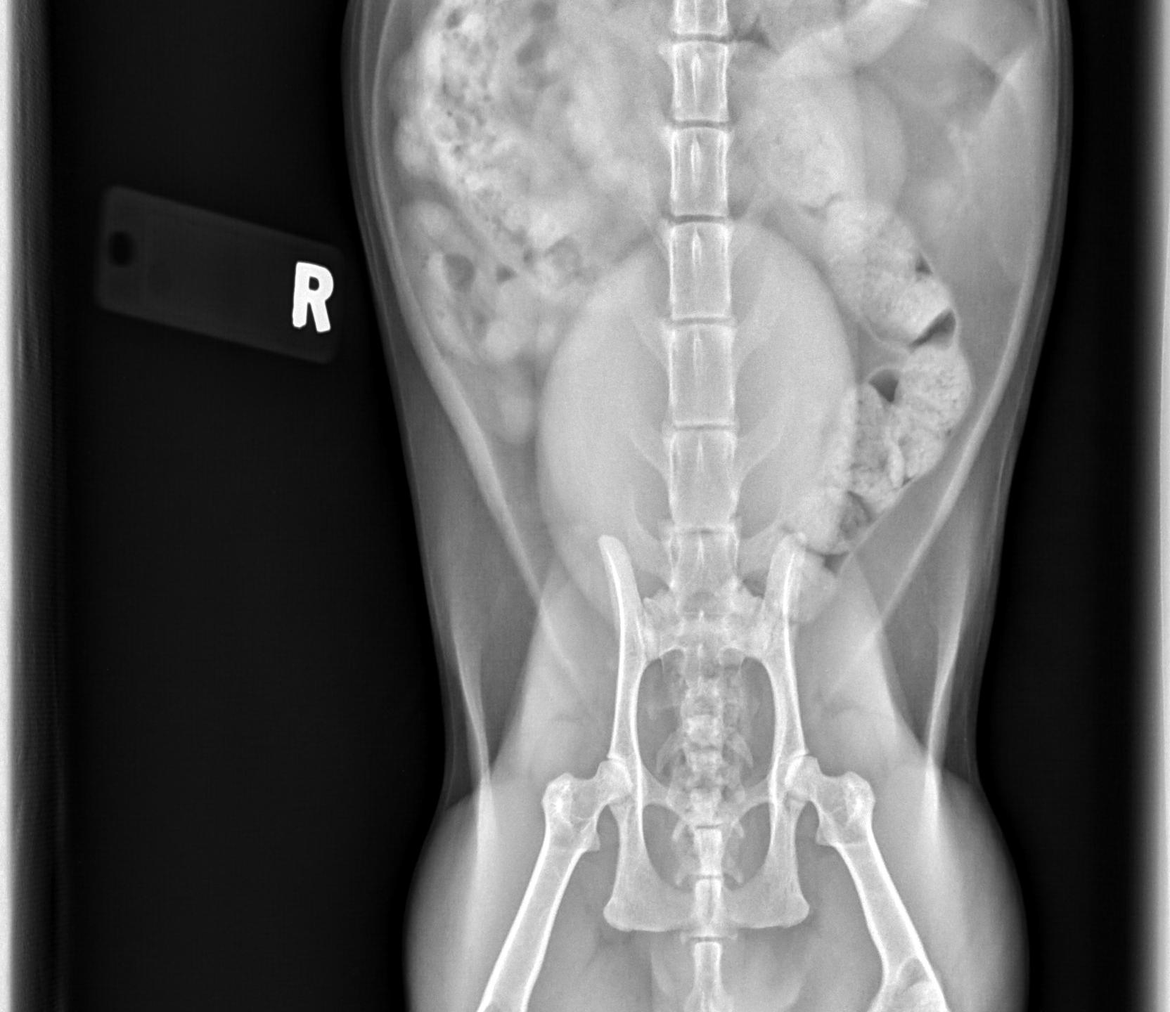
The urinary bladder is seen where in this image
ventral midline
Where is the fundus of the stomach located
dorsal and left of midline
Where is the pylorus of the stomach located
ventral and right of midline
What may shift stomach position and axis
hepatomegaly
What might be seen when the stomach has gas in it on x-ray
rugal folds
In a right lateral x-ray what part of the stomach will fill with gas
fundus
In a left lateral x-ray what part of the stomach will fill with gas
pylorus
The duodenum travels caudally on what side of the abdomen
right
Can you distinguish between the jejunum and ileum on x-rays
no
Normal SI diameter in a dog
1.5x body height of L2
Normal SI diameter in cats
12mm
Normal LI diameter in cats
<1.28x body height of L5
The descending colon is positioned on what side of the abdomen
left
The cecum appears as a gas filled structure in the right-mid abdomen. Do dogs or cats have a more developed cecum?
dogs
What species rarely has an empty LI
cats

What is causing the radioluscency in the center of the kidney
fat in the renal hilus
When is it indicated to take abdominal radiographs
acute abdominal pain, pain or distension, vomiting, diarrhea, weight loss, palpable mass, stranguria, hematuria, trauma, chemistry abnormalities suggesting liver or kidney disease
Additional abdominal radiographs
compression, horizonal beam, perineal view, special procedures
What can you use to take a compression abdominal radiograph
wooden spoon
How do you adjust mAs and kVp to increase contrast
high mAs and low kVp
Low kVp reduces what kind of scatter
Compton
When should you use the grid to minimize scatter
body depth >10 cm
Abdominal structures that are not normally visible unless enlarged or have an abnormal opacity
gallbladder, pancreas, LNs, adrenal glands, ureters, ovaries/uterus, prostate (unless intact male)
What species normally has mineralized adrenal glands
cats
Roentgen signs
size, shape, opacity, margination, location, number
Fluid has the sampe opacity as
soft tissue
Superimposition
overlapping anatomical structures or external objects in a 2D image
Border effacement
loss of clear visualization of an organ’s margin causing an inability to clearly distinguish boundaries of soft tissue structures due to contact with another organ or fluid
Summation
radiographic effect in which 2 or more superimposed structures add to the total radiopacity or radiolucency in the image
What causes summation
superimposition of 2 or more structures

This image shows generalized loss of
abdominal serosal detail
Ultrasonography is more sensitive when it ocmes to identifying fluid in the abdomen, what amount of fluid can an ultrasound detect
4 mL/kg
What amount of fluid can a radiograph detect
8 mL/kg
What are pathologic reasons for loss of serosal detail
effusion, cellular infiltration, peritonitis, carcinomatosis, mass, organomegaly, too little fat, too much fat
What are non-pathologic reasons for loss of serosal detail
age, wet fur artifact, food filled GIT, feces filled colon, full urinary bladder, gravid uterus, improper exposure

How would you describe this abdominal radiograph
pendulous abdomen with generalized peritoneal effusion

What is going on in this radiograph
normal puppy

This is an example of an xray of an overconditioned or underconditioned patient
overconditioned

This is a radiograph from an overconditioned or underconditioned patient
underconditioned

What is going on in this radiograph
Right sided cranioventral abdominal mass
Mass effect
when organs are displaced from their normal position due to a space-occupying structure
Mass effect can help localize the
origin of the mass

A: cranial
B: central
C: caudal
What organs are located in the cranial abdomen
pancreas, liver, gallbladder, stomach, duodenum
What organs are located in the central abdomen
spleen, intestines, LNs, mesentery
What organs are located in the caudal abdomen
urinary bladder, urethra, prostate, uterus, colon

This abdominal distension is most likely caused by
food bloat

What is seen in this image
splenic mass
CHANG is an acronym that can be used for
determining the type of mass
What does CHANG stand for
cyst, hematoma, abscess, neoplasia, granuloma
What can cause mass lesions
infection, inflammation, obstruction, torsion, hypertrophy, hyperplasia, hollo viscus enlargement
The retroperitoneum communicates with
mediastinum and perianal region
What organs might you see in the retroperitoneum
kidneys, adrenal glands, ureters
Causes for loss of retroperitoneal detail
hemorrhage, urine, neoplasia, abscess, foreign body granuloma
Free gas in the abdomen will increase or decrease contrast
increase
What can make it difficult to detect free gas in the abdomen
free fluid
What can cause pneumoperitoneum
post-operative, perforation of GIT, penetration of abdominal all, ruptured abscess, migrating foreign body with fistula or cutaneous communication, open skin wounds
Post-operatively you might see what in the abdomen on a radiograph that will go away overtime
gas

What is seen in these radiographs
free gas in the abdomen

Besides the GDV, wht else can be seen in these radiographs
pneumoperitoneum
Horizontal beam radiogrpahy is used for the detection of
free gas
Why do you put a patient into left lateral recumbency to take horizontal beam radiographs
so the larger part of the stomach is down and won’t fill with gas, this makes it easier to detect free gas in the abdomen
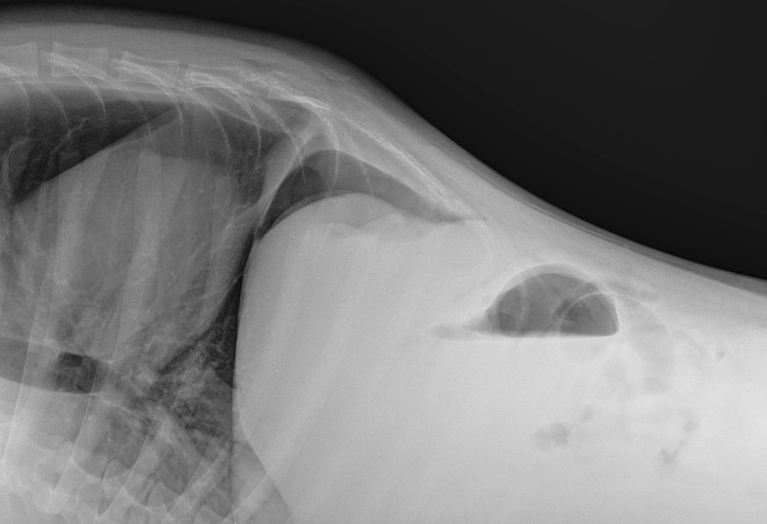
What 2 things can be seen in this horizontal beam radiograph
free gas and free fluid in the abdomen

What is seen in this x-ray
pneumoretroperitoneum
What might cause a pneumoretroperitoneum
extension of a pneumomediastinum, puncture wound, migrating foreign body with a fistula or cutaneous communication, open wound at perianal region
Generalized hepatomegaly causes what kind of displacement to the gastric axis
caudal, dorsal, left
Generalized hepatomegaly causes what displacement of the descending duodenum
left