Forebrain
 Call Kai
 Learn
 Practice Test
 Spaced Repetition
 Match
 Flashcards
 Knowt Play
1/42

There's no tags or description
Looks like no tags are added yet.
43 Terms
Diencephalon
thalamus
hypothalamus
epithalamus
subthalamus
Thalamus
largest subsection of diencephalon; collection of nuclei that selectively filter input to cerebral cortex
regulate flow if info
regulate activity level of cortical neurons
receive input from BG, cerebellum, and all sensory systems except olfactory
nuclei are divided into groups by location and function
Lateropulsion/Pusher Syndrome
lesion to posterior thalamus
pulvinar, lateral geniculate, and medial geniculate nuclei
pushing away from less paretic side
high fall risk
affects sitting, standing, walking
resistant to postural adjustment
resolves over time (-6 mo), motor recover slower
Location-based Nuclei Groups
named for their location
divided by Y-shaped lamina (white matter)
anterior, medial, and lateral groups
lateral group is further divided into dorsal/ventral


Function-based Nuclei groups: Relay nuclei
Motor: ventral anterior (VA) and ventral lateral (VL)
Sensory:
Ventral Posterolateral (VPL)- somatic sensation from body
afferent: spinothalamic tract, lemniscus pathway
efferent: somatosensory cx
Ventral Posteromedial (VPM)- somatic sensation from face
afferent: sensory nucleus, trigeminal nerve
efferent: somatosensory cx.
Medial Genticulate (MGN)- hearing
afferent: inf. colliculi
efferent: auditory cx
Lateral Genticulate (LGN)- vision
afferent: optic tract
efferent: visual cx
Lesion: interruption of ascending pathways, compromise contralateral sensation, proprioception usually affected the most
motor and sensory impairments


Functional-based: Association Nuclei (memory, sensory integration, emotion)
Anterior Nucleus (AN)- declarative memory
Lateral Dorsal (LD)- declarative memory
Midline (M)- declarative memory (hippocampus afferents, prefrontal cx efferents)
Lateral Posterior (LP)- sensory integration (reciprocal with parietal cx)
Pulvinar (P)- sensory integration (reciprocal connections with parietal, occipital, temporal cx)
Medial Group (MG or MD)- emotions (reciprocal with emotional areas)


Function-based: Non-specific Nuclei (consciousness, arousal, attention)
Intralaminar (I)- arousal, attention (ascending reticular system afferents, widespread cortical efferents)
Lesion: moderate/severe disability or vegitative state
causes: TBI, thalamic stroke, PD
Reticular (R)- adjust activity of thalamus (interconnections with other thalamic nuclei)
Midline (M)- facilitates communication between limbic structures; key role in memory and executive functions; specialized roles in visceral functions

Hypothalamus
integrates behaviors and visceral functions (eating/digestion)
homeostasis (body temp, metabolic rate, BP, water intake, excretion, digestion)
eating, reproduction, and defensive behaviors
emotional expression of pleasure, rage, fear, aversion
regulation of circadian rhythms (sleep, wake cycles)
endocrine regulation of growth, metabolism, reproductive organs
activation of sympathetic NS
regulates pituitary gland
Pituitary gland
regulates metabolism, reproduction, stress response, urine production
anterior and posterior part
pituitary hormones target the adrenal cortex, thyroid gland, ovaries/testes
Hypothalamus regulation of ant. pituitary gland
arises from outgrowth of epithelia tissue in roof of embryonic oral cavity
hormones released from hypothalamus increase/decrease hormone secretions from ant. pituitary
secretes 6 different hormones

Hypothalamus regulation of post. pituitary gland
outgrowth of inf. brain in area of hypothalamus and continuous with brain
secretions are considered neurohormones
AP in hypothalamus causes release of neurohormones from post. pituitary directly into bloodstream
Neurohormones:
antidiuretic hormone (ADH; vasopressin): maintains osmolality and volume of extracellular fluid; increases water reabsorption in kidney and prevents excretion of large amounts of urine (helps when you have low BP or dehydrated)
Oxytocin: uterus smooth muscle contractions and milk expulsion in lactation

Pituitary tumors
may compress cranial nerves 3 (oculomotor), 4 (trochlear), 6 (abducens)→ double vision, bitemporal hemianopia
hormonal hypo/hypersecretion
headaches, nausea, vomitting, irregular menses and lactation, sexual dysfunction, high BP, increased blood glucose
acromegaly- growth hormone disorder causing gigantism, enlargement of head, hands, feet
Cushing’s disease- excessive ACTH causing fatigue, abnormal deposits of facial and trunk fat
Epithalamus
Pineal gland- endocrine gland innervated by sympathetic fibers
helps regulate circadian rhythms with melatonin
influences secretions of pituitary gland, adrenals, parathyroids, and Islets of Langerhans
parathyroids- produce parathyroid hormone; maintains appropriate balance of calcium in the bloodstream and in tissues
Islets of Langerhans- cluster of cells in pancreas that produce and release hormones that regulate glucose
Subthalamus
located in sup. to substantia nigra in midbrain
facilitates BG output nuclei
involved in regulating movement
Telencephalon- Cerebral Hemisphere
most common cells in the cerebral cortex:
granule cells- small interneurons that remain in cx; process and transmit sensory info; olfactory and hippocampus
pyramidal cells- some have short axons that synapse without leaving the cx; transmit sensory and motor info, plan and execute behavior, key role in cog. processes
most travel through white matter as projection, commissural, or association fibers
75-85% of cortical neurons
projection fibers
info from subcortical structures to cerebral cx
info from cerebral cx to SC, BG, and thalamus (red lines)
travel through internal capsule
spread apart to form corona radiata

Commissural fibers
connect homologous areas of cerebral hemisphere
corpus collosum: links many areas of R and L hemisphere
ant. and post. commussure: link R and L temporal lobes

Association fibers
connect cortical regions within 1 hemisphere
short: conect adjacent gyri
long: connect lobes within a hemisphere
Long Association Fibers:
cingulum: connects frontal, parietal, and temporal lobe cx
occipitofrontal fasciculus: connects frontal, temporal, and occipital lobes
inferior longitudinal fasciculus: connects occipital and temporal-occipital areas of the brain to ant. temporal areas


internal capsule
ant. limb:
corticopontine fibers- cx and pons
frontopontine fibers- frontal lobe and pons
fibers connecting thalamic and cortical emotion/motivation areas
genu: cortical fibers projecting to CN motor nuclei and reticular formation
post. limb:
corticospinal projections
thalamocortical projections- relay somatosensory, visual, auditory, and motor info to cx
retrolenticular and sublenticular fivers- post. and inf.


Middle Cerebral Artery
lenticulostriate branches- superior and anterior portions of internal capsule
anterior choroidal artery- posterior portion of internal capsule

Brodmann’s Area
cytoarchitectural organization for cerbral cx
53 areas numbered in order
don’t need to memorize areas

Primary sensory cx (S1)
receive sensory info from ventral tier of thalamic nuclei
separate primary sensory areas for somatosensory, auditory, visual and vestibular info
lesions: impair ability to discriminate intensity and quality of stimuli (impaired senses) and conscious proprioception
Pathway:
starts with tactile and proprioceptive receptors
input received from afferent dorsal column neuron, medial lemniscus neuron, and thalamocortical neuron
crude awareness of somatosensation occurs in thalamus (VPL and VPM)
neurons in somatosensory cx identify location of stimuli and discriminate for shapes/size/texture
cortical termination of nociceptive is more widespread- not limited to primary somatosensory cx

Pathway to primary auditory cx
conscious awareness of intensity of sounds
cochlea of both ears → inf. colliculus and medial geniculate body of thalamus → cortex
auditory info has extensive bilateral projections to cortex
unilateral lesion only interferes with localization of sound
primary vestibular cx- perceives head mvmt and position
Lesion: loss of localization of sounds
Pathway to primary visual cx
specialized neurons in visual cx distinguish between light/dark, shapes, various shapes, location of objects, and movements
retina → lateral geniculate body of thalamus → primary visual cx
Lesion: contralateral homnymous hemianopia

primary sensory cx summary

Secondary sensory areas
more complex analysis of sensation and making sense of it; analyze input from thalamus and primary sensory cx
perceptual: interpretation of sensation into meaningful forms
active process; requires interaction between brain, body, environment
memory, motivation, expectations, selection of sensory info, and active search for pertinent info

Secondary somatosensory cx (brodmann area 5,7)
stereognosis: integrate tactile and proprioceptive info from manipulating an object and using memory to compare it to other objects; object identification through tactile and no visual
astereognosis: can describe object by shape, size, texture, but would not be able to recognize it through touch and manipulation
Secondary visual cx (brodmann area 18-21)
analyzes color and motion
sends info to superior colliculis→ directs visual fixation
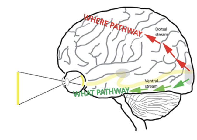
dorsal (where) action stream: adjust limb mvmt in relation to visual info
ventral (what) perceptial stream: object identification in contralateral visual field
visual agnosia: inability to visually recognize/identify objects, but able to describe shape, size, etc.
ventral stream lesion: can’t recognize object, but could reach and pick up with correct orientation of hand, fingers
dorsal stream lesion: optic ataxia; inability to direct mvmt based on visual info, but can identify and describe objects visual
prosopagnosia: unable to recognize faces but can respond/interpet emotional expressions; can recognize people from voice, mannerisms, clothes
bilateral damage to inf. secondary visual areas (ventral stream)
secondary auditory cx (brodmann area 22, 42)
compares sounds with memories of other sounds and categorizes sounds as language, music, noise, etc.
auditory agnosia: perceives but can’t recognize sound
left lesion: affects speech recognition (Wernike’s aphasia)
right lesion: interpreting environmental sounds
Motor cx Brodmann areas
primary motor cx- 4
supplementary motor area- 6
premotor area- 6
broca’s area- 44
frontal eye field- 8

primary motor area (M1)
in precentral gyrus
corticospinal and corticobulbar tracts
controls contralateral voluntary movement, particularly fine movements of hand and face
trunk and proximal limbs involve more gross motor activity and bilateral activation
Lesion: paresis, loss of fine motor control, spastic dysarthria

M1 Lesions: Dysarthria
speech disorder from spasticity or paresis of speaking muscles
Spastic dysarthria: UMN damage; harsh, awkward speech
Flaccid dysarthria” LMN (CN 9, 10, and/or 12); breathy, soft, imprecise speech
M1 Lesions: Paresis
contralateral paresis and loss of selective motor control
lower face and distal limbs affected the most
wrist and finger ext. and ankle dorsiflexion lost
M1 Lesions: Alien Hand Syndrome (Dr. Strangelove Syndrome)
involuntary, uncontrollable movement of UE
self-groping and self-oppositional behavior; extreme cases report suffocating oneself; can last days to years
damage to a variety of cortical and subcortical structures
possible that lesion to parietal cx impacts intentional planning but activation of contralateral M1
R hemisphere: use of visual feedback and sensory stimulation
L hemisphere: cognitive therapy for anxiety and anger

cortical motor planning areas
Broca’s area (L hemisphere): planning movements of mouth for speech
Broca’s aphasia: broken speech; intact comprehension but difficulty with producing speech or writing
Inferior Frontal Gyrus (corresponding area in R hemisphere): plans non-verbal communication, emotional gestures, tone of voice
Supplementary Motor Area (SMA): initiation of movement, orientation of eyes and head, planning bimanual and sequential movements
important for controlling fast, complex mvmts and anti-phase hand mvmts (R and L hand are doing different things)
Unilateral lesion causes total loss of contralateral mvmts and impairment of ipsilateral mvmt
Premotor Cx (PMC): learns goal-oriented actions, prepares, selects, initiates mvmt
Lesion: speed/automaticity of reaching/grasping, sequential mvmt, gait, posture
poorer rehab potential; persistent proximal weakness in contralateral limbs, poor motor planning

apraxia
lesions to supplementary motor area and premotor cx; difficulty with everyday mvmts and gestures
“motor agnosia”- sensation, coordination, muscle strength, and understanding of task are intact, but how to do the task is lost
consistently inconsistent; may use objects correctly in rehab but forget at home
motor perseveration
more associated with amount of neural damage than site of damage
uncontrollable repetition of movmenet
Association cx
located in frontal, anterior temporal, and temporoparietal cx
not directly involved with sensation or movement
controls behavior, interprets sensation, and processes emotions and memories
responsible for complex cognitive functions

Flow of info from primary sensory cx to motor output

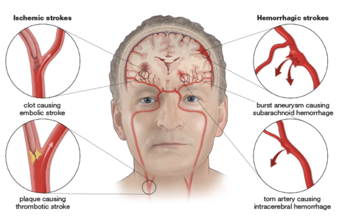
cerebral stroke
infarct: thrombus (blood clot) or embolus (any blockage) lodges in a vessel, obstructing blood flow
hemorrhage or edema can compress vital structures and affect oxygenation
tumors
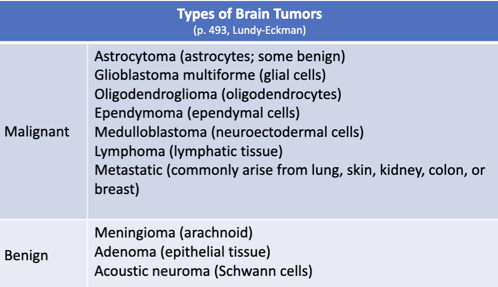
spontaneous, abnormal growth of tissue forming a mass, causing compression
symptoms based on location and size of tumor
epilepsy
sudden attacks of excessive cortical neuronal discharge interfering with brain function
involuntary movement, disruption of autonomic regulation, illusions, hallucinations
generalized seizures affect entire cx, partial affect only some areas
absence seizure: brief loss of consciousness without motor manifestation
tonic-clonic: tonic contraction of skeletal muscles followed by alternating contraction and relaxation for about 1 min. each, followed by confusion and no memory of seizure
causes: tumor, infection, stroke, TBI, neurodegeneration disease, febrile seizures
medical emergency if: cause is unknown, diabetic, injured, pregnant, lasts longer than 5 min., 2nd seizure begins, consciousness does not return, occurs in water
Basal Ganglia
functions: decision-making, judgement, prioritizing info, emotional processing and responses, learning, eye movements, spatial attention, movement
NTs:
glutamate = excitatory
GABA = inhibitory
dopamine = modulatory
striatum = head of caudate and putamen
nucleus accumbens- main component of ventral striatum
pallidum = GPe, GPi, GPe
Lentiform nucleus = putamen, GPi, GPe
subthalamic nucleus = subthalamus
substantia nigra pars compacta (dopamine)
motor, reward, motivation
substantia nigra pars reticularis (GABA)
movement
thalamus
main output to thalamus- globus pallidus internus (GBi) and substantia nigra reticularis (SNr)
