43. camelid medicine 2
 Call Kai
 Learn
 Practice Test
 Spaced Repetition
 Match
 Flashcards
 Knowt Play
1/29

There's no tags or description
Looks like no tags are added yet.
30 Terms
what are the 4 mite genera of relevance? how are they transmitted?
sarcoptes, chorioptes, psoroptes, demodex
sarcoptes scabei var auchenia and chorioptes bovis
most widespread & problematic
co-infections with both species can occur
transmission: horizontal spread by direct contact & fomites from environment
sarcoptic mange — clinical presentation
alopecia, hyperkeratosis, and severe pruritus
often begins on ventral abdomen and chest, axillae, and groin
gradual extension to medial thighs, prepuce, perineum, legs, interdigital spaces, face, and pinnae
secondary bacterial infection common in severe cases
severe sarcoptes → entire head, body, and limbs involved
cause of weight loss & decreased fiber production ± death
sarcoptic mange — diagnosis
deep skin scrapings (negative does not rule out)
mites burrow into skin
histopathology
superficial eosinophilic interstitial dermatitis, marked parakeratotic, hyperkeratosis
mites in surface keratin and crusts or within epidermis
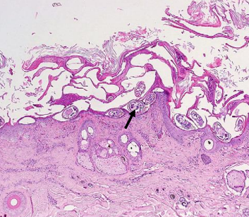
sarcoptic mange — treatment
topical lime sulfur (best, but not popular)
combined topical and parenteral ivermectin may have some efficacy
other treatments (benzene, amitraz) are not environmentally sound / approved
zoonotic risk
chorioptic mange — clinical presentation
absent or mild pruritus
first lesions usually seen on distal limbs
varying degrees of alopecia, erythema, scaling, crust, and lichenification
may extend from feet and legs to trunk, perineum, neck, ventral abdomen, and head
in crias, lesions may be restricted to tips of ears
chorioptic mange — diagnosis
superficial skin scrapings from animals with or without skin disease
sample all in group
dorsal interdigital area & axilla → best sites for scraping
mites are large and can be seen with naked eye when present in large numbers
chorioptic mange — treatment
pour on & topical ivermectins of limited efficacy
topical application of 1 mL of 1% ivermectin in 60 mL of water with 10 mL DMSO weekly — anecdotally works
lime sulfur (2%) weekly for 6-8 wks
fipronil (frontline spray) — repeated application (expensive)
not zoonotic risk
trimming fighting teeth
upper I3 + upper & lower canines
tools
obstetric wire
cutting wheel attached to hand-held rotary tool
leave 2-3 mm of tooth above gum line
complications (rare)
gingivitis
ascending infection
thermal damage of sensitive structures at root
which teeth are more likely to get tooth root abscesses?
mandibular >> maxillary
cheek teeth >> incisors or canines
which ages are affected by tooth root abscesses?
all ages, but most are present by 5 years of age (many cases are young mature animals, not aged animals)
what is the most common pathogen isolated from tooth root abscesses?
trueperella pyogenes
tooth root abscess — clinical presentation & diagnosis
hard bony swelling over affected tooth
reactive bone & osteomyelitis
degree of swelling generally indicative of chronicity of disease
± draining tract
most animals eat normally; usually without pain
± pain when eating, anorexia, weight loss possible
maxillary teeth: malodorous nasal discharge if maxillary sinus involved
definitive diagnosis: radiology or CT
tooth root abscesses — treatment
medical treatment
systemic antimicrobials (long-term)
florfenicol
penicillin
ceftiofur
anti-inflammatories
flunixin meglumine
± surgery to remove nidus of infection (tooth / tooth root)
post-op complications: reinfection, chronic draining tract, osteomyelitis
tooth root abscess — prognosis
higher mortality rate associated if animal is thin or emaciated at presentation
routine dental care
points and overgrown incisors
typically not needed; only if malocclusive problems noted
normally have some points, but should not be causing a problem
malocclusion of incisors may require careful trimming
what is the #1 cause of neurologic disease in camelids in the US?
parelaphostrongylus tenuis (deer meningeal worm)
(snail = intermediate host)
p. tenuis — clinical signs
depend on migration of parasite — CNS signs
incoordination, ataxia, hypermetria
stiffness, muscle weakness
posterior paresis, paralysis, inability to rise
head tilt, circling, or drifting gait
torticollis, arching neck
blindness
signs may be asymmetric
± cases year-round; more cases occur during grazing season
p. tenuis — diagnosis
history, clinical signs
CSF: eosinophilic pleocytosis
p. tenuis — treatment
fenbendazole + ivermectin
flunixin meglumine
± DMSO
oral vitamin D
p. tenuis — prevention
prophylactic treatment with ivermectin or fenbendazole
may be ineffective + ↑ resistance in other nematodes
attempt to deer-proof pasture with fencing (not easy!)
(molluscicide use – rarely done because of the possible environmental impact of the chemicals used)
non-parasitic causes of paresis in alpacas
trauma → vertebral fractures, cervical luxations / subluxations
young intact males using neck to fight
discospondylitis — degenerative or infectious
vertebral body abscesses / pathologic fractures
cranial / brain diseases giving vestibular or paretic signs
severe otitis, listeriosis, polioencephalomalacia, brain abscesses, viral meningocencephalitis (WNV, EHV, EEE)
rye grass staggers, botulism
potential causes of colic in camelids
GI
ulcers, forestomach obstruction, C1 impaction, intestinal obstruction, peritonitis (often from C3 ulcer rupture; sepsis)
urogenital
uterine torsion, dystocia, urolithiasis, UTI
signs of abdominal discomfort — young camelids (< 6 months)
more subtle
lethargy or depression; lying in abnormal positions (ex. lateral)
(kicking at abdomen, rolling, thrashing — rare)
signs of abdominal discomfort — mature animals
restlessness: active colic
getting up & down
frequent stretching of legs
rolling from sternal to lateral recumbency & back, with legs extended
kicking at abdomen
indications for GI surgery
continuous & intractable pain → poorly or only briefly responsive to NSAIDs or alpha agonists (ex. xylazine) / opioids
persistent low-grade pain despite supportive therapy
persistent or increasing abdominal distention
U/S or radiography demonstrate highly suspect surgical lesion
abnormal peritoneal fluid
failure to pass feces for >24 hrs
failure to urinate for 6-8 hrs
heat stress — clinical signs
muscle exertion exacerbates / accelerates pathology
dehydration & multiple organ dysfunction can be rapid
hypophagia, weakness, ataxia, myopathy, neurologic signs
recumbency, CV collapse, scrotal edema (negative effects on fertility)
heat stress — treatment
cool environment
clip
increase heat loss, fan
cool oral and IV fluids
cool enema / ice
correct acid/base & electrolyte abnormalities
heat stress — prevention
pasture during cool part of day
shade
fans in barn
clip
BVDV — disease in camelids
alpacas primarily affected; rarely reported in llamas
PI condition in crias due to in utero exposure during early gestation (very similar to cattle)
unthrifty, chronic poor doers, usually die as juveniles with mucosal disease-like signs
may survive and reproduce (like cattle)
transient infection of immunocompetent animals also reported with BVDV-like signs
BVDV — screening in camelids
can screen for PI via PCR of EDTA blood samples in juveniles → repeat test positives 3 wks later to r/o transient infection
can screen herd by PCR also