Exam 1: Chapter 5
 Call Kai
 Learn
Learn Practice Test
 Spaced Repetition
 Match
Match Flashcards
 Knowt Play
1/43

There's no tags or description
Looks like no tags are added yet.
Name | Mastery | Learn | Test | Matching | Spaced | Call with Kai | Chat |
|---|
No analytics yet
Send a link to your students to track their progress
44 Terms
Integument system
The largest system of body
Sixteen percent of body weight
Made up of two parts
Cutaneous membrane (skin)
Outer epidermis (superficial epithelium)
Inner dermis (connective tissue)
Hypodermis (subcataneous layer)
Accessory structures (originate in the dermis)
Extend through the epidermis to skin surface
Hair
Nails
Multicellular exocrine glands
Functions of skin (100%)
PROTECTION
Production of keratin (make)
Synthesis of vitamin D3
Storage of lipids
Detection of touch, pressure, pain and temperature (sensory)
Connections
Cardiovascular system
Blood vessels in the dermis
Nervous
Sensory receptors for pain, touch, and temperature
Cells of epidermis
Keratinocytes
Contain large amounts of keratin
Are the most abundant cells in the epidermis
Epidermis: Thin and Thick skin
Thin skin (missing stratum lucidum)
Covers most of the body
Has four layers of keratinocytes
Thick skin
Covers the palms of the hands and soles of the feet
Has five layers of keratinocytes
Epidermis: Stratum Basale (germanous) and cells
The deepest layer, is attached to basement membrane by hemidesmosomes
Forms a strong bond between epidermis and dermis
Forms epidermal ridges (project downward towards dermis) (basis of fingerprints)
Dermal papillae (project upward towards epidermis) (tiny mounds)
Cells
Merkel
Found in hairless skin
Respond to touch receptors (trigger nervous system)
Melanocytes
Contain the pigment melanin
Scattered throughout stratum basale
Epidermis: Stratum spinosum
The “spiny layer”
Produced by division of stratum basale
Eight to ten layers of keratinocytes bound by desmosomes
Cells shrinks until cytoskeletons stick out (spiny)
Epidermis: Stratum granulosum and cells
The “grainy layer”
Stops dividing, starts producing: Keratin
A tough, fibrous protein
Makes up hair and nails
Mitosis starts here
Cells
Produce protein fibers
Where cells dehydrate and die
Epidermis: Stratum lucidum
The “clear layer”
Found only in thick skin
Covers stratum granulosum
Epidermis: Stratum corneum
The “horn layer”, most superficial layer
Exposed surface of skin
15-30 layers of keratinized cells
Water resistant, not water proof
Shed and replaced every two weeks
Keratinization
The formation of a layer of dead, protective cells filled with keratin
Occurs on all exposed skin surfaces except eyes
Skin life cycle
Perspiration (100%)
Insensible perspiration
Interstitial fluid lost by evaporation through the stratum corneum. Not perceived by the body, submerged in salt water (hypertonic solution)
Sensible perspiration
Water excreted by sweat glands
Dehydration
Perceived by the body, evidence by sweating
If a person is this we want to give isotonic solution, will prevent water in and out (equilibrium) (never give hypertonic solution, 350 miliosmos)
Pigments for skin: Carotene
Orange-yellow pigment
Found in orange vegetables
Accumulates in epidermal cells and fatty issues of the dermis
Pigments for skin: Melanin and functions
Yellow-brown or black pigment
Produced by melanocytes in stratum basale
Functions
Melanin protects skin from sun damage
Ultraviolet (UV) radiation
Causes DNA mutations and burns that lead to cancer and wrinkles
How to determine cancer and the two types
A - Asymmetry of border (is the border different?)
B - Border (are there any irregulars?)
C - Color (is the color even?)
D - Diameter (is it larger than 6mm/quarter of an inch)
E - Evolving (did it get bigger over time?)
Two types
UVA causes cancer
UVB burns, can cause cancer
Cancer: Basal cell carcinoma
Most common type
Looks benign but not
Does not follow ABCDE
Light pink, pearly bumps
Found in sun exposed skin
Treatment is Mohs surgery or cyro therapy
Survival rate if caught early is 99%
Squamous cell carcinoma
2nd most common
Follows ABCDE
Has a different color and crusts over
UV exposure and tanning beds
Found in people with fair skin
Treatment is Mohs surgery or cyro therapy
Survival rate if caught early is 99%
Melanoma
Follows ABCDE
Tanning beds and UV radiation
If caught before it metastasizes its a 99% survival rate, if not 5 year survivable rate only at 35%
Hard to see in dark skin
Capillaries and skin color
Oxygenated red blood contributes to skin color
Blood vessels dilate from heat, skin reddens
Blood flow decreases, skin pales
Cyanosis
Blush skin tint
Caused by severe reduction in blood flow or oxygenation
Lack of oxygen to the skin
Illnesses and skin color
Buildup of bile produced by liver
Yellow color
Pituitary tumor
Excess MSH
Addison’s disease
A disease of the pituitary gland
Skin darkening
Vitiligo
Loss of melanocytes
Loss of color
More noticeable on dark skin (assumed to be an autoimmune reaction)
Vitamin D3 (100%)
Epidermal cells produce cholecalciferol (vitamin D3)
In the presence of UV radiation
Liver and kidneys convert vitamin D3 into calcitriol
Insufficient vitamin D3 → causes rickets
The dermis
Located between epidermis and subcutaneous layer
Anchors epidermal accessory structures (hair follicles, sweat glands)
Consist of two components
Outer papillary layer (areolar tissue)
Deep reticular layer
Layers of the dermis: Papillary and Reticular layer
Papillary
Consists of areolar tissue
Contains smaller capillaries, lymphatics, and sensory neurons
Has dermal papillae projecting between epidermal ridges
Reticular
The deeper layer of the dermis
Dermatitis
An inflammation of the papillary layer
Caused by infection, radiation, mechanical irritation, or chemicals
Atopic dermatitis (eczema) - IGE reaction (immune hypersensitivity)
IGG reaction id delayed hypersensitivity
Skin damage: Sagging and wrinkles
Sagging and wrinkles (reduced skin elasticity)
Caused by
Dehydration
Age
Hormonal
UV exposure
The Hypodermis
The deepest layer
Stabilizes the skin
Made of elastic areolar and adipose tissues
The site of subcutaneous injections using hypodermic needles
Deposits of subcutaneous fat
Distribution patterns determined by hormones
Reduced by cosmetic liposuction
Hair in the human body
Palms
Soles
Lips
Portions of external genitalia
Functions of hair
Protects and insulates
Guards opening against particles and insects
Is sensitive to very light touch
Structures of hair
Hair follicle
Located deep in dermis
Produces nonliving hairs
Arrector pili
Always next to hair follicle
Involuntary smooth muscle
Causes hairs to stand up
Produces “goosebumps”
Sebaceous glands
Lubricate the hair
Control bacteria
Always next to a hair follicle

Regions of hair
Hair root
Lower part of the hair
Attached to the integument
Hair shaft
Upper part of the hair
Not attached to the integument
Bulb
Where the arteries and veins are
Hair production and color
Begins at the base of a hair follicle, deep in the dermis
The hair papilla contains capillaries and nerves
The hair bulb produces hair matrix
Color
Produced by melanocytes at the hair papilla
Determined by genes
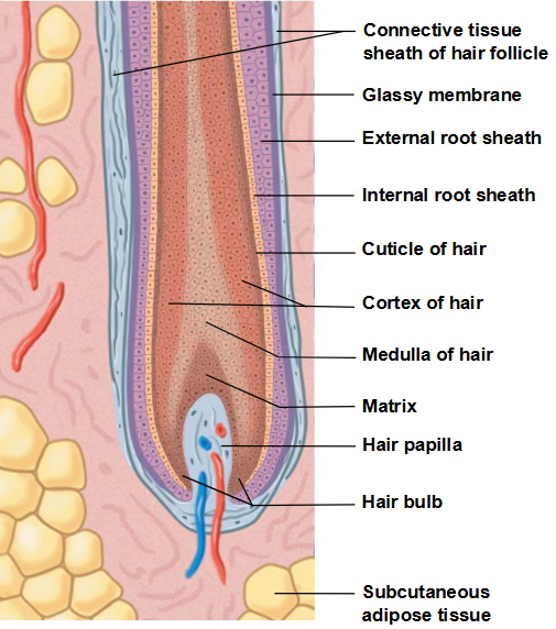
Hair shaft structures
Medulla
The central core (deepest part)
Cortex
The middle layer (outer part)
Cuticle
The surface layer
Keratin
As hair is produced, it is keratinized
Medulla contains flexible soft keratin
Cortex and cuticle contain stiff hard keratin
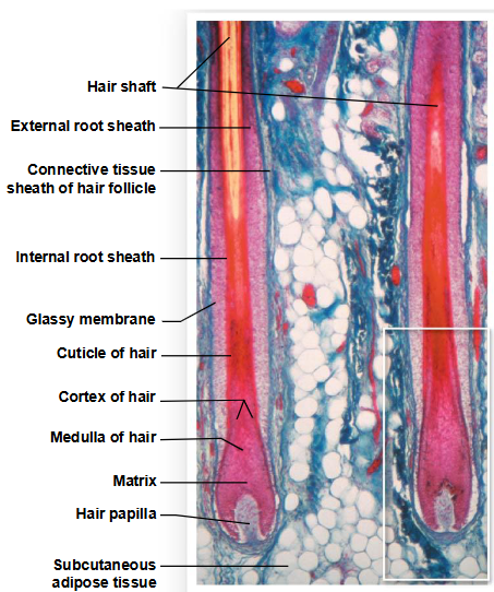
Layers in the follicle
Internal root sheath
The inner layer
Contacts the cuticle in lower hair root
External root sheath
Extends from skin surface to hair matrix
Glassy membrane
A dense connective tissue sheath
Contacts connective tissues of dermis
Hair growth cycle
Growing hair
Firmly attached to matrix
Club hair
Not growing
Is attached to an inactive follicle
Vellus hairs
Soft, fine
Cover body surface
Terminal hairs
Heavy pigmented
Head, eyebrows, and eyelashes
Other parts of body after puberty

Exocrine glands: Sebaceous glands
Oil glands
Its a holocrine gland
Types
Simple branched alveolar glands - associated with hair follicles
Sebaceous follicles - discharge directly onto skin
Sebum
Contains lipids and other ingredients
Lubricates and protects the epidermis
Inhibits bacteria
Sweat glands: Apocrine glands
Found in armpits, around nippl*s, and groin
Secrete products into hair follicles
Break down and causes odor, increase after puberty
Sweat glands: Merocrine (eccrine) glands
Widely distributed on body surface
Especially on palms and soles
Discharge directly onto skin surface
Sensible perspiration
Functions
Cools skin
Excretes water and electrolytes
Flushes microorganisms and harmful chemicals from skin
Other integumentary glands
Mammary glands (over breast)
Produce milk
Ceruminous glands
Produce cerumen (earwax)
Protect the eardrum
Nails and nail production
Protects fingers and toes
Made of dead cells packed with keratin
Metabolic disorders can change nail structure
Nail production
Occurs in a deep epidermal fold near the bone called the nail root
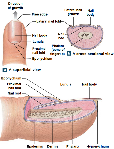
Structure of a nail
Nail body
The visible portion of the nail
Covers the nail bed
Lunula
The pale crescent at the base of the nail
Sides of nails
Lie in lateral nail grooves
Surrounded by lateral nail folds
Skin beneath the distal free edge of the nail
Is the hyponychium (onyx=nail)
Visible nail emerges
From the eponychium (cuticle)
At the tip of the proximal nail fold
Repair of the skin
Bleeding occurs
Mast cells trigger inflammatory response
A scab stabilizes and protects the area
Germinative cells migrate around the wound
Macrophages clean the area
Fibroblasts produce scar tissue
Inflammation decreases, clot disintegrates
Fibroblasts strengthen scar tissue
A raised keloid may form
The rules of the 9s