Cardio microanatomy (lectures 1 and 2)
 Call Kai
 Learn
 Practice Test
 Spaced Repetition
 Match
 Flashcards
 Knowt Play
1/115

There's no tags or description
Looks like no tags are added yet.
116 Terms
structure of vessel wall
vessels larger than capillaries have 3 concentric layers, intima, media, and externa
tunica interna
endothelium and its BM with small amount of underlying CT, internal elaxtic membrane, has gaps to allow for communication of endothelium
tunica media
smooth muscle arranged in curcular patter, thickness varies, has collagen fibers and related CT matrix, may have elastic laminae, external elastic membrane seen in larger vessels
tunica externa
type 1 collagen and elastic fivers, blends into surrounding CT, has small nerves and vessels, may contain smooth muscle
elastic vessels
conduits for moving large volumes of fluid, tunica media is modified to contain many layers of fenestrated elastic laminae, making these arteries “stretchy”, maintains even flow and constant bp between heart beats
muscular vessels
axillary and femoral arteries, walls will contract and expand to distribute blood, tunica media is modified to contain many layers of smooth muscle, regulate volume delivery in response to peripheral demand
arterioles
resistance vessels, tunica interna present with thin internal elastic membrane, smallest arteries with reduced layers of smooth muscle, controls distribution of blood to capillaries through vasoconstriction and dilation
capillaries
are endothelial tubes that consist of endothelium and BM, no tunica media, 3 basic types (continuous, fenestrated, sinusoidal)
lymphatic capillaries
very irregular shape, have valves, appear similar to venules but should have proteinaceous lymph fluid residue in the lumen
venules
post-capillariy in position, similar in size to capillaries but larger in diameter, thin tunica media
small to medium veins
constitute most of the anatomically named veins, smooth muscle in tunica media, but always less than in companion artery, collagen and elastic fibers in adventtia
large veins
may have internal elastic membrane, less smooth muscle than companion artery
endocardium
inner layer of heart, continuous with the tunica intima of vessels, comprised of endothelial lining and subendothelial CT, heart valves are endocardial folds
myocardium
middle layer and thickest layer, continuous with tunica media of vessels, cardiac muscle, CT, and purkinje cells and fibers
epicardium
pericardium, outer layer, continuous with the tunica externa of vessels, serous membrane
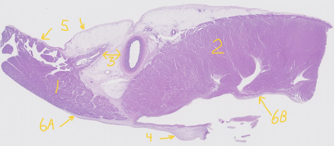
what is number 1?
atrium
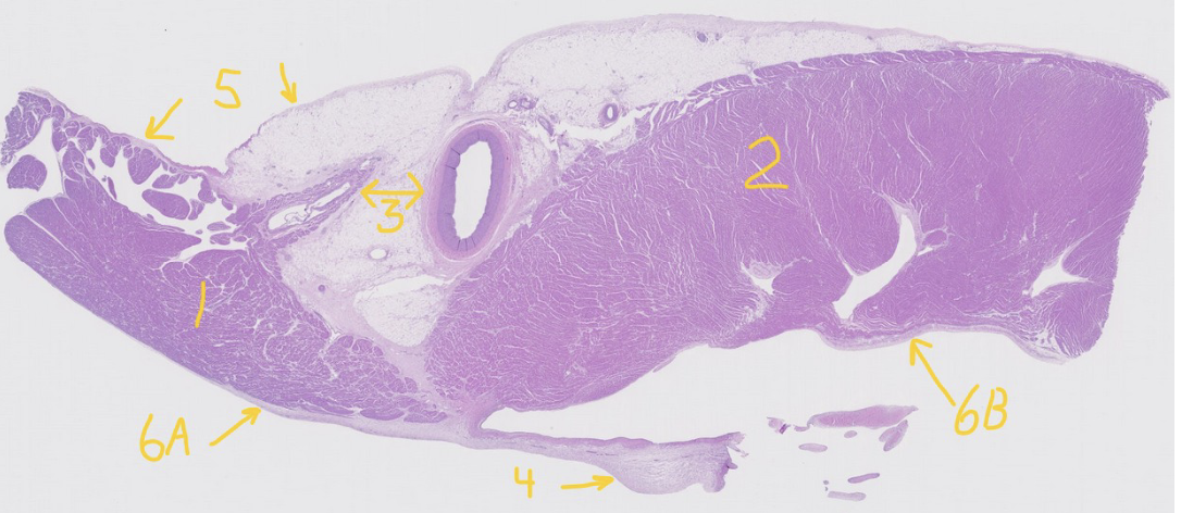
what is number 2?
ventricle
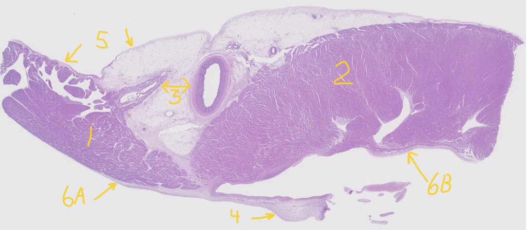
what is number 3?
coronary vessels (artery on right and vein on left)

what is number 4?
atrioventricular valve (AV)

what is number 5?
epicardium/pericardium
what term do we use when referencing simple squamous epithelium lining the outer surface of the heart?
mesothelium

what is 6a and 6b?
endocardium/endothelium

what part of the heart is 6b?
ventricle
what kind of fibers are in the ventricle?
purkinje

is this a muscular or elastic artery?
muscular

what is number 1?
tunica interna

what is number 2?
tunica media

what is number 3?
tunica externa

what is the pink line running through the tunica interna?
internal elastic membrane

is this vessel a muscular artery or an elastic artery?
elastic

is this elastic or muscular artery?
elastic

what is 1?
artery

what is 2?
vein
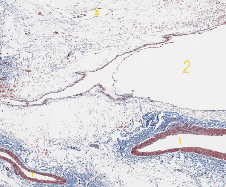
what is 3?
lymphatic vessel
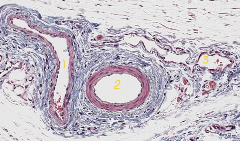
what is 1?
vein

what is 2?
artery

what is 3?
lymphatic vessel

what are the yellow arrows pointing at?
capillaries
hilus
indentation where blood vessels enter and exit the node, and where efferent lymphatic vessels exit
parts of stroma
capsule, trebeculae, reticular CT network
capsule
dense irregular CT
trabeculae
continuous with the capsule, penetrate the parenchyma, may contain smooth muscle
reticular CT network
reticular cells and their secreted fibers (type 2 collagen), supportive scaffolding for lymphocytes
modular cortex
mostly B-lymphocytes, have primary and secondary follicles
primary follicle (nodule)
low level of any mitotic activity, unstimulated or resting condition
secondary follicle
high level of mitotic activity, centrally located germinal center surrounded by mantle
germinal center of secondary follicle
clonally dividing B lymphocytes
mantle of secondary follicle
non-proliferating B lymphocytes
diffuse paracortex
diffuse lymphoid tissue, surrounds cortical follicles and separates cortex fromm medulla, lymphocytes found in region are mostly T cells, venules, HEV
HEV
high endothelial venules, where lymphocytes exit the blood circulatory system and enter the paracortex
medulla
divided into the medullary cords and sinuses
medullary cords
rich in plasma cells, T and B lymphocytes and macrophages
medullary sinuses
part of sinus system of the lymph node
sinuses of lymph node
channels through which lymph fluid flows, less densely populated with lymphocytes, reticular cells form structural scaffolding, lymph fluid flows
subscapular sinuses
less cellular spaces deep to the capsule
cortical sinuses
less cellular spaces within diffuse paracortex that tend to be around trabeculae, connect the subscapular to medullary
medullary sinuses
less cellular spaces within the medulla, drain the different lymphatic vessel at the hilus of node
lymph flow in node
lymph enters from afferent lymphatic
flows through subscapular sinus
flows through cortical sinus
flows through medullary sinus
flows out of node through efferent lymphatics at hilus
spleen
largest of lymphoid organs, only organ specialized in filtering blood
hilus of spleen
long indentation where splenic blood vessels enter and exit
capsule of spleen stroma
dense irregular CT, varying amount of smooth muscle
trabeculae of spleen stroma
continuous wit hthe capsule, trabecular artery here, varying amount of smooth muscle
reticular CT network of spleen stroma
reticular cells and their secreted fibers (type 3 collagen), supportive scaffolding for red and white pulp
white pulp
refers to both diffuse and nodular lymphoid tissue, periarterial lymphatic sheath and splenic follicles
periarterial lymphatic sheath (PALS)
diffuse lymphoid tissue surrounding central arteries, mainly T cells but can include APCs (macrophage and dendritic)
splenic follicles
lymphatic follicles, dense accumulation aggregates of B-cells located in margins of PALS, may have germinal centers, and receive blood from branches of central artery (nodular artery)
red pulp
splenic sinuses and cords
splenic sinuses
lined by endothelium with discontinuous BM, RBCs must be flexible enough to pass from cords into sinuses, if inflexible become trapped and are phagocytized by macrophages which recycle iron from hemeglobin
splenic cords
located between splenic sinuses, highly cellular, contain macrophages filled with hemosiderin deposits
hemosiderin deposits
storage form of iron derived from the breakdown of red blood cells
marginal zone
located between red and white pulp, important site for antigen trapping
sequence of flow in spleen
splenic artery
trabecular artery
central artery
various arterioles
capillaries
splenic cords or sinuses
trabecular veins
splenic veins
function of spleen
immunological defense via filtration of blood for detection of pathogens, temporary storage of RBCs, removal of old RBCs by macrophages and recycle iron
MALT
mucosal associated lymphoid tissue, common in most mucous membranes, tonsils, BALTs GALTs,
tonsils
aggregates of lymphoid tissue nodules in the pharynx
palantine tonsil
mucosa of oropharynx, lined by stratified squamous epithelium, composed of both diffuse and nodular lymphoid tissue
tonsillar crypts
some species have theirs, indentations of epithelium into lymphoid tissue to increase surface area
BALT
bronchus associated lymphoid tissue, aggregates of nodular and diffuse lymphoid tissue in the bronchi and bronchioles
GALT
gut associated lymphoid tissues, diffuse and nodular lymphoid tissue in GI tube
peyers patches
lymphoid tissue found in lamina propria of small intestine

what type of immune organ is this?
lymph node, secondary lymphoid organ

what part of the lymph node is 1?
medulla

what part of lymph node is 2?
cortex

what are the yellow arrows pointing to?
capsule

what are the blue arrows pointing to?
trabeculae

what is the yellow circle?
subscapular sinus

what is the green circle?
cortical sinus

what CT is encircled in yellow?
dense irregular

what is the scaffold-forming cell encircled in green?
reticular cell

what is the cortical structure encircled in orange?
follicle/nodule

what is 1
medullary sinus

what is 2?
medullary cords

what is 3?
blood vessel
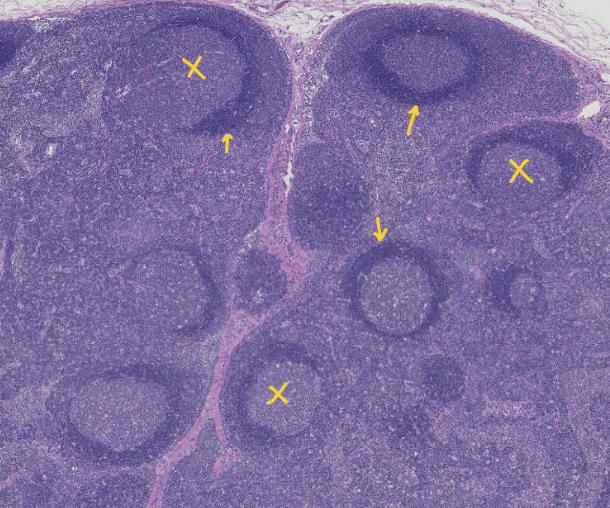
what is indicated by the X
germinal centers
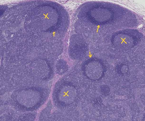
what are the arrows pointing at?
mantles

what lymphocyte type is mainly in the orange circle?
B cells concentrated in the nodules

what lymphocytes are in the green circle
T cells, concentrated in the diffuse paracortex

what structure is indicated by the black circle?
white pulp

what structure is indicated by the orange X?
red pulp