chap 3 (protein structure and function)
 Call Kai
 Learn
Learn Practice Test
 Spaced Repetition
 Match
Match Flashcards
 Knowt Play
1/50

There's no tags or description
Looks like no tags are added yet.
Name | Mastery | Learn | Test | Matching | Spaced | Call with Kai | Chat |
|---|
No analytics yet
Send a link to your students to track their progress
51 Terms
components of an amino acid
central carbon, carboxyl group, amino group, R side chain
ph 7: both amino and carboxyl groups are ionized —> neutral charge

basic side chains
positive charge at physiological pH
histidine, arginine, lysine (HRK)
acidic side chains
negative charge at physiological pH
aspartic acid, glutamic acid (DE)
nonpolar side chains
alanine, valine, methionine, tryptophan, leucine, isoleucine, glycine, cysteine, proline, phenylalanine
(AVMWLIGCPF)
2 cysteine side chains form disulfide bonds
uncharged polar side chains
neutral charge at physiological pH
asparagine, glutamine, serine, threonine, tyrosine (NQSTY)
where are there steric limitations?
on the bond angles in a protein
there is flexibility, but there is also steric hinderance
3 types of NONcovalent bonds to help proteins fold
electrostatic attractions - b/w positive and negative charges of side chains
hydrogen bonds - between H to N,O,F of side chains
van der Waals - b/w nonpolar side chains
protein 3D conformation
hydrophobic core (nonpolar side chains) + hydrophilic exterior (polar side chains form bonds with water)
denature + renature (reversible) based on temperature, pH, ionic solvents
what kind of structure (1, 2, 3, 4) involved R-group bonding?
tertiary
beta sheet structures
anti-parallel
parallel

coiled coil
multiple alpha helices
proline + glycine common

protein domains
modular units from which larger proteins are built
form from a singular polypeptide domain
a conserved part of a given protein sequence (w/ its own tertiary structure)
can function independently from the rest of the protein
*can live on its own if separate domains broken apart*
domain shuffling
some protein domains found in many different proteins
—> even with different function (evolutionary history)

three common protein domains
immunoglobulin module
fibronectin type 3 module
kringle module

increasing complexity… (in terms of number of domains)
= increasing domains
≠ increasing genome size
quarternary structure
larger protein molecule often have MORE THAN ONE polypeptide chain
each subunit = individual polypeptide chain
example: heme

some globular proteins form long helical filaments
dimer —> tetramers —> octamers
ex: vementin, actin

many protein molecules have what kind of shape?
elongated, fibrous shapes

covalent cross-linkages stabilize extracellular proteins
interchain disulfide bond = b/w 2 chains
intrachain disulfide bond = w/in 1 chain
oxidation —> 2 cysteines bond —> a disulfide bond
reduction —> a disulfide bond breaks into 2 cysteines
individual protein molecules often serve as subunits for the assembly of larger structures
ex: protein coat of virus, tubes (microtubules), sheets

proinsulin (inactive form) —> insulin (active form
specific folding stabilized by INTRAchain disulfide bonds
connecting peptide removed —> 2 chain insulin molecule
reduction irreversibly separates 2 chains

amyloid fibrils
form from many proteins —> lead to disease
many copies of the protein stack together (beta strand of each line up) held together by hydrogen bonds —> hydrophobic zipper —> amyloid fibril
2 monomers of alpha-synnuclei pair up

prion diseases
neurodegenerative diseases
sheep: scrapie
humans: creutzfeld jacob, kuru
cattle: bovine spongiform encephalopathy (mad cow disease)
—> can lead to alzheimer’s disease
amyloid structures can perform useful functions in cells
help in release of soluble peptide hormones (glucagon + calcitonin) in mammals
create bacteria biofilms (protection from attacks) to protect individual bacteria
cAMP is…
a ligand
—> has to bind to an active site
surface conformation of a protein determines its chemistry and binding
3 ways a protein can find to each other:
surface-string
helix-helix
surface-surface

equilibrium constant (K)
measures binding strength
depending on how well the surfaces of 2 molecules match, it will change the strength of the bonds that they can form
dissociation rate, association rate, and equilibrium constant equations

relationship b/w standard free-energy differences (°ΔG) and equilibrium constants (37°C)
larger value = stronger binding b/w A and B = binding more favorable
for every ↓ 5.9kJ °ΔG = ↑ K * 10

general enzymes

enzyme kinetics graph + value definitions
Vmax = maximum value that can be reached
all substrate binding sites on enzyme are occupied
Km = substrate concentration at which reaction is 0.5Vmax
measures how tightly substrate binds to enzyme

enzymes speed up chemical reactions by lowering the activation energy (graph)
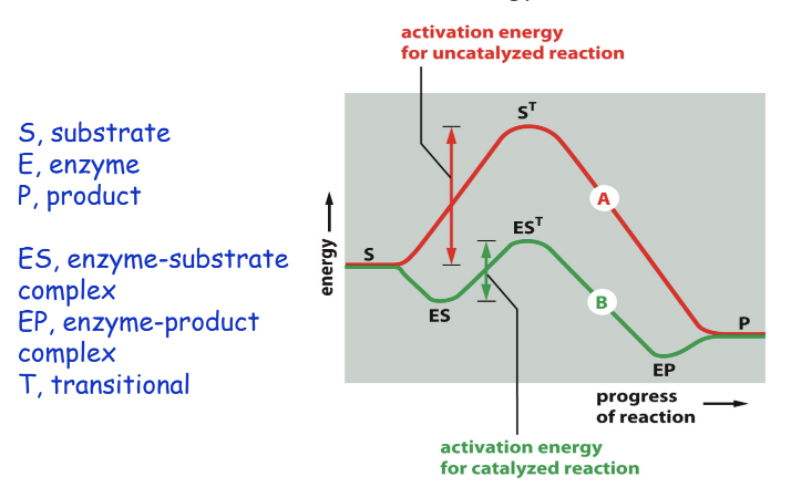
enzymes can use simultaneous acid and base catalysis

reaction catalyzed by lysozyme
catalyzes cutting of a polysaccharide chain
vitamin derivatives function as…
coenzymes
tightly bound small molecules…
add extra functions to proteins
ex: retinal - light sensitive molecule attached to rhodopsin in the eye
ex: heme - tightly bound to each of the 4 polypeptides of hemoglobin
the cell regulates the catalytic activities of its enzymes through…
positive and negative feedback
positive feedback - the binding of one molecule affects binding of another by changing the 3D structure of the enzyme
negative feedback - binding of either ligand interferes with binding of the other through change in 3D structure
enzyme activity vs. conc. inhibitory ligand (graph)
↑ complexity = ↓ inhibitor concentration needed

cooperative allosteric transition…
enzyme that has 2 identical subunits
disrupting an already disrupted enzyme = easier

kinases and phosphatases
kinases - phosphorylate —> usually activate
phosphatases - dephosphorylate —> usually inactivate
plays a big role in signal transduction pathways
—> regulates acitivation of genes —> regulates proteins being made
^^similar —> descended from a common ancestor
a protein can function as a microprocessor
ex: regulation of the src protein kinase

how does src protein kinase act as a signal integrating device?
integrates several inputs to decide whether its kinase activity should fully turn on

GTP-binding proteins (GTPases)…
act as molecular switches that activate (P) + inactivate (de-P)
ex: ras protein
2 major signaling intracellular pathways in eukaryotic cells
GEF - guanine nucleotide exchange factor
GAP - GTPase activating protein

ubiquitilation
when proteins can be regulated by the covalent addition of other proteins
ubiquitin - small 76aa protein that is added to other proteins by ubiquitin ligases
—> regulates their activity

ubiquitinylated proteins are often marked for… which amino acid does ubiquitin bind to…
proteosomal degradation
lysine side chain of target protein
names of ubiquitination enzymes (E1-E3)
E1 (ubiquitin-activating enzyme)
—> ubiquitin binds
—> E2 (ubiquitin-conjugating enzyme) binds
—> ubiquitin transfers from E1 to E2
—> E2 + E3 (ubiquitin ligase) moves ubiquitin from E2 to amino group of lysine side chain on target protein

motor proteins produce large movements in cells (image)

intrinsically disordered protein sequences = versatile binding sites

bringing molecules close together helps them interact more efficiently
scaffold proteins - a platform that holds proteins near each other
multienzyme complexes - an assembly line
biomolecular condensates - concentrated gathering of moelcules
computationally designed proteins
designed so that each protein would move into a specific 3D conformation in its lowest energy state