Methods of Data Acquisition-Part 1
 Call Kai
 Learn
 Practice Test
 Spaced Repetition
 Match
 Flashcards
 Knowt Play
1/42

There's no tags or description
Looks like no tags are added yet.
43 Terms
Name the 3 methods of data acquisition
Preliminary (Localizer) Scanning
Axial Scanning
Helical (Spiral Scanning)
Are localizers cross-sectional?
They are NOT cross-sectional in nature
How do localizer scans compare to conventional radiography?
They are similar to conventional radiography
Localizer scans have slightly poorer/worse image quality compared to X-ray
Localizer scans deliver approximately equal radiation dose to the patient as X-ray
What tube positions acquire which localizer?
The X-ray tube does not rotate during their acquisition
Position of the X-ray tube determines orientation of the image
Example: if the tube is positioned above the patient. the resulting localizer scan will be an AP view whereas positioning the tube to the side of the patient will result in a lateral view
How many localizers are normally taken?
In all routine studies, at least 1 localizer is taken
Why are localizer scans important?
It includes all areas to be scanned and ensures that anatomy to be imaged has been placed within the range of the system
Correct positioning on the table
Mis-centering on the Z axis
The Z-axis is head to foot
Patient outside of the scan range at the bottom or top (clipped)
This would happen if the patient is positioned too close to the end of the scanning table
Mis-centering on the X-axis
Right to left
If the patient were to not be centered appropriately in the gantry
The patient would be lying more to one side of the table
This can result in out-of-field artifact
Mis-centering on the Y-axis
Anterior to Posterior
Occurs when the technologist sets the table too low or too high within the gantry
Can also result in out-of-field
What software uses the localizer, using proper centering imperative for its use
Automated Tube Current Modulation (ATCM)
What 2 things can be negatively affected if the localizer scan is not properly centered
Image Quality
Radiation dose to the patient
What could happen if directional instructions are not input correctly by the technologist
It will incorrectly annotate all subsequent images in the study and can result in a misdiagnosis and serious medical errors
How can a technologist check for location accuracy if a structure is hard to see on a localizer?
The technologist should take 1 slice through an area to check for accuracy
What 2 things are set using the localizer
DFOV and Image Center
What type of data do you need in order to change DFOV and Image Center retrospectively
Raw data
What is it called when the scout image is used to locate what level the slice is at
Reference Image
What table and tube movement happens during an axial scan
The table moves to the desired location and remains stationary while the x-ray tube rotates within the gantry, collecting data
What are axial images perpendicular to?
Produces images that are perpendicular to the z-axis AND parallel to every other slice
What are the advantages to Axial Scanning
High image quality due to the image being acquired perpendicular to the Z-axis (not slanted like helical scans)
Can be programmed 3 ways:
Contiguous (gapless slices)
Non-contiguous (gapped slices)
Decreases patient dose
You miss some information
Used for high resolution chest CTs
Overlapped (Cine or Dynamic)
Done Rarely
Increase patient dose
What are the disadvantages of Axial Scanning
Brief pause between slice acquisition adds to the scan time
Brief delay is significant when giving contrast (contrast does not stay in vessel long)
Significant when it comes to motion from breathing (motion artifact)
Misregistration happens when you stop and start the exam in the middle so the patient can take a breath
Diaphragm movement may cause the anatomy to be at a different level
Limited reconstruction options
What are gapless slices called
Contiguous
What are gapped slices called
Non-contiguous
What is it called when slices are overlapped
Cine or Dynamic
What does SDCT stand for?
Single-Detector Row Systems
Used 1 row of detectors
What does MDCT stand for?
Multi-Detector Row Systems
Multiple rows of detectors
What does dual-source CT use
Uses 2 X-ray tubes and 2 detector assemblies
What factor is controlled by opening or closing the collimator in axial SDCT
Slice thickness
What problems come from opening the collimator beyond the detector element size
Opening the collimator more than 15 mm could not increase slice thickness since there is no detector element beyond that point, therefore the largest allowable slice thickness is set less than the detector width-usually 10 mm
If opening the collimation beyond the detector element size was allowed, it would
Increase dose to the patient
Increase the amount of scatter radiation
How do you calculate anatomy covered for contiguous SDCT exam
Number of slices X slice increment
How do you calculate anatomy covered for non-contiguous SDCT exam
Number of Slices + gap X slice increment
How does MDCT scanning work
A single rotation produces multiple slices
What are the 2 different types of arrays that can be used for MDCT axial scanning
Fixed array- parallel rows are equal size
Variable array- Rows not equal in size
What determines slice thickness in MDCT
Determined by a combination of the x-ray beam width (controlled by the collimators) and the detector configurations
What are the 3 advantages of MDCT axial scanning when compared to SDCT axial scanning
Reduces motion artifact
Enhances low contrast detectability
Optimizing iodinated contrast enhancement

Which of these diagrams is a fixed array
A and B
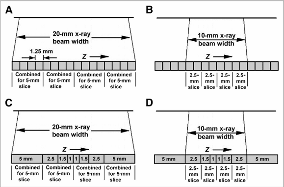
Which of these diagrams is a variable array
C and D
Why did the 4th generation CT design not work with MDCT
Because you would need as many as 4,800 detectors for a single row detector, so for a 64-slice scanner MDCT, you would need as many as 307,200 detectors
This design of scanner would need A LOT of maintenance
What is temporal resolution?
The time that it takes to acquire the data
What are the advantages of dual-source CT
Temporal resolution is halved in dual-source CT scanning
Does not increase patient dose
Good for cardiac studies-they may not have to give the patient beta blockers to slow the heart rate
Can use 2 different energy settings (kVp’s) can be used
Known as Dual Energy CT or Spectral Imaging
What is the other name for dual-energy CT
Spectral Imaging
What is the difference between dual-source and dual-energy
Dual-Source imaging uses 2 set of X-ray tubes with 2 corresponding detector arrays
Dual-Energy imaging uses 2 different kVp settings simultaneously to acquire data (does not have 2 tubes and detectors)

Which image is Dual Source
A

Which image is Dual-Energy
A and B