Chapter 19 The Cardiovascular System: Blood Vessels
 Call Kai
 Learn
 Practice Test
 Spaced Repetition
 Match
 Flashcards
 Knowt Play
1/45

There's no tags or description
Looks like no tags are added yet.
46 Terms
Velocity: How fast blood flows
Wider = Faster, but no gas exchange
Narrower = Slower, but has gas exchange
Capillaries
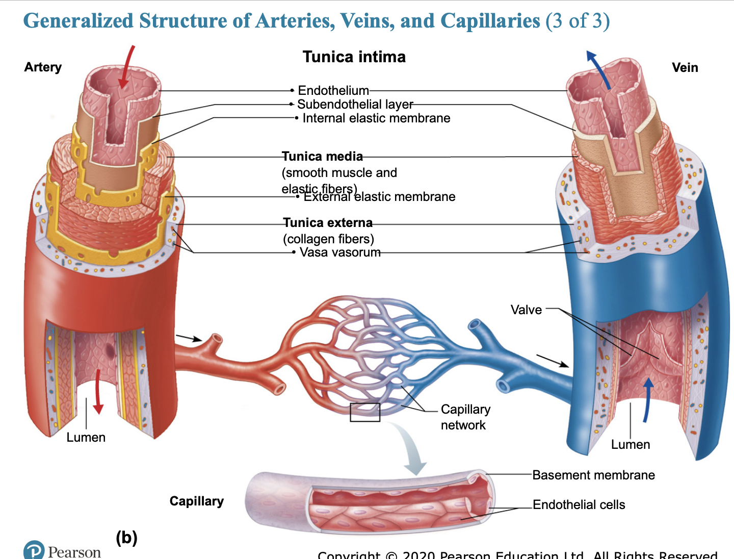
Function: The sites of exchange between the blood and surrounding tissue cells. They directly serve cellular needs by delivering oxygen, nutrients, and hormones, and picking up waste products like carbon dioxide.
Structure: Extremely thin walls—consisting of just a single layer of endothelial cells (one cell thick). This microscopic width allows materials to easily diffuse across the barrier.
Blood Flow & Velocity: Blood flow slows down significantly here, which provides the necessary time for gas and nutrient exchange to take place.
Lumen: Extremely narrow (often only wide enough for a single red blood cell to pass through in single file).
Valves: No valves.
GAS EXCHANGES
via simple diffusion
Three Types
Continuous: skin, muscle, lungs, and CNS
Fenestrated: active filtration (kidneys), absorption (intestines), or endocrine hormone secretion.
Sinusoidal: liver, bone marrow, spleen, and adrenal medulla.
Capillary Beds: Interwoven network of capillaries between arterioles and venules. MICROCIRCULATION
Vascular Shunt: channel that directly connects an arteriole with venule
Precapillary Sphincter: cuff of smooth muscle surrounding each true capillary that branches off a metarteriole; acts as a valve regulating blood flow into the capillary bed.

What are the three layers of the vessels, except capillaries
Tunica Intima: Simple squamous epithelium.
Tunica Media: Middle layer, smooth muscle, vasoconstriction and vasodilation.
Tunica Externa: collagen fibers that protect and reinforce wall and anchor it to surrounding structures.

Arteries
3 groups
elastic: Thick-walled with a large, low-resistance lumen, e.g., the aorta; a pressure reservoir that expands and recoils as blood is ejected.
muscular: deliver blood to body organs
arterioles: smallest, control flow into capillary beds; e.g., efferent arteriole to peritubular capillaries.
No valves

Veins
50% if blood supply
No pressure
Venous Return: muscle pumping, exercise, and respiratory return
Systemic Veins
Supply all of the body except the lungs, are distensible, contain a large portion of blood volume, and are called capacitance vessels or blood reservoirs.
Adaptations
Venous valves (spider veins) and sinuses
Varicose Veins
Dilated and painful veins due to incompetent (leaky) valves
Heredity and conditions that hinder venous return, e.g., prolonged standing, obesity, or pregnancy.
Elevated venous pressure, like strain from birth or bowel movements (hemorrhoids)

Anastomoses
Vascular Anastomoses: Interconnections of blood vessels
Arterial Anastomoses: alternate pathways to ensure continuous flow, even if one artery is blocked.
Arteriovenous anastomoses: shunts in capillaries
Venous anastomoses: so abundant that occluded veins rarely block blood flow
In simple terms, an anastomosis (plural: anastomoses) is a connection or joining together of blood vessels that usually branch apart.
Think of it like a detour, backup route, or alternative highway system for your blood:
The Problem: If a blood vessel (like an artery) gets blocked by a blood clot or plaque, the tissue downstream normally wouldn't get any blood and could die.
The Solution: Anastomoses act as natural cross-connections or bridges between blood vessels. If the main road is blocked, blood can seamlessly reroute through these alternate paths to keep oxygen and nutrients flowing to your tissues.
As seen on your Flashcard Set - Edit | Knowt, there are a few main types:
Arterial Anastomoses: Alternate pathways between arteries that ensure continuous blood flow even if one artery gets blocked (common around joints, the heart, and the brain).
Venous Anastomoses: Connections between veins. They are very common, which is why blocked veins rarely stop blood flow entirely.
Arteriovenous Anastomoses: Direct shunts (shortcuts) that connect an artery straight to a vein, skipping the capillary bed entirely.

Blood Flow
Volume of blood flowing through a vessel, organ, or the entire circulation in a given period.
ml/min = CO
Increased in arteries
Blood Pressure (BP)
Force per unit area exerted on the wall of a blood vessel by blood.
mmHg
Measured as systemic arterial BP in large arteries near the heart.
The pressure gradient provides the driving force that keeps blood moving from higher- to lower-pressure areas.
^ Adipose/Subcutaneous Fat = Longer vessels = Increase BP
Resistance (peripheral resistance)
Opposition to flow
Measurement of the amount of friction blood encounters with vessel walls, generally in peripheral (systemic) circulation.
SOURCES OF RESISTANCE
Blood Viscosity: ^RBC
Total blood vessel length: ^weight/height
Blood vessel diameter: ^sympathetic* greatest influence on resistance.
Systemic Blood Pressure
The pumping action of the heart generates blood flow.
Pressure results when flow is opposed by resistance.
Systemic pressure is highest in the aorta and declines throughout the pathway, drop in the arterioles.
Arterial Blood Pressure
Elasticity and volume.
Blood pressure near the heart is pulsatile. Rises and falls with each heartbeat.
Systolic: Pressure exerted in the aorta during ventricular contraction
Diastolic: lowest level of aortic pressure when the heart is at rest
Pulse Pressure: Systolic-Diastolic
Pulse: Throbbing of arteries due to the difference in pulse pressure
MAP Mean Arterial Pressure
Pressure that propels blood to tissues. In simple terms, Mean Arterial Pressure (MAP) is your body's average blood pressure throughout one complete heartbeat cycle.
Pulse pressure phases out near the end of the arterial tree.
Flow is nonpulsatile with a steady MAP.
*deasolic pressure + 1.3 pulse pressure
ex) BP 120/80 PP 120-80 = 40
80 +(1/3)*40 = 93 mmHG
systemic vs arterial blood pressure
Here is the super short and simple breakdown:
Arterial Blood Pressure: The specific pressure of blood inside your arteries (the big pipes carrying blood away from the heart). It goes up and down with every heartbeat.
Systemic Blood Pressure: The overall pressure across the entire body loop (from the heart, through the whole body, and back).
Capillary Blood Pressure
Ranges from 35 mmHg to 17 mmHg
Low pressure is desirable because high BP would rupture thin-walled capillaries.
Venous Blood Pressure
Muscular Pump
Respiratory Pump
Sympathetic Vasoconstriction
Regulation of Blood Pressure
Maintaining blood pressure requires cooperation of the heart, blood vessels, and kidneys. *All are supervised by the brain.
3 Main Factors
CO Cardiac Output
PR Peripheral Resistance
Blood Volume
CO = SV * HR
MAP = CO x R
MAP = SV x HR x R
short-term regulation: neural controls and hormonal controls
Long-term regulation: renal controls
Major Factors That Increase MAP
CO ^ = SV ^ & HR ^
PR ^ = ^ diameter, blood viscosity, and blood length
MAP = CO & PR

Short-Term Regulation
Neural Control: MAP is maintained by adjusting blood vessel diameter, thereby altering blood distribution to organs in response to demand.
Cardiovascular center of the medulla, baroreceptors, chemoreceptors, and higher brain centers.
Baroreceptors are pressure sensors in your major arteries (like your neck and heart) that detect when your blood pressure goes too high or too low.
They instantly send signals to your brain, which adjusts your heart rate and blood vessel width to quickly bring your blood pressure back to normal.
Chemoreceptors watch for changes in the chemical composition of your blood—specifically monitoring levels of oxygen, carbon dioxide, and pH (acidity).
Here is how they work:
Where they are: Located in major blood vessels near the heart and neck (the carotid and aortic bodies) as well as directly inside the brainstem.
What they do:
If your carbon dioxide goes up (or your blood becomes too acidic), or if your oxygen drops drastically, these receptors immediately fire signals to your brain's respiratory and cardiovascular centers.
The Response: They tell your heart to speed up and your breathing rate to increase so you can flush out excess carbon dioxide and take in more oxygen.
If MAP is low
Reflex vasoconstriction is initiated, increasing CO and blood pressure
Example: upon standing, BP falls and triggers:
– Carotid sinus reflex: baroreceptors that monitor BP to ensure enough
blood to brain
– Aortic reflex maintains BP in systemic circuit
Baroreceptors are ineffective if altered blood pressure is sustained
– Become adapted to hypertension, so not triggered by elevated BP levels

Hormonal Controls
Epinephrine/Norepinephrine increase CO and cause vasoconstriction
Angiotensin II & ADH : stimulate vasoconstriction
Atrial Natriuretic peptide: decreases BP by antagonizing aldosterone
Long-Term Mechanisms: Renal Regulation
Baroreceptors quickly adapt to chronic high or low BP, so they are ineffective for long-term regulation.
Long-term by altering blood volume via the kidneys
Direct renal mechanism
Increased BP/BV causes elimination of more urine, thus reducing BP
Decreased BP/BV causes kidneys to conserve water, and BP rises.
Indirect renal mechanism (RAAS)
Renin-angiotensin-aldosterone mechanism
Decreased arterial blood pressure causes release of renin from the kidneys.
Renin catalyzes angiotensinogen from liver to angio I
Angiotensin-converting enzyme from lungs converts angio I to agio II.
angio II stabilizes arterial BP, and ECF stimulates aldosterone secretion, causing ADH release, hypothalamix thirst center, and vasoconstriction to increase BP.

The goal of blood pressure regulation is to keep blood pressure high enough to provide adequate tissue perfusion, but not so high that blood vessels are damaged
– Example: If BP to brain is too low, perfusion is inadequate, and person loses consciousness
– If BP to brain is too high, person could have stroke
Homeostatic Imbalances in BP
Transient elevations in BP occur during changes in posture, physical exertion, emotional upset, and fever.
Age, sex, weight, race, mood, and posture
Hypertension
Sustained elevated arterial pressure of 140/90mmHG or higher
Prehypertension is when values are elevated but not yet in the hypertension range.
Prolonged hypertension is a major cause of heart failure, vascular disease, renal failure, and stroke
Heart must work harder, myocardium enlarges, weakens, and becomes flabby.
Hypotension
Low blood pressure below 90/60 mm Hg
Usually not a concern unless it causes inadequate blood flow to tissues.
Associated with long life and lack of cardiovascular illness
Orthostatic hypotension: temporary low BP and dizziness when suddenly rising from a sitting or reclining position.
Chronic hypotension: hint of poor nutrition warning sign for Addison’s disease
Acute hypotension: important sign of circulatory shock.
Circulatory Shock
A condition in which blood vessels are inadequately filled and can’t circulate blood normally. Inadequate blood flow can’t meet tissue needs.
Hypovolemic shock: large-scare blood loss
Vascular shock: extreme vasodilation and decreased peripheral resistance.
Cardiogenic shock: an inefficient heart can’t sustain adequate circulation.

Control of Blood Flow
Tissue Perfusion:
Delivery of O2
Gas Exchange
Absorption of nutrients (digestive tract)
Urine formation (kidneys)
Extrinsic Control: Sympathetic nervous system and hormones
Intrinsic Control: Autoregulation (local) control of blood flow uses paracrine factors or properties of muscle tissue.
Ex) At rest, skeletal muscles receive about 20% of total blood flow in the body, but during exercise, skeletal muscle can receive over 70% of blood flow. Intrinsic: skeletal muscle arterioles dilate, increasing blood flow to muscle. Extrinsic: decreases blood flow to other organs, such as the kidneys and digestive organs.
Autroregulation: Intrinsic Regulation of Blood Flow
Autoregulation: local (intrinsic) conditions that regulate blood flow to that area – Reactive hyperemia: increased blood flow to an area due to intrinsic factors
Two types of intrinsic mechanisms that determine the final autoregulatory response
Metabolic control uses chemical signals ("We are working hard and need oxygen!") to widen blood vessels and let more blood in.
Myogenic control uses physical stretch ("The pressure just spiked!") to squeeze blood vessels shut and protect downstream tissues.
Long-term Autoregulation
Occurs when short-term autoregulation can’t meet tissue nutrient requirements. Angiogenesis and existing vessels enlarge. People in high altitudes.
Active/Exercise Hyperemia
During muscle activity, blood flow increases in direct proportion to metabolic activity. Local controls override sympathetic vasoconstriction, increasing flow.
Oxygen is the fuel your tissues need, while carbon dioxide and hydrogen ions are the waste products produced when cells work hard.
When CO2 and H+ build up O2 they act as chemical alarms that trigger chemoreceptors and local blood vessels to widen, increasing blood flow and breathing so your body can deliver more oxygen and flush out waste.
Brain
Blood flow to the brain is constant.
Increased MAP causes cerebral vessels to constrict.
MAP below 60mmHg causes syncope (fainting). MAP above 160mmHg can result in cerebral edema.
Skin
Functions of blood flow through the skin.
Supplies nutrients, helps regulate body temperature, and provides a blood reservoir.
Regulated by sympathetic nervous system reflexes.
Lungs
Arteries/arterioles are more like veins/venules
Autoregulatory mechanisms are opposite. Allows O2 to flow to the lungs.
Arterial resistance and pressure are much lower than in systemic circuit
Heart
Heart – Blood flow through heart is influenced by aortic pressures and ventricular pumping – During ventricular systole, coronary vessels are compressed
§ Myocardial blood flow ceases § Stored myoglobin supplies sufficient oxygen – During diastole, high aortic pressure forces blood through coronary circulation
Heart – At rest, coronary blood flow is ~250 ml/min
§ Control is probably via myogenic mechanisms – During strenuous exercise, coronary vessels dilate in response to local accumulation of vasodilators § Blood flow may increase three to four times § Important because cardiac cells use 65% of O2 delivered – Other cells use only 25% of delivered O2 – Increasing coronary blood flow is only way to provide more O2
Velocity of Blood Flow
Velocity of flow changes as blood travels through the systemic circulation
Fastest in the aorta, slowest in the capillaries, and then increases again in the veins.
Vasomotion
Intermittent flow of blood through capillaries due to the on/off opening and closing of precapillary sphincters.
vasomotion is the slow, rhythmic opening and closing of precapillary sphincters (tiny muscular valves), which causes blood to flow through capillaries in intermittent "waves" rather than all at once.
Because your body doesn't have enough blood to fill every single capillary in your entire body at the same time, vasomotion allows your body to route blood only to the specific areas that need it most at that moment.
Capillary Exchange/Transport
In simple terms, capillary exchange is the process where oxygen, nutrients, and hormones pass out of your blood and into your body's tissues, while waste products (like carbon dioxide) move out of the tissues and back into the blood.
This happens continuously through tiny gaps in the capillary walls via diffusion, filtration, and reabsorption. DFR

Fluid Movements: Bulk Flow
Fluid is forced out clefts of capillaries at arterial end, and most return to blood at venous end.
Bulk fluid flow
Hydrostatic Pressure (HP) — The Push:
What it is: The physical pressure exerted by fluid against the inside walls of a blood vessel.
How it works: Driven by the pumping action of the heart, hydrostatic pressure acts like a water hose—it pushes fluid out of the capillaries and into the surrounding tissues (especially at the arterial end of a capillary bed).
Colloid Osmotic Pressure (COP) — The Pull:
What it is: The osmotic pressure created by large proteins (like albumin) stuck inside the bloodstream that cannot escape through capillary pores.
How it works: Because these proteins cannot cross the wall, they act like a sponge, creating an osmotic pull that draws fluid back into the capillaries (especially at the venous end).
Edema
Abnormal increase in the amount of interstitial fluid. Caused by an increase in outward pressure or a decrease in inward pressure.
Capillary hydrostatic pressure accelerates fluid loss from blood due to incompetent venous valves, localized blood vessel blockage, congestive heart failure, or high blood volume.
Inflammation
An increase in interstitial fluid osmotic pressure can result from an inflammatory response. Inflammation increases capillary permeability and allows proteins to leak into interstitial fluid.
Hypoproteinemia
Low blood protein (especially albumin), which lowers colloid osmotic pressure and causes fluid to leak out of capillaries, resulting in swelling (edema).

Vascular System
Pulmonary circulation: short loop that runs from the heart to the lungs and back to the heart
Systemic circulation: long loop to all parts of the body and back to the heart
Circulatory Pathways
Arteries run deep, whereas veins are both deep and superficial.
– Venous pathways are more interconnected § Unlike arterial pathways, venous pathways have more interconnections – Veins can have more than one name, making venous pathways harder to follow – The brain and digestive systems have unique venous drainage systems § Brain contains dural venous sinuses § Venous system of the digestive system drains into hepatic portal system,
Developmental aspects of blood vessels
Ductus venosus bypasses luver
Congenital vascular problems are rare.
Vessel formation occurs to support body growth, support wound healing, or rebuild vessels lost during menstrual cycles.
With aging, varicose veins, atherosclerosis, and increased blood pressure may arise.