Normocytic anemias
 Call Kai
 Learn
 Practice Test
 Spaced Repetition
 Match
 Flashcards
 Knowt Play
1/16

There's no tags or description
Looks like no tags are added yet.
17 Terms
Causes of hemoglobinopathies
A single AA substitution in (usually) the beta chain → Sickle Cell anemia
Abnormal synthesis of one of the AA chains→ Thalassemias
Fusion of Hemoglobin chains→ HGB Lepore (δβ)
Extension of AA chain → Hgb Constant spring (α chain)
Sickle cell anemia genetics
β chain substitutions on chromosome 11
Autosomal codominant abnormality (both alleles expressed simultaneously instead of one masking the other)
Occurs 1:375 of African Americans
Predominant in malaria areas: Central & West Africa, parts of Mediterranean, Asia, India
Trait: 8-10% of African Americans
Anemia: 0.3-1.3% African Americans
4 haplotypes, depending on amount of HgbF (higher HgbF= less severe S/S):
Asian
Senegal
Benin
Bantu
Sickling gene
Glutamate substituted by Valine at position 6 from the N Terminus (total 146 AAs)
Glu is hydrophilic and Val is hydrophobic
Creates sticky patch that cause Hgb to stiffen

Sickle cell shape
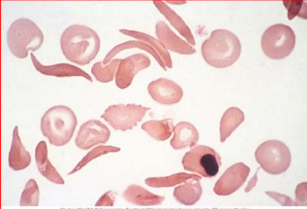
HgbS forms liquid tactoids (elongated)/ stiff rod fibers beneath membrane
Stiffens RBCs in low O2 areas: spleen, liver, kidney, joints, extremities
Sickle/ boat shape obstruct small blood vessels, adheres to vascular endothelium
Less O2 delivered to tissues
pH decreases
Sickling induced by: hypoxia, acidosis, dehydration, fever, cold exposure
Repetitive sickling→ RBC life is 10-20 days
Sickle cell S/S
Basic S/S:
Chronic hemolytic anemia
Recurrent painful attacks
Bacterial infections
Deterioration of tissues/ organ fxn
Shortened life expectancy
Dactylitis also present (sickle cells in microvasculature→ swollen hands and feet)
Diagnosed between 6-24 months
Protected with high levels of Hgb before then

Sickle cell indices and peripheral smear
normocytic, normochromic anemia
Hgb 6-8 g/dL (normal: 12-16M 10-14F)
Decreased: RBC count, hematocrit
Decreased Hpt: indicates extravascular hemolysis
Increased Bilirubin, LDH
Increased Reticulocytes→ mildly elevated MCV
HgbS precipitates→ Positive sickle solubility test in high-molarity buffered phosphate solutions
HgbS also present in hgb electrophoresis
Sickle cell trait + population
Heterozygous
60% HgbA, 40% HgbS
US: 2.5M people
Africa: 25-30% where Malaria is endemic
Circumstances that cause crisis: unpressurized cabin, high altitudes, low oxygen
HgbC disease gene + population
Substitution from the 6th position of the N-Terminal of the beta chain where glutamic acid is substituted by Lysine
Considered a sickling disorder
HgbC Disease: two abnormal genes
HgbC trait: one abnormal gene (asymptomatic)
Heterozygous: HgbAC: 60% HgbA, 40% HgbC
Population:
17-28% in West Africa (or Northern Ghana)
2-3% African Americans have the trait
0.02% have the disease

HgbC indices
normochromic, normocytic
MCHC mildly elevated due to dense HgbC
RBC lifespan 38 days
HgbC peripheral smear
C for crystal/ “Bars of gold”
obstructs microvasculature, melts in spleen
Codocytes predominant: 50-90%
Electrophoresis has wide band of HgbC in same position as HgbA2


Hgb electrophoresis
HgbC and HgbA2 on same level
Based on charge as they move from diff charges

HgbSC
Heterozygous combination of HgbC and HgbS
RBC lifespan: 28days
Less severe than homo HgbS but more severe than homo HgbC
Considered a sickle disease
Electrophoresis reveal 50% HgbS and 50% HgbC (according to Dr. Nocera)
But according to The Blood Project, it’s 50% HgbS and 45% HgbC


HgbSC indices/ peripheral smear
Increased MCHC
normocytic, normochromic
Hct >25%
Some Hgb off-center
Codocytes and sickle cells present

HgbD
Punjab and LA variants
but high incidence in Britain
β121 Glu→ Gln mutation
Migrates with HgbS in electrophoresis
Does not sickle and has normal solubility
Could exist hetero with HgbS
Produces aggregates

HgbE
2nd most prevalent hemoglobinopathy worldwide
Mostly in SEA
Nearly 50% in Thailand
Cambodia and Laos also have high numbers
β26 Glu→ Lys
Unstable under oxidant stress
Oxygen dissociation shifted to right (less affinity)
Hypochromic, microcytic but asymptomatic
Hgb-Phila
Occurs in those of African descent
α68 Asn→ Lys
HgbO Arab
β121 Glu→ Lys
Rare in African Americans