Neuraxial Anesthesia

0.0(0)
Studied by 0 people
call kaiCall Kai
learnLearn
examPractice Test
spaced repetitionSpaced Repetition
heart puzzleMatch
flashcardsFlashcards
GameKnowt Play
Card Sorting

1/226

flashcard set

Earn XP

Description and Tags

POA EXAM 4

227 Terms

1
New cards
<p>Vertebral Canal extend from ______ to __________?</p>

Vertebral Canal extend from ______ to __________?

foramen magnum to tip of the coccyx

2
New cards

Total number of vertebrae in the human spine

33

3
New cards
<p>Which spinal region has a concave curvature? </p>

Which spinal region has a concave curvature?

Cervical and lumbar

4
New cards
<p>Which spinal region has a convex (posterior) curvature? </p>

Which spinal region has a convex (posterior) curvature?

thoracic and sacral

5
New cards

Number of cervical vertebrae

7

6
New cards

Number of thoracic vertebrae

12

7
New cards

Number of lumbar vertebrae

5

8
New cards

Number of sacral vertebrae

5

9
New cards

Number of coccygeal vertebrae

4

10
New cards

Anatomical opening containing the spinal cord and nerve roots

vertebral foramen

<p>vertebral foramen</p>
11
New cards

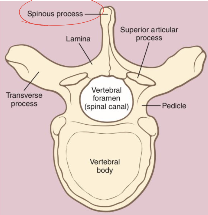
Poster vertebral structure that defines the anatomic midline + bony projection used as landmark

spinous process

<p>spinous process</p>
12
New cards

Paired structures connecting the vertebral body to the lamina

pedicles

<p>pedicles</p>
13
New cards

Paired posterolateral bony projections of a vertebra

transverse processes
<p>transverse processes</p>
14
New cards

Paired posterior bony plates that fuse to form the spinous process

laminae

<p>laminae</p>
15
New cards
Anatomical direction of cervical and thoracic spinous processes

inferiorly (downward); longer and more acutely angled

<p>inferiorly (downward); longer and more acutely angled</p>
16
New cards
Anatomical structure formed by stacked vertebral notches and articulating surfaces
intervertebral foramina
17
New cards
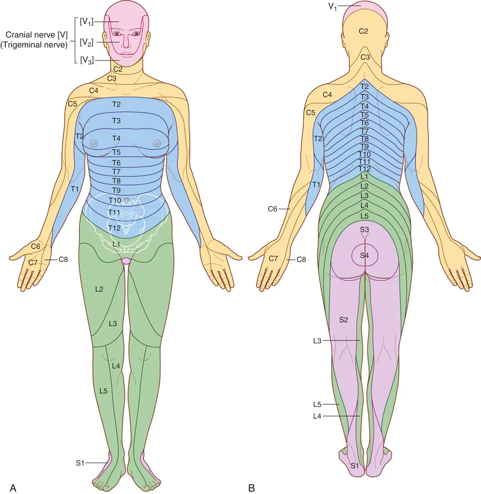
Primary function of the intervertebral foramina
protects and provides passage for spinal nerves
18
New cards
Structures that innervate the facet joints
branches of adjacent spinal nerves
19
New cards

Region of skin innervated by single spinal nerve, clinical manifestation of facet joint injury-related pain

dermatomal distribution
20
New cards

Group of muscles innervated by single spinal nerve, clinical manifestation of facet joint injury-related muscle spasms

myotomal distribution
21
New cards

Lumbar vertebra characteristics (size, spinous process shape, overlap)

largest size, shorter/broader spinous processes, least overlap

<p>largest size, shorter/broader spinous processes, least overlap</p>
22
New cards
<p>Anatomical structure that typically hinders needle access to an upper thoracic interspace (e.g., T5 space) </p>

Anatomical structure that typically hinders needle access to an upper thoracic interspace (e.g., T5 space)

inferior portion of the superior spinous process (e.g., T4)

<p>inferior portion of the superior spinous process (e.g., T4)</p>
23
New cards

Anatomical structure formed by the incomplete lamina of the last sacral vertebra (usually at the S3-S4 vertebral level)

sacral hiatus (key clinical landmark for accessing the epidural space)
24
New cards
Bony horn-like projections that guard the sacral hiatus
sacral cornua (cornu)
25
New cards
Primary clinical purpose of palpating the sacral cornua
landmarks for caudal anesthesia
26
New cards
Clinical concern prompting decreased use of pediatric caudal blocks noted in the handwritten text
risk of spinal injury
27
New cards

Total number of spinal nerve pairs and their anatomical range

31 pairs (C1 to S5)

28
New cards

Bundle of lower spinal nerves extending beyond the terminus of the spinal cord

cauda equina

29
New cards
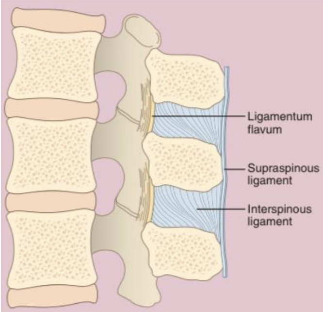
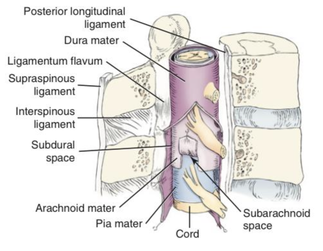
Three major posterior spinal ligaments serving as neuraxial landmarks
supraspinous, interspinous, and ligamentum flavum
30
New cards
Spinal ligament that connects the apices of the spinous processes
supraspinous ligament
31
New cards
Three structural layers of the supraspinous ligament
superficial, middle, and inner
32
New cards
Spinal ligament that runs directly between adjacent spinous processes
interspinous ligament
33
New cards
Spinal regions where the interspinous ligament is poor/absent versus very thin

cervical: absent/poor, lumbar: very thin

34
New cards
Strongest posterior spinal ligament, composed of yellow elastic tissue
ligamentum flavum
ligamentum flavum
35
New cards
Anatomical structures joined together by the ligamentum flavum
adjacent laminae (vertebral arches)
36
New cards
Average thickness of the ligamentum flavum at the L2-3 interspace
3 to 5 mm
37
New cards
Clinical/tactile significance of the ligamentum flavum during neuraxial blocks
thick, distinct tactile feel (indicator to slow down)
38
New cards
Superior anatomical boundary of the spinal cord
medulla oblongata
39
New cards
Inferior spinal level terminus of the spinal cord in most adults vs in children
L2 - L3 vertebra (adults) & L3 vertebra (children)
40
New cards
Length range of the adult spinal cord
42 to 45 cm
41
New cards
Common pediatric neuraxial block levels
L3-L4
42
New cards
Terminal tapered tip of the spinal cord
conus medullaris
43
New cards
Two regions where the spinal cord is physically enlarged

C4 to T1 and L2 to S3 (supply the upper and lower limbs)
44
New cards
True or False: Spinal cord levels directly correspond with vertebral levels
FALSE
45
New cards
Vertebral body levels spanning the adult lumbosacral enlargement (L2-S3)
T11 to L1
46
New cards
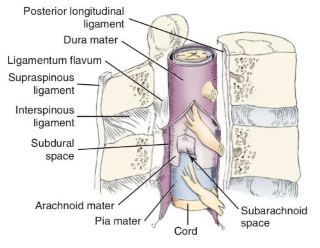
Outermost, thickest, and most protective spinal meningeal layer
dura mater
dura mater
47
New cards
Tactile sensation expected when a needle punctures the dura mater
pop (leather feel)
48
New cards
Middle, delicate, nonvascular, spiderweb-like spinal meningeal layer
arachnoid mater
arachnoid mater
49
New cards
Anatomical structure where the arachnoid mater ends inferiorly
S2
50
New cards
Potential space located between the dura mater and the arachnoid mater
subdural space
subdural space
51
New cards
Anatomical space located between the arachnoid mater and the pia mater (Primary target space for a spinal anesthesia injection)
subarachnoid space
subarachnoid space
52
New cards
Four major structures contained within the subarachnoid space
CSF, spinal nerves, blood vessels, support ligaments
53
New cards
Anatomical range of the subarachnoid space
brain to S2
54
New cards
Innermost, delicate, and highly vascular spinal meningeal layer. (Meningeal layer in thin, direct contact with the outer surface of the spinal cord)
pia mater
pia mater
55
New cards

Anatomical boundaries of the epidural space & Anatomical range of the epidural space

outside the dural sac but inside the vertebral canal from the base of the cranium to the base of the sacrum

<p>outside the dural sac but inside the vertebral canal from the base of the cranium to the base of the sacrum</p>
56
New cards
Average width of the epidural space
~5 mm
57
New cards
Five major structures contained within the epidural space
epidural veins, fat, lymphatics, segmental arteries, and nerve roots
58
New cards
Clinical complication risk associated with the vascular structures in the epidural space
bleeding and hematoma formation
59
New cards
Key physiological characteristic of epidural veins
valveless
60
New cards
Two patient populations where epidural veins become engorged and swollen
obese and pregnant patients
61
New cards
Clinical risk associated with utilizing a lateral approach for an epidural block
increased risk of hitting blood vessels
62
New cards
Clinical presentation of an accidental local anesthetic injection into the subdural space
patchy and oddly high blocks
63
New cards

True or False: The subdural space is NOT a desirable target for intentional anesthetic injection

TRUE

64
New cards
Primary goal of neuraxial anesthesia
block pain transmission from areas of injury, disease, or surgery
65
New cards
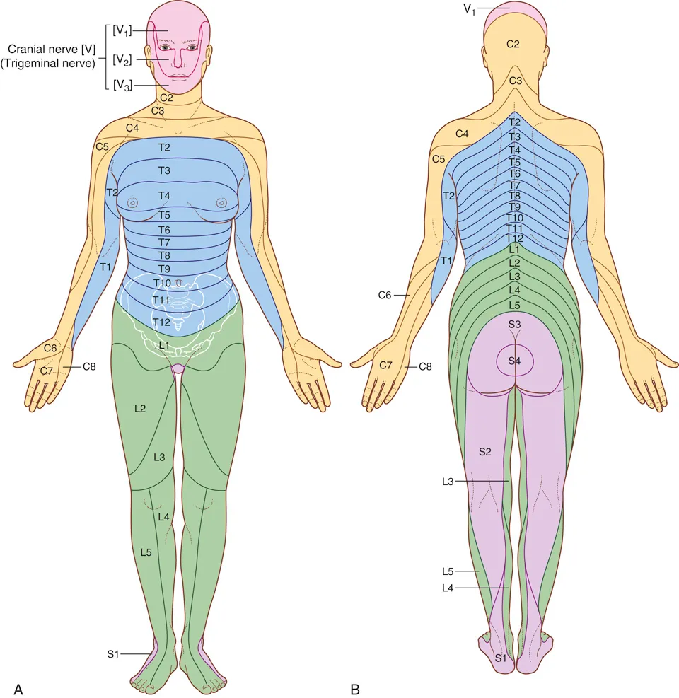
Spinal nerve levels providing cutaneous innervation to the umbilical area
T7 to T11
66
New cards
Vertebral body level located directly posterior to the umbilical area
L3 vertebra
67
New cards
Clinical methods used to assess the level of a neuraxial block
motor or sensation tests
68
New cards
Target ion channel and specific state required for local anesthetic binding
Blocks sodium (Na+) channels on the cytoplasmic (inner) surface of the nerve. LA selectively binds to channels in the Activated Open and Inactivated Closed states.
69
New cards
Relationship between nerve firing rate and local anesthetic blockade sensitivity
faster firing increases blockade (frequency-dependent)
70
New cards
Number of consecutive nodes of Ranvier required to be blocked for myelinated axons
3 adjacent nodes
71
New cards
Anatomical definition of a differential block
interruption of autonomic nerve transmission without sensory or motor blockade
72
New cards
Dermatomal level relationship: Sympathetic block vs. Sensory block
sympathetic is 2 to 6 dermatomes higher
73
New cards
Dermatomal level relationship: Motor block vs. Sensory block
motor is 2 dermatomes lower
74
New cards
Why are smaller, lightly myelinated/unmyelinated nerve root fibers blocked first?
located closer to the nerve root surface
75
New cards
Why are larger, myelinated motor and sensory fibers harder to block
located deeper within the nerve bundle
76
New cards
Nerve fiber type possessing the highest susceptibility to local anesthetic blockade (++++ sensitivity)
B fibers (preganglionic autonomic)
77
New cards
Nerve fiber type that is largest in diameter, lowest in sensitivity (+), and hardest to block
A-alpha (motor)
78
New cards
Nerve fiber types corresponding to the second wave of contact and blockade
C and A-delta fibers
79
New cards
Specific temperature sensation lost first during local anesthetic onset
cold before hot
80
New cards
Nerve sensation that is rarely lost due to its profound resistance to blockade
deep pressure
81
New cards
Initial anatomical site of action where epidural local anesthetics act first
dural cuffs (sheath)
82
New cards
Structures epidural local anesthetics must diffuse through after longitudinal spread to reach nerve roots
meninges and dural cuffs
83
New cards
Three clinical variables that can be increased to achieve a denser epidural block
concentration, volume, and time
84
New cards
Primary mechanism of initial drug distribution in the epidural space vs. the spinal space
bulk flow (epidural) vs. diffusion (spinal)
85
New cards
Order of Nerve Fiber Blockade (Onset):

B→ C → A-delta → A-gamma→ A-beta → A-alpha

(Autonomic → Sensory → Motor);

But Can Delta Give Better Actions?

86
New cards
Order of Blockade by Clinical Sensation
superficial pain > sympathetic (T1-L2) > Temperature > Motor > touch > proprioreception > deep pressure
87
New cards
Blockade Assessment correlates with sympathetic block
loss of temperature (ice test)
88
New cards
Bloackade Assessment used to determine the sensory level of a block
distinguishing sharp vs. dull sensations (blunt needle, sharp object)
89
New cards
Spinal levels evaluated by assessing a patient's ability to raise their knees
L2-L3
90
New cards
Spinal levels evaluated by assessing a patient's ability to tense their abdominal muscles
T6-T12
91
New cards
Spinal levels evaluated by assessing a patient's ability to dorsiflex their foot
S1-S2
92
New cards
Dermatome level corresponding to the nipple line
T4
T4
93
New cards
Dermatome level corresponding to the xiphoid process landmark
T6
T6
94
New cards
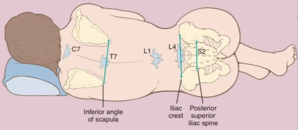
Dermatome level corresponding to the inferior angle of the scapula
T7
T7
95
New cards
Dermatome level corresponding to the umbilicus landmark
T10
T10
96
New cards
Dermatome level corresponding to the inguinal ligament
L1
L1
97
New cards
Dermatome level corresponding to the iliac crest landmark (Tuffier's line)
L4
L4
98
New cards
Required sensory level for a hemorrhoidectomy
S2-S5
99
New cards
Required sensory level for foot surgery
L2-L3 (knee)
100
New cards
Required sensory level for lower extremity surgery
L1-L3 (inguinal ligament)