Biomedical Histology Quiz 2
 Call Kai
 Learn
Learn Practice Test
 Spaced Repetition
 Match
Match Flashcards
 Knowt Play
1/50

There's no tags or description
Looks like no tags are added yet.
Name | Mastery | Learn | Test | Matching | Spaced | Call with Kai | Chat |
|---|
No analytics yet
Send a link to your students to track their progress
51 Terms
what is purpose of connective tissue (CT)
physical and chemical support of other tissue and organs
around/between other tissue types
cells do not form junctions with each other
Extracellular matrix (ECM) is usually the most abundant component
ECM usually gives the tissue its characteristic properties
conduit for blood vessels and nerves
what are the components of ECM
Fibres
Proteins that polymerize into long or short fibrils and fibres
Primarily collagen and elastin
Ground substance
‘hydrated space’ (binds to water) with many proteins, multiadhesive glycoproteins (bind more then 1 thing) and proteoglycans (have more sugars then protein)
Glycosaminoglycans =GAGs (hyluronan): polymers of modified sugar residues
sequesters water bind to hematoxylin
what are the 2 types of cells in Connective tissue
Resident (fixed) cells
Synthesize and maintain ECM
Fibroblasts and members of the fibroblast family
Transient Cells
Cells of the immune system: leukocytes
Secondarily take up residents in CT (macrophages) and/or traffic between CT and immune organs (lymphocyte)
Examples of transient CT cells
Macrophages
Derived from blood monocytes
Phagocytose microorganisms and damaged cells
Long-term or short term residents
Mast Cells
Similar to blood basophils
Prominent granules contain vasoactive substances
involved in allergic reactions

How do we classify fibrous CT
Loose (areolar) CT
Abundant ground substance, few and relatively sparse fibres
thin collagen 1 fibre bundles (may not be obvious in LM)
Reticular Connective tissue
Fibres form 3D network of Type III collagen in a loose disposition
Inside loose CT or in lymphoid organs where it forms scaffold to support cells
Dense CT
Abundant collagen 1 fibres in large/prominent fibre bundles, comparatively less ground substance, few cells
Dense irregular CT = collagen bundles randomly oriented
Dense Regular CT = collagen bundles in regular orientation (aligned with fibroblasts)
How does Reticular Connective tissue stain
H and E
collagen type III doesn’t stain
Reticular cells are fibroblasts that make this collagen
other cells are immune system cells
Silver stain - for reticular fibres

which is dense irregular and regular
left = dense irregular
Right = dense regular
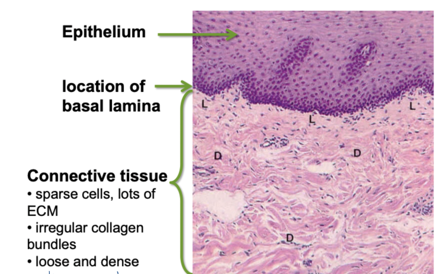
what does loose and dense mean how is it shown
loose is the lighter pink, dense is darker pink (thinker fibres)
this is because it is rich in collagen type 1 which is + so binds to eosin
What are ECM fibres made up of
Collagen
approx 90% CT fibers are collagen
protein polymers that make long straight fibres and meshworks
Tensile strength (can withstand being stretched and pulled)
very eosinophilic
Elastin
Approx 10% CT fibers
stretch and recoil
poorly eosinophilic, light refractile, binds special stains
NOTE: most fibers see in LM will be collagen
what collagen made of/ collagen polymerization
25 different types
all are made up of 3 polypeptides, arranged as triple helix
outside cell, these collagen molecules polymerize into long fibrils
molecules polymerize into striped fibrils
fibrils grouped into larger fibres
fibres may further aggregate into fibre bundles
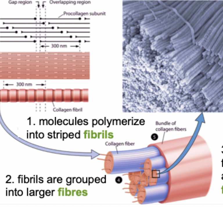

what each pointing at
Fibril
Fibre
Fibre bundle
porcollagen molecule
what are main types of collagen

Collagen Type I
whole procollagen molecule is all triple helix
makes lingest/largest and strongest fibers of body
very strong in tensile strength but not stretchy

Collagen Type IV
Procollagen molecules has globular/non helical regions at either end
Form short segments of lateral association with multiple other type IV molecules (meshes)
meshwork of collagen type IV interacts with laminin, fibronectin and other basal lamina proteins, collagens and ground substances in ECM

Collagen Diseases
Proper collagen arrangement in ECM requires
many steps and enzymes both inside and outside cells to form correct fibrils
several collagens and other molecules to form fibers and fiber bundles and other arangements
Collagenopathies due to mutations in collagen genes
osteogenesis imperfecta =fragile bones
Collagen damage by imflammation
rheumatoid arthritis
Defective collagen production/processing
scurvy (vit C deficiency) = collagen goes to RER, vit C is a cofactor for collagen to be hydroxylated to help form proper connective tissue
What is elastin, what made of
random coiled elastin molecules attached covalently together
when fibre is stretched the coiled straighten but molecules dont pull apart; recoil upon release of stretch
produced by fibroblasts and vascular smooth muscle cells
two proteins in elastic fibres: core of elastin, surface network of fibrillin
Fibrillin network acts as scaffold for assembly of elastic fibres
mutations in fibrillin lead to Marfan Syndrome
general CT defects, elongated ones, hyperflexible joints, aortic aneurysm

More ground substance details
water (60-90%) - can sequester
Carbohydrate polymers
Glycosaminoglycans (GAG) - neg charge
Proteoglycans
Multiple other soluble proteins and glycoproteins, including fibronectin, laminin and growth factors
stains poorly with H and E
provides turgor pressure (resistance to compression)
occupies large volume and resists compression because of bound water
Can be watery (loose CT) to very thick gel (cartilage) depending on GAG concentration
gels inhibit cell movement depending on amounts of GAGs
water in gels is the medium of diffusion through CT
GAGs and PGs characteristics
GAGs: long, straight, polymers of sulphated or carboxylated sugars
PGs: multiple GAG molecules attached to a protein core
Both carry net negative charge (bind water and basic dyes)

what adipose tissue made up of and found?
Made up of adipocytes (fat cells)- found as single cells or in small groups in loose CT
differentiate from mesenchymal/fibroblast like precursor
Adipose tissue highly cellular (unlike most CT)

Adipose tissue types
Unilocular (white)
triglycerides stored as single lipid droplet
rim of cytoplasm with flattened nucleus
energy homeostasis and hormone secretion; padding and insulation when large numbers
Multilocular (brown)
triglycerides stored in multiple droplets
round nucleus
obvious cytoplasm with lots of mitochondria
lipid metabolized in cell for heat production
animals that hibernate, babies as they grow disappear slowly
EXTENSIVE BLOOD SUPPLY

which is which
left = unilocular
Right = multilocular
Blood details
plasma - tissue fluid (like ground substance minus fibres)
Fibres of blood are soluble; proteins become fibrous upon activation of coagulation
View blood using cell smear
eosin and 2 basic dyes but basophilic still blue even though hematoxylin not used
what are components of blood
“Formed elements” = Erythrocytes and platelets
No nuclei or organelles
Fragments of cells
Leukocytes (white blood cells)
Granulocytes
Nucleated blood cells with specific granules or storage vesicles
Agranulocytes
Nucleated blood cells without specific granules
What is the origin of blood cells
Hemopoiesis
Red bone marrow = makes blood cells
yellow bone marrow as get older red bone marrow replaced by fat
RBC lose their nucleus as they mature

what are Erythrocytes
red blood cells
O2/CO2 exchange and pH balance
most abundant “formed element” in blood (40% volume)
Homogeneous eosinophilic cytoplasm - 30% hemoglobin
present in most tissue sections

erythrocyte morphology
anuclear bi-conclave discs for maximim surface area
very eosinophilic due to high hemoglobin content
single mutation in hemoglobin gene leads to sickle cell generation
What is a leukocyte
Nucleated cells or blood are larger than erythrocytes
not really involved in functionality of blood directly
Travel from site to site and surveillance (defence)
Associated with responses to tissue homeostasis, damage, infection, immunity, allergy

cells in blood vs CT vs both

Granulocytic Leukocytes
Eosinophils - stain pink because granules are eosinophilic
basophils - stain hematoxylin purple
Neutrophils - little granuals don’t have significant colour
common features:
from common bone marrow precursor
Multilobed nucleus
azurophilic granules (1degree granules; lysosomes)
Unique features
specific granuals (2 degree granules)
unique functionality for each cell type
Neutrophils (PMNs) details and function
most abundant granulocyte
multi lobed nucleus, immature has band like nucleus
pale, poorly stained cytoplasm
specific granules not abundant or large, so best seen in TEM
Function
usually first immune cell to enter site of inflammation/infection
recruited to specific sites by mediators and cytokines in underlying CT
kill microorganisms by phagocytosis and secretion of antibacterial enzymes/peptides/ROS
Most die in process; accumulated dead and dying PNMs create pus

Eosinophils details and function
mid-sized and fairly abundant leukocytes in blood
large bi-lobed nucleus
pale, poorly stained cytoplasm
large prominent eosinophilic specific granules
granules contain rod shaped inclusions visible in TEM
function:
Kill endo-parasites
contents of specific granules able to perforate parasite membranes
eosinophil products modulate mast cell activity
trigger histamine release
promote survival
intensify allergic inflammation
Play important role in Asthma

Basophils details and function
very rare <0.5% of all leukocytes
large bi-lobed nucleus
pale, poorly stained cytoplasm
large, deeply stained basophilic granules, may obscure nucleus
granules stain metachromatic (cause colour shift) with Toluidine Blue
Function
cells releared from bone marrow and circulate for short time
modulate allergic reactions: trigger rxn
immediate reaction to second stimulus by antigen
Degranulation causes vasodilation, edema, mucus secretion, bronchiole constriction
Can be life-threatening: systemic
Mast cells are related cell type found in tissue- usually a more localized response but can also be systemic response
Agranulocytic Leukocytes
Lymphocytes
Monocytes
Directly related to immune responses
Adaptive immunity (lymphocytes)
Intrinsic immunity (monocyte/macrophage)
General Features
no obvious specific granules
azurophilic granules as all leukocytes; may or may not be obvious in LM
nucleus multilobed
relatively less cytoplasm
Lymphocytes details and function
Smallest nucleated cell in blood; not much larger than RBC
very little cytoplasm; usually basophilic
Nucleus with dense heterochromatin; nucleus is often only prominent feature, especially in sections
Function
involved in targeted immunity; antigen specific, highly regulated and part of complex system
3 major types
B lymphocytes (B-cell): Ig (antibody) production
T-lymphocytes (Thymus): recognition and regulation, cytotoxicity
NK cells (Natural Killer Cells): killing of abnormal cells
All look alike so need immunostaining to differentiate based on surface molecules
Freq transit between blood, CT, lymph/lymphoid organs
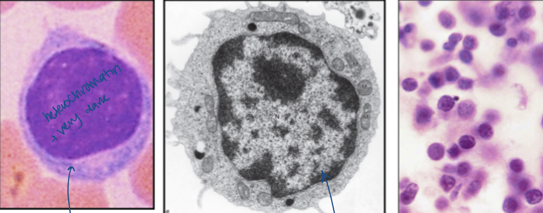
Plasma cells
Heterochromatic nucleus with obvious heterochromatin clumps
nucleus eccentric; more extensive cytoplasm than lymphocyte
Cytoplasm basophilic with light staining Golgi zone
Differentiated daughters of B lymphocytes; only see in CT and lymphoid organs
Clonally selected and specialized for secreting antibody
limited lifespan

Monocytes
largest blood cells
indented nucleus, often kidney, ‘U’ or horseshoe-shaped
No specific granules but azurophilic granules often prominent


Macrophages
Differentiate from monocytes after exit blood
ones that exit as a response to inflam/tissue damage look similar to monocytes but with more phagocytic vesicles
ones that become permanent specialized residents look very different and unique for each organ of residence
best way to identify macrophages is via presence of phagosomes
LM sometimes can see engulfed pigments (especially with haemorrhage)
Paraffin sections
only dependable ID is through presence of phagosomes eg. uptake of particulate dyes or blood pigment
TEM
abundance of lysosomes, phagocytic vesicles and residual bodies
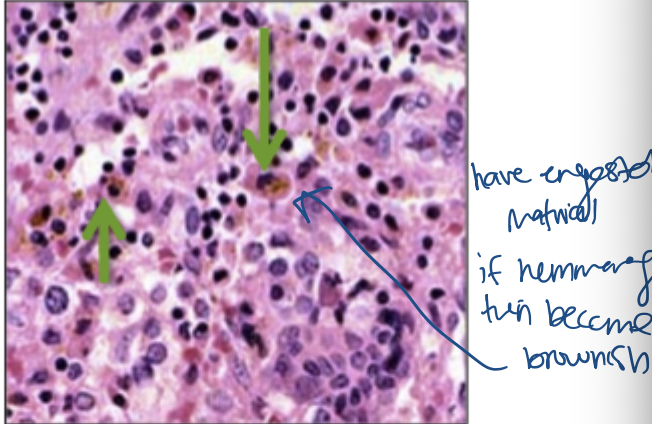

what is function of macrophage
Inflammation
major cell types in sites of inflammation after PMN/neutrophils
kill bacteria via cytotoxic secretions
Phagocytosis
engulf debris, old cells, foreign cells/molecules tagged by antobodies
Immune response
Coordinate immune activities via production of lymphokines and cytokines
antigen presenting cell
Platelets (thrombocytes)
2-3 um diameter formed elements
are plasma membrane-enclosed cytoplasmic fragments of megakaryocytes
no nuclei, but contain other organelles
Function: clot formation (hemostasis) and other signalling cascades
extensive network of membrane channels
numerous granules that contain vasoactive substances, factors for clotting and clot dissolution, growth factors
microtubule and actin network regulate platelet shape and adhesion

coagulation
tissue damage or wounding triggers platelet activation
adhesion characteristics are altered; platelets clump together adhere to damaged tissue and degranulate
platelet factors activate clotting cascade leading to conversion of plasma fibrinogen to insoluble strands of fibrin
fibrin network traps RBSs and forms clot to prevent further blood loss
other platelet factors trigger repair/healing
Cartilage function and location
specialized type of CT; still, pliable resilient and gel-like
resists mechanical stresses, absorbs shock, slides on surfaces
avascular: no blood supply, limit size, has poor capacity for repair, low, anaerobic metabolism
Function: supports soft tissue, facilitates bone movement, guides bone development
Location
articular surfaces of bones
ears and nose
airways and lungs
growth plates of bones
components of cartilage
Extracellular Matrix
Cells
Extracellular matrix made up of
accounts for ~95% volume; solid and firm but pliable
resists compression
Ground substance (80%)
40%-80% glycosaminoglycans (GAGs, proteoglycans, glycoproteins)
rest H2O molecularly bound to GAGs - gives nutrition and ox because tissue not vascularized
Fibers
generally collagen Type II - limits compressive deformation, resists tension
Elastin (in elastic cartilage) and collagen Type I (in fibrous cartilage)

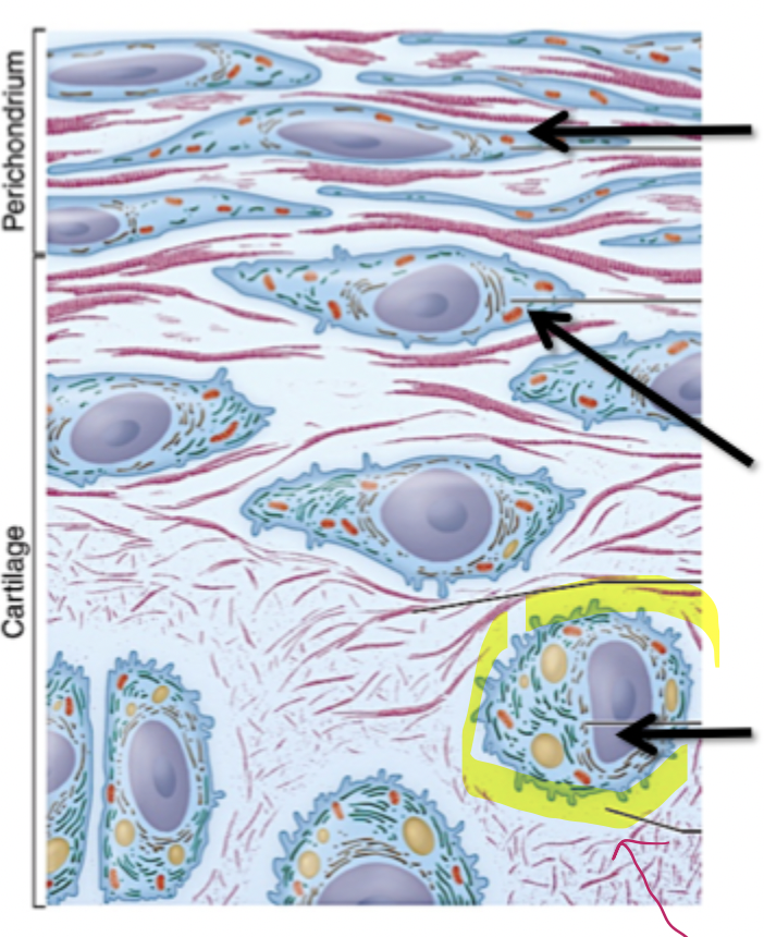
Cells in cartilage made up of
Fibroblast-like cells
edge of cartilage from a dense CT layer called perichondrium
proliferate to produce chondroblasts
Chondroblasts
acts as progenitors
proliferate and differentiate into chondrocyte
Chondrocyte
mature cartilage cells
produce and are trapped in ECM
reside in cavity called lacuna
overview classification of cartilage

Hyaline Cartilage
Chondrocytes sit individually or in small clusters with viable amount of ECM
usually considerable cell shrinking so lacuna seems only partly filled (artifact)
ECM of hyaline cartilage either poorly stained or basophilic (sulfated GAGs)
region of ‘territorial matrix’ (higher GAG, lower collagen) around each chondrocyte
hyaline cartilage chondrocyte actively secreting ECM contains extensive Golgi and RER and mitochondria
Nucleus mostly euchromatin
region ECM immediately adjacent to cell rich in GAG and PGs with collagen type IV
territorial matrix richer in collagen type II; less basophilic in LM
these 2 regions always distinguishable in LM- use territorial matrix for the wall stained area adjacent to cell

Elastic Cartilage
it is basically hyaline with elastin in ECM
Elastin fibres form dense meshwork around chondrocytes
looser network of elastin sheets throughout ECM
elastin cartilage is yellowish when fresh; elastin is not readily stained with H and E (seed specual orcein stain)
elastic cartilage very flexible; springs back into place
unlike hyaline cartilage, it does not calcify

Fibrocartilage
Essentially a mingling of hyaline cartilage and dense fibrous CT
can be distinct layers of each or less organized
Cartilaginous regions contain chondrocytes and sparse matrix; fibrous regions contain fibroblasts
fibrous regions rich in collagen type I: tissue is much more eosinophilic than other cartilage
Cushioning and tensile strength; resist tearing
fibrocartilage chondrocytes contain extensive RER but not much Golgi; not producing much GAGs and PGs
Nucleus is mostly euchromatin
Region of ECM around chondrocytes fairly typical for cartilage
also large prominent bundles of collagen in multiple direction - mostly collagen type I