Clinical Methods Orthopedic Tests
 Call Kai
 Learn
 Practice Test
 Spaced Repetition
 Match
 Flashcards
 Knowt Play
1/106

There's no tags or description
Looks like no tags are added yet.
107 Terms
What Does Adam’s Sign Test for?
Checking For Curvature of the Spine.
Diff. Whether scoliosis is functional or Structural
In Adam’s Sign, if a standing curvature of the spine is no longer present when the patient bends over, is this an example of Functional or Structural Scoliosis
This is A negative sign, indicating a functional scoliosis
In Adam’s Sign, if a standing curvature of the spine is no longer present when the patient bends over, is this an example of Functional or Structural Scoliosis
This is A negative sign, indicating a functional scoliosis
What Is the Adam’s Test
In this test you ask the patient to bend forward at the hip and examine their back to determine if there is an imbalance/curvature that remains when the spine is flexed forward

What is The Belt test?
The Doctor will stabilize a patient’s hips and ask them to bend forward

What does the Belt test test for?
Differentiate between lumbar or sacral lesions
In the Belt test, if a patient reports pain when leaning forward Is this a positive sign or a negative sign?
This is an example of a positive test if pain is reproduced. Indicative of a lumbar lesion
In the Belt test, if a patient does not report pain when leaning forward Is this a positive sign or a negative sign?
This is a negative test. Indicative of a Sacral Lesion
What does the Iliac Compression Test test for
This Test assesses for a fracture of the ilium or sacroiliac lesion, sprain, subluxation, etc
How does the Iliac Compression Test work?
The patient lays on their side and the doctor, from behind the patient, places hands on the patient’s hip, between the iliac crest and greater trochanter, and exerts downward pressure on the pelvis
What would result in a positive result for the Iliac Compression Test?
If the patient experiences SI joint pain when the doctor pushes down on their hips.
What is the Degerine Sign? What indicates if this sign is present for a patient?
The Dejerine Sign is a test that suggests a space occupying lesion in the patients spine (Thoracic + Lumbar) if the patient experiences pain when coughing, straining, and sneezing
What might be implied if a Patient experiences radicular pain with a Positive Degerine Sign?
There may be neurological compression present- also helps to identify a more specific location of SOL
What is The Valsalva Maneuver?
This is a test that works similarly to the Dejerine sign, encouraging a patient to increase their interthecal/abdominal pressure and reproduce their pain.
What ways can you explain the Valsalva Maneuver to a patient?
Exhale with pursed lips and nose closed
Simulate blowing up a balloon
Bear Down as if simulating a bowel movement
What are the Significant Findings of the Valsalva Maneuver?
Local Pain indicates the possibility of a SOL (herniation, tumor, infection)
Radicular pain indicates SOL causing Nerve Root Compression
What does the Bechterew Sitting Test test for?
This test attempts to reproduce a patients lower back pain, to identify if it originates from the lumbar region, and if the pain radiates.
How does the Bechterew Sitting Test Work?
The patient is seated and the doctor will extend one of the patients legs and apply downward pressure on the thigh. (Resisting Hip Flexion). Test with other leg, and then have Patient extend both legs

What is an Important note about the Bechterew Sitting Test?
If the patient experiences pain at any time, or exhibits the Tripod sign during the test, the test should be stopped and a positive sign has been exhibited.
What are the Positive signs for the Bechterew Sitting Test?
LBP Reproduced: Target tissue may have a spasm, disc lesion, or subluxation
Radiating Leg Pain Reproduced: Disc lesion, nerve root compression
Unable to Perform (Tripod Sign): Disc lesion/Sciatic Nerve Involvement
What does the Kemp Test test for
This test differentiates between different types of pain (Radiating, radicular, local) and target tissues (Nerve Root, Strain, Sprain)
How does the Kemp Test Work?
There is both a seated and standing version, but in both the Doctor will place an arm across the patient’s shoulders and stabilize a side of the lateral lumbar spine with a fist. (The side with the fist is what is being tested) The doctor moves the patient’s upper body in this order: Lateral flexion away, forward flexion, and lateral flexion towards
Why would the Doctor want to do the standing version of Kemp’s Test?
The standing version may emphasize facet joint pain more
In Kemp’s Test, if radiating pain is produced, what might this be a sign of?
This is the Positive finding, and would be indicative of a nerve root compression
In Kemp’s Test, what would it mean if the Patient experienced pain when leaning away from the area being tested
This is a provocative finding that indicates a muscle sprain or strain
In Kemp’s test, what might be the issue if the patient experiences pain when in extension and rotation (When leaning towards pain at the end of test)
This is a provocative finding that would be likely due to facet issues
If a patient has a lateral disc protrusion/SOL, what direction of flexion will relieve their pain? Towards or away from the Lesion?
Leaning away from this kind of Lesion will lesson the nerve compression and reduce pain
If a patient has a medial disc protrusion/SOL, what direction of flexion will relieve their pain? Towards or away from the Lesion?
Leaning towards this kind of lesion will reduce nerve compression and reduce pain
What does the Straight Leg Raising Test test for?
This test aims to differentiate between different caucuses of lumbosacral pain that radiates into the leg (SOL, SIJ inflammation, etc)
What are the target Tissues of the Straight Leg Raising Test?
Sciatic Nerve, Nerve Roots, Dura
How do you perform the Straight Leg Raising Test?
Patient lays on their back and the doctor will lift the leg that exhibits radiating pain. (Make sure to support knee and Ankle)
If pain is reproduced, the doctor will ask the patient to touch their chin to their chest (In order to further increase traction to the nerve)
What are the Significant Findings of the Straight Leg Raising Test?
Raising Leg reproduces pain
Pain from 0-35°: Indicates a SIJ Lesion, Lumbosacral Issues, or subluxation
Pain from 35°-70°: Indication of Dural adhesions or a SOL deforming the nerve root (disc protrusion, osteophyte, tumor)
Pain above 60°-70°: Pain is likely due to movement of a Lumbar joint
(Note that additional pain from traction in the neck is further evidence of nerve root issue)
What does the Well Leg Raise test for?
This test is to differentiate between medial and lateral SOL/Disc Protrusion
How does the Doctor Perform the Well Leg Raise Test
The Patient will lie on their back (Supine) and the doctor will raise the leg that does not exhibit radiating pain (Make sure to support knee and ankle in extension).
If the patient experiences pain with this movement, the Doctor will lower the leg just before pain is reproduced and perform Bragard and Sicard Sign.
What are the Significant Findings of the Well Leg Raise test?
If Radiating leg pain is reproduced when lifting the unaffected side, it is indicative of a Medial disc protrusion, SOL, or dural adhesion
How do you perform Bragard Sign Test
This test is done if the patient experiences pain during the Well Leg Raise test
Doctor will raise the Well leg to just below reproduced pain and dorsiflex the ankle

How do you perform Sicard Sign Test?
This test is done if the patient experiences pain during the Well Leg Raise test
Doctor will raise the Well leg to just below reproduced pain and dorsiflex the great toe

What should you do between performing Bragard and Sicard sign?
Allow the patient to rest their ankle on the table before lifting their well leg again to perform the other test
What is the purpose of Bragard and Sicard Sign tests?
These tests will further confirm that nerve root tension is the pain generator
What is/are the significant findings of Bragard and Sicard signs
Pain reproduced with dorsiflexion of the ankle or great toe are positive findings indicative of Sciatic radiculopathy from a SOL or dural adhesion
What is the Purpose of the Milgram Test?
This test assesses for the presence of a SOL/Disc protrusion
How do you perform Milgram test?
Patient lies supine and is instructed to lift their feet 6 inches off the table (You may lift the patients legs and let go rather than purely instruction)
Have the patient hold this position as long as they can (30 seconds normal), and see if this reproduces their pain
What are the Significant Findings of the Milgram Test
The positive finding for this test is when the patient experiences their lower back pain- indicates a SOL
What are the provocative findings of the Milgram test
Patient may not be able to hold position due to abdominal weakness
Pain can also be produced if the patient has a lumbar sprain or strain (can affect lumbar ligament, muscles, or joints)
What is the purpose of Patrick/FABERE test?
This test assesses the hip joint for mobility and pain.
How do you perform Patrick/FABERE Test?
Patient lies supine and the doctor will hold the patient’s ankle and flex their knee (taking ankle to the opposite leg, either proximal leg or distal thigh- do not rest patient’s ankle on their knee)
Then stabilize the patient’s ASIS on the opposite side and apply downward pressure to the knee

What are the Positive Significant Findings of the Patrick/FABERE Test?
Hip pain is reproduced for the patient- This would indicate a hip joint pathology (Osteoarthritis, Rheumatoid arthritis, etc)
What are the Provocative Significant Findings of the Patrick/FABERE test?
SIJ pain is reproduced- can indicate sacroiliitis, SIJ subluxation, SIJ sprain
Can assess tightness in muscles surrounding the hip joint
What is the purpose of the Thomas Test
This is a mobility test for flexion and tightness of the iliopsoas. This test is not for reproducing pain.
How do you perform Thomas Test?
The patient lies supine and the doctor makes a note of the space between the patients knee and the table (measure with fingers/hand)
Have the patient hug their knee to their chest (the leg being tested is the leg lying straight not the bent one), and measure if the knee on the patient’s. Straight leg lifts higher off the table

What are the positive Significant Findings of the Thomas Test?
The Lumbar spine maintains a lordosis or lordosis curvature increases as they hug their knee
The space between the patient’s knee and the table increases- indicating tight iliopsoas muscles
What does the Goldthwait sign test for?
The purpose of this test is to differentiate between pain in the SIJ or the Lumbar Spine
How did you perform Goldthwait Sign?
The patient lies supine on the table and the doctor will (ask to) slide their hand under the patient’s back and palpate L4 & L5 and S1/S2 Tubercles
Doctor will slowly raise the patient’s affected leg first (Be sure to be aware of what part of the spine is moving as you raise the patient’s leg)
Repeat process with unaffected leg

When testing for the Goldthwait Sign, if the patient experiences pain with minimal leg lifting (<25°) what area is likely affected?
This is Indicative of pain originating from the Sacral Region (SIJ)
When testing for the Goldthwait Sign, if the patient experiences pain with more significant leg lifting (>25°) what area is likely affected?
This is Indicative of pain originating from the Lumbar spine
What are other Significant findings to be aware of during the Goldthwait Sign test?
If the patient experiences pain at roughly the same height in both the affected and unaffected leg when lifting then this is indicative of a lumbosacral lesion
If the unaffected leg can be raised higher than the affected leg without pain, this indicates sacroiliac involvement on the affected side
What is the Purpose of Gaenslen Test?
This test aims to assess and test for sacroiliac pathology using Pelvic Torsion
How do you perform Gaenslen Test?
Patient is lying supine for this test, the side that is tested is closest to the edge of the table (test unaffected side first) Then ask the patient to hug their knee of the leg not being tested (the Doctor may also hold the patient’s leg for them)
Then instruct the patient to slide their other leg off of the side of the table, ensure that their foot is not touching the ground.
The doctor will slowly hyperextend the hip by pushing downwards on the top of the knee of the leg being tested
Repeat for the affected side
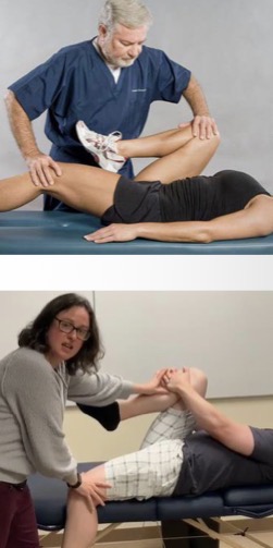
What is/are the significant findings for Gaenslen Test?
If pain is reproduced in SIJ or referred down the thigh then this indicates a sacroiliac pathology
What is the purpose of Sacroliliac Stretch Test?
This is the Most Specific Test. It assesses for a sprain of the anterior sacroiliac ligaments
How do you perform the Sacroiliac Stretch Test?
The latent will lie supine with legs extended. With crossed (or uncrossed) arms, the doctor will place their hands on the patient’s ASIS (be aware to use the soft sides of your hands to be as comfortable to the patent as possible)
The doctor will gently and slowly apply downward lateral pressure bilaterally for up to 30 seconds.

What are the Positive Significant Findings of the Sacroiliac Stretch Test
Unilateral gluteal pain in this test is indicative of a sprain of the anterior sacroiliac ligament
What are the Provocative significant findings for the Sacroiliac Stretch Test
This test can be uncomfortable on the patient’s ASIS, and cause pain to the SIJ
What is the purpose of the Thigh Thrust Test?
This test is the most Sensitive Test. It will assess for involvement of the sacroiliac joint in the patient’s pain
How do you perform the Thigh Thrust Test?
Patient is supine on the table and the Doctor will stand on the side of the patient being tested. The doctor will place a hand between the table and the patients sacrum to stabilize the sacrum. With the other hand, the Doctor will flex the patient’s knee.
The doctor will apply vertical force through the line of the femur and do the same with the knee positioned slightly superior and inferior.
Then the doctor will abduct the knee (away from median) and apply similar force, posterior, superior and inferior. And do the same three more times with the Knee slightly adducted.
This. Test ends when any pain is produced.

What are the positive significant findings of the Thigh Thrust Test?
Reproduction of SIJ pain, indicating SIJ involvement
What is the purpose of the Sacral Thrust Test?
This test assesses for issues with the sacroiliac joint
How do you perform the Sacral Thrust Test?
The patient will lie prone for this test. The doctor will place the heel of their hand on the base of the sacrum to start, with fingers pointed superiorly, and apply a small amount of downward pressure. If there is still no pain at this point, the doctor can lightly spring on the joint. These actions are then repeated on the apex of the sacrum, being sure to use less force here as it is more sensitive.

What are the Significant findings for the Sacral Thrust Test?
If SIJ pain is reproduced then this test indicated that there is SIJ involvement
What is the Purpose of the Nachlas Test?
This test determines any issues with the sacroiliac joint and/or the lumbosacral area
How do you Perform the Nachlas Test?
The patient is prone for this test. The doctor will test the unaffected side first. Stand opposite of the side you are testing and start by stabilizing the sacrum with one hand and lifting the patient’s opposite knee from the ankle to 90°. Then the Doctor will bend the leg further, try to get the patient’s ankle to their buttcheek of the same side. Then repeat this for the patient’s symptomatic side

What are the significant findings for the Nachlas Test?
If localized pain in either the SIJ or lumbosacral area is produced then this indicates a disorder or issue of those areas
This test will also stretch the patient’s anterior thigh
What is the Purpose of the Hibbs Test?
This Test assesses for anterior sacroiliac involvement
How do you perform the Hibbs Test?
Similarly to the Nachlas test, the patient is Prone and the doctor will first test the unaffected side by stabilizing the pelvis over the side nearest to the doctor (the side opposite of the leg being tested) and lifting the patient’s leg. Then bend the heel towards the patient’s buttock. From this position, slowly push the patient’s leg laterally to cause internal rotation of the femoral head. Repeat this process for the affected side.

What is the Positive Significant Findings of the Hibbs Test?
If SIJ pain is reproduced then this would be indicative of an issue involving a sacroiliac lesion
What is the Provocative Significant Findings from the Hibbs Test?
If hip pain is produced then this would be an indicator that there is a hip pathology
What is the Purpose of the Ely Sign Test?
The purpose of this test is to assess for lumbar radicular involvement or femoral nerve inflammation
How do you perform Ely Sign Test?
The patient will lie prone and the doctor will test the unaffected side first. Stand to the side of the patient opposite of which leg you are testing with. Apply firm pressure to the SIJ of the side being tested to stabilize the pelvis. Lift the leg to 90° and then slowly lower the patient’s heel to the contralateral buttock (opposite side). This causes external rotation of the femoral head. After this step the thigh can be hyperextended (lift leg off table slightly). Repeat for the affected side.

What are the Significant Findings of the Ely Sign Test?
If the Patient experiences pain on their Anterior Thigh then this indicates that there is inflammation of lumbar nerve roots relating to the femoral nerve. (L1-L3 Disc Herniation)
If the Patient experiences upper lumbar pain, this would be the result of stretching lumbar nerve adhesions
What is a provocative Significant Finding of the Ely Sign Test?
If the patient experiences pain in their SIJ, this would indicate a sacroiliac joint lesion
What is the Purpose of the Yeoman Test?
This test assesses for an anterior sacroiliac ligament injury
How do you perform the Yeoman Test
The patient will lie prone for this test. The doctor will stand opposite of the side they are testing and work on the unaffected side first. The doctor will apply firm pressure over the nonaffected SIJ to stabilize the pelvis. In the other hand, the doctor sill flex the patient’s knee and lift the knee off of the table. Repeat for the symptomatic side.

What is the Significant Finding of the Yeoman Test
Patient will feel increased pain in their sacroiliac joint.
This test will also stretch the quad and hip flexors.
What is the purpose of Schepelmann Sign?
This test assesses for costal and intercostal tissue integrity
How do you perform Schepelmann Sign?
Seated or Standing
Have the patient laterally flex their body in either direction and identify what side they experience pain on

What is the significant finding for Schepelmann Sign test if the patient feels pain on the Convex Side?
Pain on this side indicates Myofascial/intercostal strain
What is the significant finding for Schepelmann Sign test if the patient feels pain on the Concave Side?
Pain on this side indicates Intercostal Neuritis
What is the purpose of the Chest Expansion test?
This test is an early indicator for involvement of costovertebral joints in ankylosing spondylitis
How do you perform Chest expansion chest (Measured Version)
Doctor will place the tape measure at T10 level and wrap around to the back. Have the patient exhale fully and measure this range. Then have the patient fully inhale and measure again at the end range of motion.
The difference between inhalation and exhalation should be 1.5-3 inches

How do you perform Chest Expansion test (Approximated Thumbs Version)
Patient is sitting for this test. Doctor will place thumbs on the inferior angles of the scapula and allow the patient to exhale. During the exhale squeeze your thumbs together before the patient inhales, and observe the distance your thumbs travel and look for symmetrical ribcage movement

What are the Significant findings of the Chest Expansion Test?
Limited movement is an indicator of Ankylosing Spondylitis. Could also be Due to poor diaphragmatic breathing or COPD
What is the Purpose of the Passive Scapular Approximation test?
This test will assess for a T1 or T2 nerve root problem
How do you perform the Passive Scapular Approximation Test?
Have the patient be seated and stand behind them. The doctor will passively adduct the scapulae bilaterally (Squeezing them together)

What is the significant findings of the Passive Scapular Approximation Test?
If the patient feels a sharp pain in between their scapular, then this would indicate a T1 or T2 nerve root irritation
Note that a provocative finding for this test is stretching the patient’s pecs if they are tight.
What is the Purpose of the Sternal Compression Test?
This test assesses for a possible rib fracture
How do you perform a Sternal Compression Test?
The patient may be seated or lying supine for this test. The patient creates a barrier over their chest with their hands over their sternum. From here, the doctor can place a hand over their sternum patient’s hands and their back (if seated) and apply Anterior to posterior pressure. The doctor will then apply lateral pressure on the ribcage.

What are the significant findings of the Sternal Compression Test?
If the patient experiences rib pain during this test, that would be indicative of a rib fracture. AP pressure often exacerbates a lateral rib fracture and vice versa for lateral rib cage pressure.
Rib Pain can also be indicative of a rib sprain or subluxation
What is the purpose of the Soto-Hall Sign test?
This test will assess for a cervical (sometimes thoracic) subluxation, IVD lesion, strain, sprain, SOL, meningitis, etc
How do you perform the Soto-Hall Sign test?
Have the patient lie supine and place their hands over their sternum. The doctor may place their hands over top of the patient’s to stabilize the sternum. Then the doctor will hold the back of the patient’s head, supporting fully, and lift their head off the table.

What is the significant findings of the Soto-Hall Sign test?
If the patient experiences localized cervicothoracic pain, this would be indicative of a sprain, strain, subluxation, SOL, or other. This test is not super specific.