Anatomy Lab Practical #2
 Call Kai
 Learn
Learn Practice Test
 Spaced Repetition
 Match
Match Flashcards
 Knowt Play
1/61

Earn XP
Description and Tags
Integumentary System, Bones and Skeletal Tissue, The Skeleton (Axial and Appendicular)
Name | Mastery | Learn | Test | Matching | Spaced | Call with Kai | Chat |
|---|
No analytics yet
Send a link to your students to track their progress
62 Terms

Stratum Corneum
Most superficial layer of the epidermis.


Identify Keratinocytes
Keratinocytes produce keratin and make up the majority of the epidermis, primarily located in the stratum spinosum and stratum corneum.


Identify Melanocytes
Melanocytes produce melanin, the pigment responsible for skin color, and are primarily located in the stratum basale of the epidermis. They protect against UV radiation.


Identify Dendritic Cells
Dendritic cells are immune cells that help detect and respond to pathogens. They are primarily located within the stratum spinosum of the epidermis.


Identify what is attached the the Merkel cell (tactile epithelial cell)
Sensory nerve ending is attached to the tactile epithelial cell in the stratum basale.


Identify cell type (blue colored cell)
Tactile epithelial cell, also known as Merkel cells, are specialized cells involved in the sensation of touch.
They are primarily located in the stratum basale of the epidermis, close to sensory nerve endings.


Identify the structure
Dermal Papillae
It is located in the papillary dermis, superficial to the reticular dermis and accounts for 20% of the dermis.
It’s peglike projections indent the overlying epidermis.
They contain capillary loops, pain and touch receptors (tactile corpuscles, meissner’s corpuscles).

Reticular Dermis
Composed of dense irregular connective tissue,
deep to papillary dermis (80% of thickness of dermis),
well vascularized.

Identify the structure.
Sebaceous Oil Gland
Simple branched alveolar glands found all over the body except thick skin of palms and soles.
Secrete sebum that lubricates hair and skin while being bactericidal.
Are attached to hair follicles.


Identify the structure
Hair Bulb
Deep end of the follicle.


Identify the structure.
Arrector Pili Muscle
Smooth muscle fibers attached to hair follicles.
Pull hair follicle to release sebum and cause goosebumps in response to cold temps or fear.

Papillary Layer Connective Tissue is?
Loose Areolar Connective Tissue
Reticular Layer Connective Tissue is?
Dense Irregular Connective Tissue
7 Functions of Bones
Support,
Protection,
Anchorage,
Mineral Storage,
Blood Cell Formation (hematopoiesis),
Triglyceride storage,
Hormone Production

Identify the structure.
Osteon
Structural unit of compact bone.
Elongated cylinder shape oriented parallel to long axis of the bone.
Looks like tree rings.

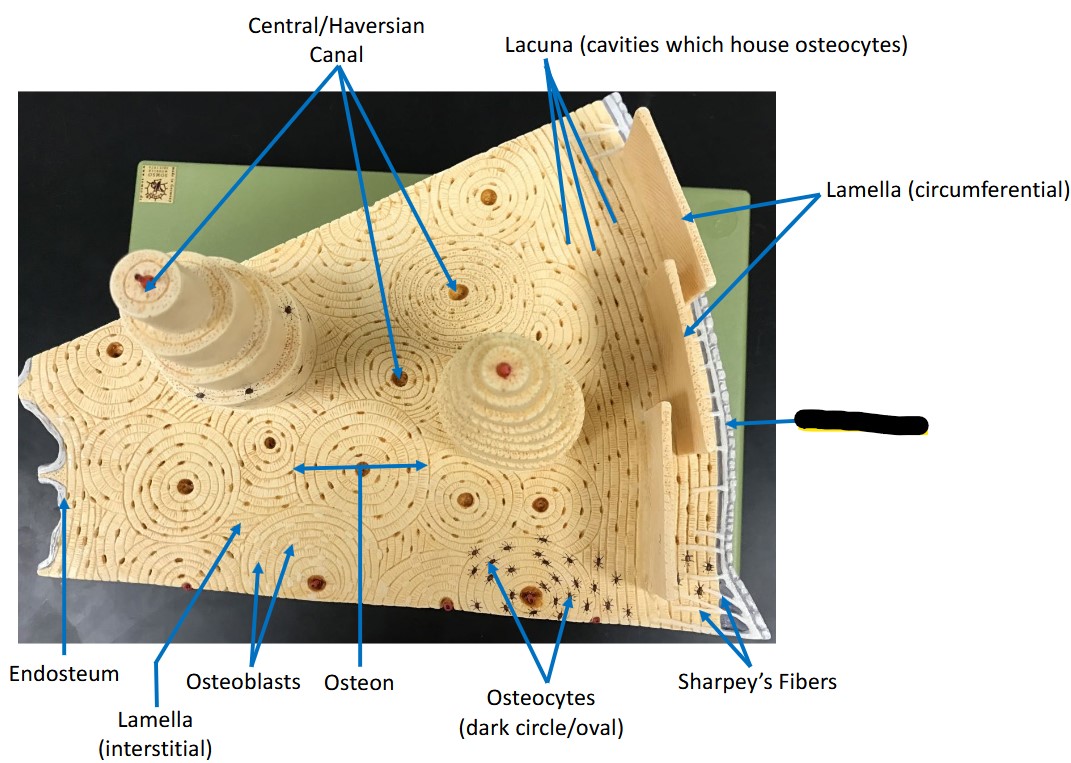
Identify the structure.
Periosteum
Forms on the superficial layer of bone.


Identify the canal.
Haversian/central canal, contains small blood vessels and nerve fibers.
Run parallel to bone axis.


Identify the canal.
Volmann’s/perforating canals, connect blood and nerve supply of medullary cavity to central canals. Lie at right angles or “perforate” through lamella.


Identify the cell type
Osteocytes - Mature bone cells, monitors mineralized bone matrix, occupy lacunae.

Articular Cartilage
Hyaline cartilage that covers ends of bones at movable joints, providing smooth, lubricated surface for articulation.

Identify the structure.
Epiphyseal line - remnant of epiphyseal plate, located at epiphysis.


Identify the cavity.
Medullary Cavity - central, hollow space inside the diaphysis of long bones.


Identify the bone landmark.
Distal epiphysis - rounded, bulbous end of long bone farthest from point of attachment to body trunk.

Long bone shape and amount in adult body.
Longer than they are wide, contain a medullary cavity.
90 long bones in human adult body.
All limb bones except patella, wrist and ankle bones are long.
Short bone shape and amount in adult body.
Roughly cube shaped.
28 short bones in human adult body on average.
Bones of wrist and ankle are short.
Flat bone shape and amount in adult body.
Thin, flattened, usually curved a bit.
36 flat bones in human adult body.
Sternum, ribs and most cranial bones are flat.
Sesamoid bone shape and amount in adult body.
Shaped like a sesame seed.
4 sesamoid bones in human adult body.
Patella is a sesamoid bone.
Irregular bone shape and amount in adult body.
Complicated shapes that fit no other class.
48 irregular bones in human adult body.
Vertebrae and hip bones are irregular.

Identify the bone.
Parietal bone - two total, most of superior and lateral aspects of the skull


Identify the bone and landmarks
Temporal Bone - landmarks: Mastoid Process, Zygomatic Process, External Acoustic Meatus

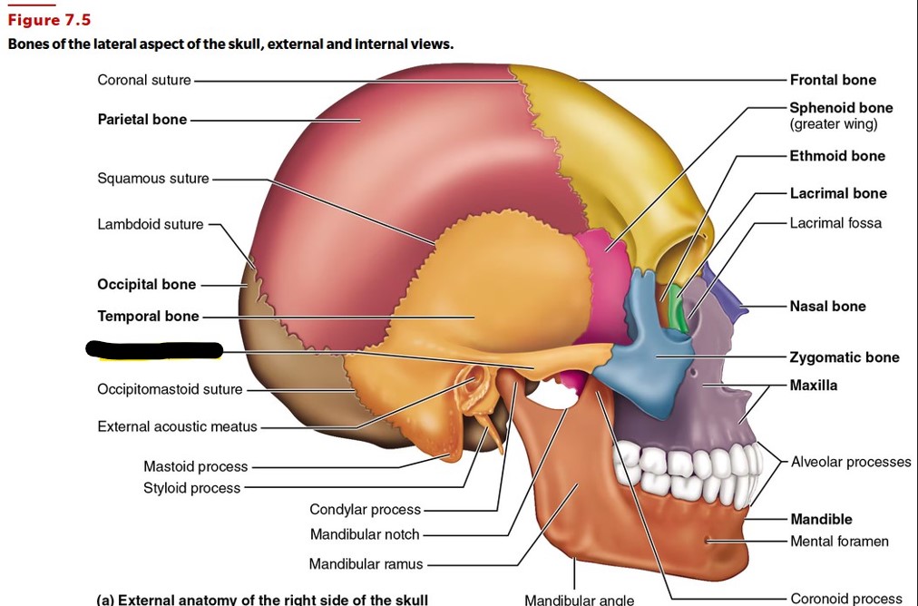
Identify the bone landmark.
Zygomatic process - Contributes to zygomatic arch, forms cheekbones.


Identify the bone landmark.
Mastoid process - Attachment site for several neck muscles.


Identify the landmarks
Occipital bone - landmarks: Occipital Condyles, Foramen Magnum


Identify the landmark.
Jugular Foramen - Passageway for internal jugular vein and cranial nerves IX, X and XI.

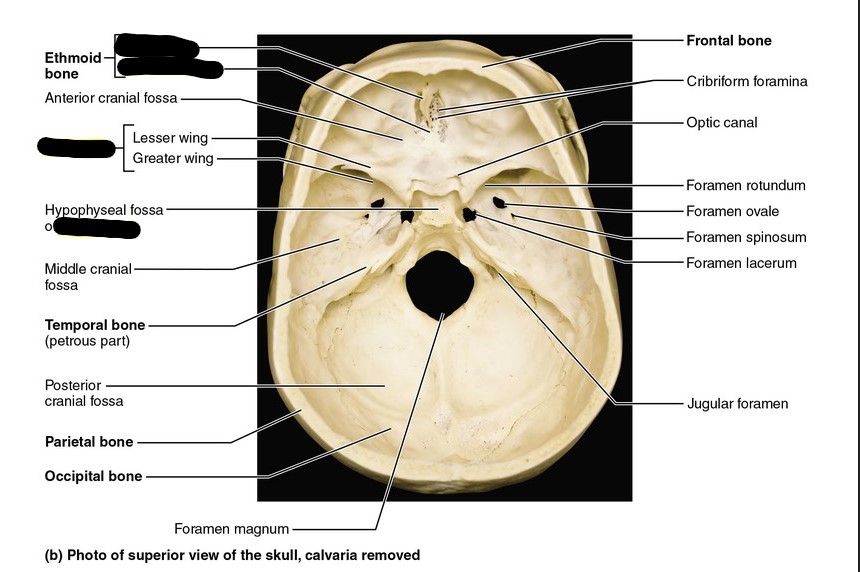
Identify bone and landmark.
Sphenoid bone landmark: Sella Turcica (houses pituitary gland)
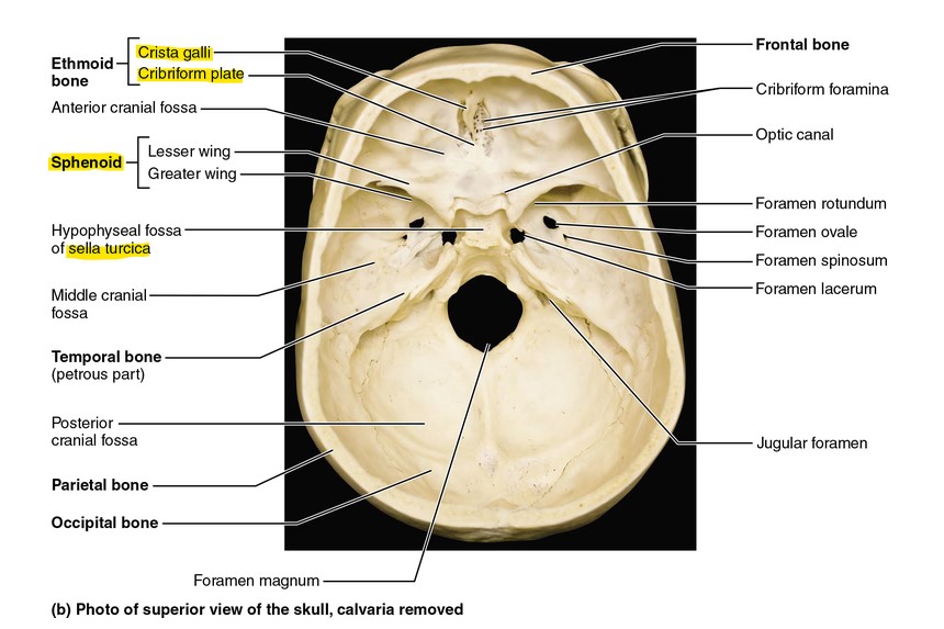

Identify the Ethmoid bone landmarks.
Cribriform Plate, Crista Galli


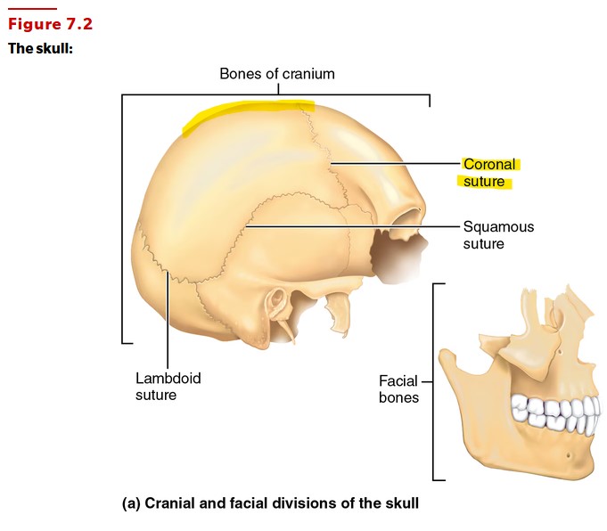
Identify the sutures (including superior highlight)
Sagittal and Coronal (sagittal suture not pictured but it runs down midline of skull posterior from coronal suture)

Identify the paranasal sinuses
Frontal, Ethmoid, Sphenoid, and Maxillary Sinuses (FESM). Clustered around nasal cavity.
They help warm and humidify inhaled air while lightening skull and enhancing voice resonance.


Identify the facial bones and landmarks (pt. 1)
Mental Foramen of Mandible
Mandible
Coronoid Process of Mandible
Condylar Process of Mandible
Maxilla


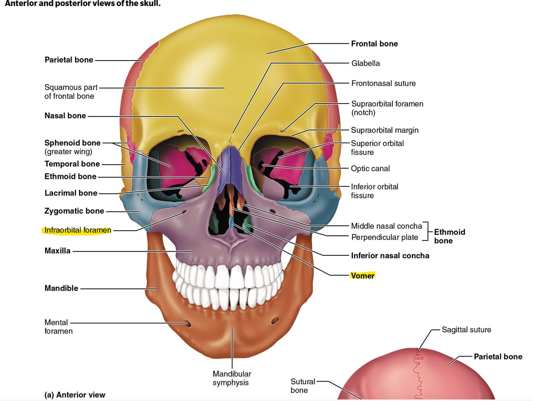
Identify the facial bones and landmarks (pt. 2)
Vomer, Infraorbital Foramen

Identify the bones and landmarks (pt. 3)
Hyoid Bone
Palatine Bone
Foramen Lacerum
Greater Horn of Hyoid Bone
Auditory Ossicles: Malleus, Incus, Stapes


Vertebral Column - Number of bones in each section and their names.
Cervical (C1-C7) (7 total);
Thoracic (T1-T12) (12 total);
Lumbar (L1-L5) (5 total)


Vertebral Column - Distinguish each type of vertebrae with evidence.
Cervical: Smaller, have transverse processes (protects, wide opening.
Thoracic: Medium size, spinous process projects downward.
Lumbar: Large size, big body, small opening.


Cervical Vertebrae - C1 and C2 and their importance
C1 - Atlas (no spinous process, superior facets receive the occipital condyles, allow you to nod “yes”).
C2 - Axis (has dens projecting superiorly, allows you to nod side to side for “no”).


Vertebrae - Identify and distinguish landmarks
Body
Spinous Processes
Transverse Processes
Foramen
Pedicle


Vertebral Column - What are the names of the most inferior bones?
Sacrum: 5 fused bones as adult.
Coccyx: 3 fused as adult (AKA tailbone).


Thoracic Cage - Names and amounts of each type of rib
True Ribs (connected directly to sternum): 7 pairs, (1-7).
False Ribs (not directly connected to sternum): 5 pairs (8-12).
Floating Ribs (connect only to vertebral column): 2 pairs (11-12)


Rib - Identify landmarks and type of bone
Flat bone, tubercle and shaft (way to remember proper rib orientation: the flat head of a rib articulates with costal facets on thoracic vertebrae).


Sternum - Identify landmarks
Jugular Notch (superior part of sternum)
manubrium (just inferior to JN)
Body
Xiphoid Process (most inferior part of sternum)
(sternum looks a lot like a tie)


Scapula - Identify landmarks
Supraspinatus fossa
vertebral border
acromion process (highest point)
glenoid fossa (head of humerus articulates here).

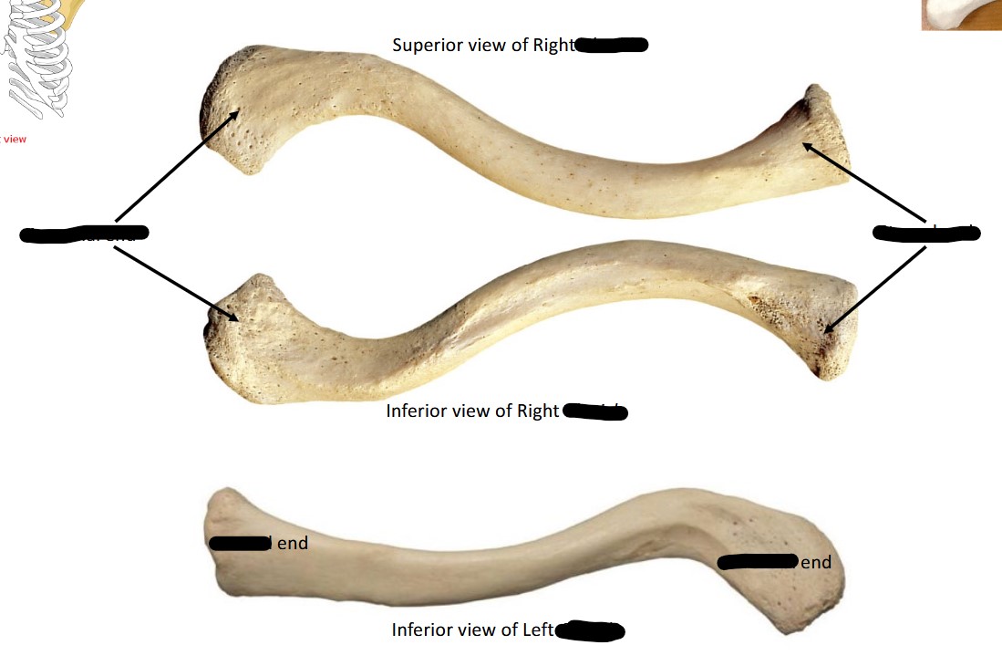
Clavicle - Identify Bone and landmarks
Acromial end: points towards scapula, is broad, thin and flat, features a tubercle (small bump).
Sternal end: points toward sternum, large, thick and rounded.


Humerus - Identify Bone and Landmarks
Greater tubercle (proximal end of humerus)
capitulum (distal end)
Olecranon fossa


Radius and Ulna - Identify each bone and their landmarks
Olecranon Process - Ulna
Trochlear notch - Ulna
Styloid Process of Ulna.
(way to remember: “U”lna has the U shaped notch, the other bone is the Radius, their styloid processes can be differentiated by the size of the epiphysis (distal end))


Identify the Radius
Differences of the radius compared to the fibula: Radius is thicker, has a flat head, has a styloid process and a wider distal end (epiphysis).


Wrist and Hand - Identify name and number of palm bones and wrist bones
Wrist Bones (3 that are need to know): Pisiform, Lunate, Scaphoid.
Metacarpals: (5 total, start from thumb) I,II,III,IV,V


Pelvic Girdle - Identify Landmarks
Iliac crest
greater sciatic notch
ischial spine
acetabulum (deep, point outward, head of femur articulates here)


Male vs. Female Pelvis
Male Pelvis: Thick and heavy, Pubic Arch <90 degrees, Pelvic Brim is narrow and heart shaped.
Female Pelvis: Light and thin, Pubic Arch >90 degrees, Pelvic Brim is wide and more oval.


Femur - Identify bone and landmarks
Greater trochanter
Linea Aspera (on shaft/diaphysis)
Medial Condyle (most distal)
Right Femur (most likely to be used in practical as well as other disarticulated bone examples)


Identify this bone and its shape
Patella (kneecap), largest sesamoid bone in the body.


Identify these bones and their landmarks
Tibia and Fibula (compare tibia to fibula, tibia is thicker)
Tibial tuberosity (roughened area)
Anterior crest (border) (compare to linea aspera on posterior of femur)
Medial malleolus (distal end of tibia, surrounds tarsal bones)


Ankle and Foot Bones - Identify bones (4)
Talus (ankle bone)
Calcaneus (heel)
Navicular (looking down at foot, is in front of talus)
Cuboid (part of lateral border of foot, posterior to phalanges IV and V)
Way to remember:
Tall Camels Never Consume Cubes (Tall = Talus, Camels = Calcaneus, Never = Navicular, ConsumeCubes = Cuboid)


Fetal Skeleton - Identify fontanelles
Fontanel are “soft spots” on cranium, turn into sutures after 2 years
Anterior Fontanel
Sphenoidal/anterolateral Fontanel
