BIOS3220 Exam 1 Material
 Call Kai
 Learn
 Practice Test
 Spaced Repetition
 Match
 Flashcards
 Knowt Play
1/194

There's no tags or description
Looks like no tags are added yet.
195 Terms
List 4 examples of cellular microbes
1) Fungi (yeasts + molds)
2) Protists (algae, protozoa, slime molds)
3) Bacteria (E. coli)
4) Archaea (methanogens)
List 4 examples of acellular microbes
1) Viruses (Protein + nucleic acid)
2) Viroids (RNA)
3) Satellites (nucleic acid, often RNA)
4) Prions (Protein)
Name the 3 domains of life
Bacteria, Archaea, Eukarya
(Based on comparison of ribosomal RNA genes)
Information about Bacteria
Mosty single celled
Majority have peptidoglycan in cell wall
Most lack membrane bound nucleus
Cyanobacteria produce lots of O2
Information about Archaea
Lack peptidoglycan in cell walls
Have unique membrane lipids
Some have unusual metabolic characteristics
Many live in extreme environments
Types of Protists (eukaryotic)
Generally larger than bacteria/archaea
Algae: photosynthetic
Protozoa: may be motile, hunters/grazers
Slime molds: Two life cycle stage
Water molds: devastating disease in plants
Types of Fungi (eukaryotic)
Yeast - unicellular
Mold - multicellular
3 types of Acellular infectious agents
1) viruses
2) Viroids and satelites
Prions
what are viruses composed of
smalled of microbes, requires host
What are Viroids and sateites composed of
Infectious agent composed of RNA
What are Prions composed of?
Infectious proteins
What is the earliest molecule in life?
RNA
Has protein and hereditary function
Ribozymes
RNA that can perform cellular work while also holding genetic information.
Earliest cells were like RNA surrounded by lipids
ATP is a ribonucleotide
What is endosymbiosis theory?
Bacteria were engulfed by larger cells, which explains the complexity of cells that now have a mitochondria/chloroplast
What is 16s rRNA
1500 nucleotide
Has structural role in small subunit of ribosome
has conserved and variable regions (good for mapping phylogenetics)
What is LUCA?
last universal common ancestor; common ancestral cell from which all cells descended
Horizontal vs Vertical gene transfer
Vertical: Cells replicate and divide, mutations cause evolution
Horizontal: microbes release and share DNA which recombine with genomes to greatly enhance diversity
Who was Antony van Leeuwenhoek
First person to see bacteria under a microscope 1670
What is spontaneous generation theory?
organisms arise spontaneously from non-living matter
Disproved by Louis Pasteur (thus pasteurization) via swan neck flask experiment
What is Miasma theory?
Disease caused by foul air and bad smells
What did Joseph Lister do?
Provided evidence that microbes caused disease
Started the development of sterile technique which reduced infection in his patients
Who was Robert Koch?
Established relationship between Bacillus anthracis and anthrax
Koch's Postulates
How to prove relationship between microbe and disease:
1) Microbe must be present in disease and absent in healthy case
2) Suspected microbe must be isolated and cultured
3) Same disease must occur when culture is used to inocculate healthy indiv.
4) Same microbe must be isolated from diseased host
Koch's Postulates limitations
ethics, asymptomatic carriage, co-infections, microbe imbalance diseases (dysbiosis)
Nomenclature: binomial system
Genus species
How to name a gene
3 lower case, followed by 1 upper case
italicized
abcD
How to name a protein
First and last of 4 letters capitalized
not italicized
AbcD
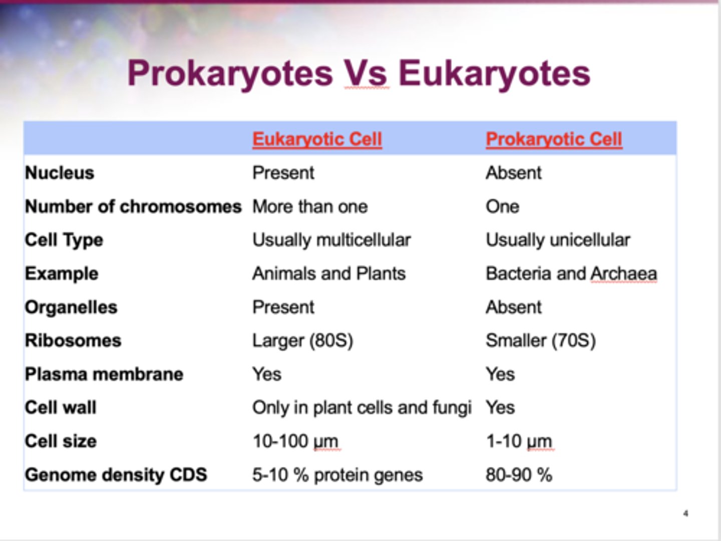
Prokaryotes vs Eukaryotes Table
What is the shape of cocci?
Spheres
How do diplococci arrange themselves?
In pairs
How do streptococci arrange themselves?
In chains
How do staphylococci arrange themselves?
In grape-like clusters
What is the arrangement of tetrads?
4 cocci in a square
What is the configuration of Sarcina?
Cubic config of 8 cocci
What is the shape of Bacilli/bacillus
rods
Coccobacilli: very short rods (cocci-like)
Streptobacilli - Chains of rods
What shape is a Vibrios
Curved rod, comma shaped
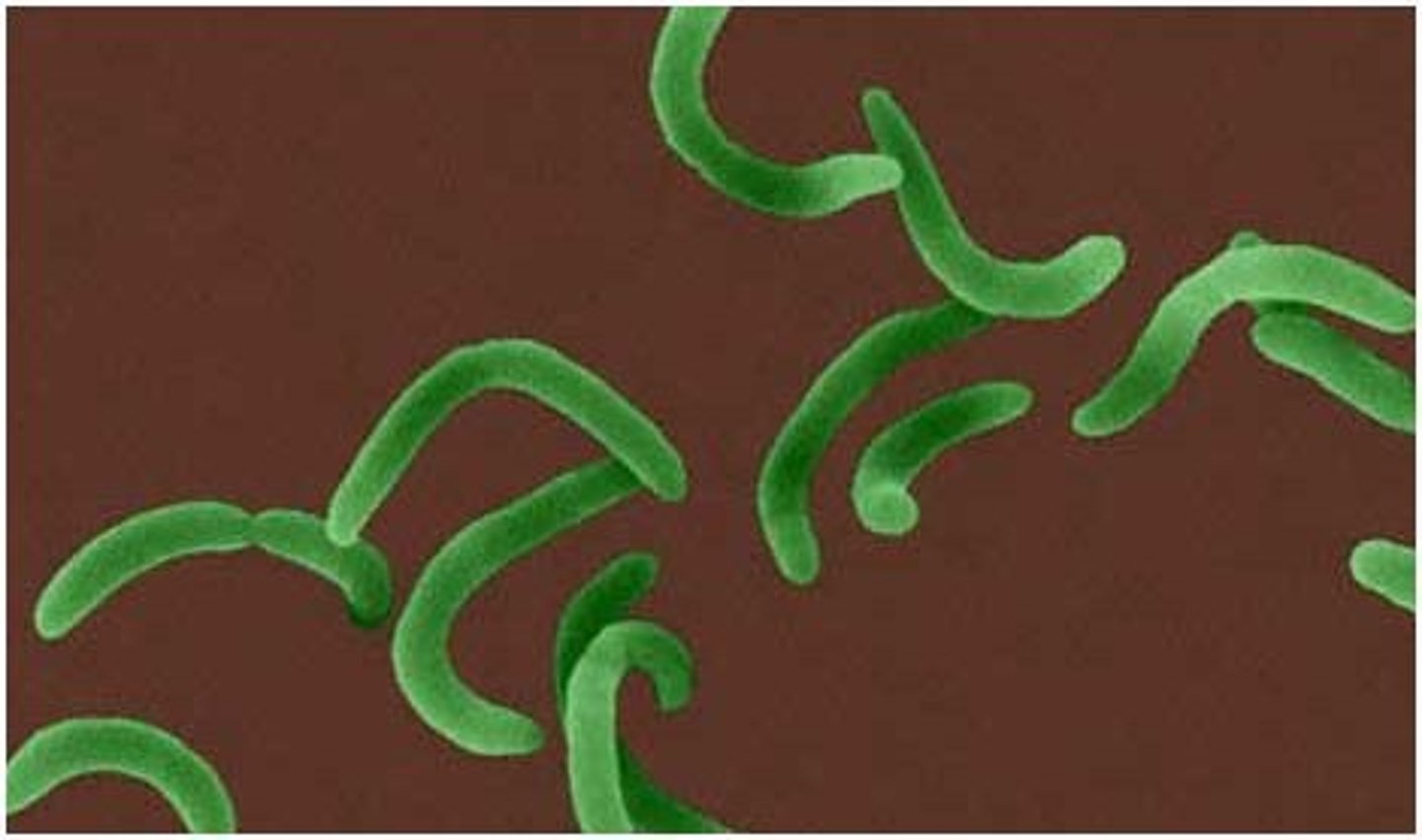
What shape is a spirilla
Rigid helice, spiral shaped
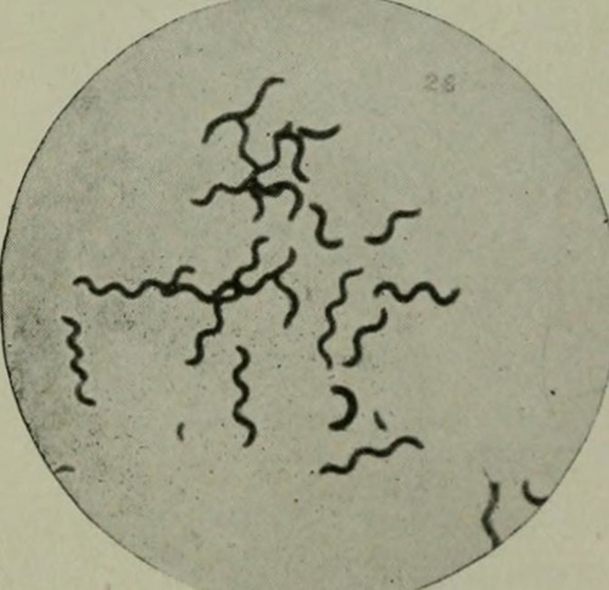
What shape is a spirochete
A flexible bendy helice, like a long spirilla
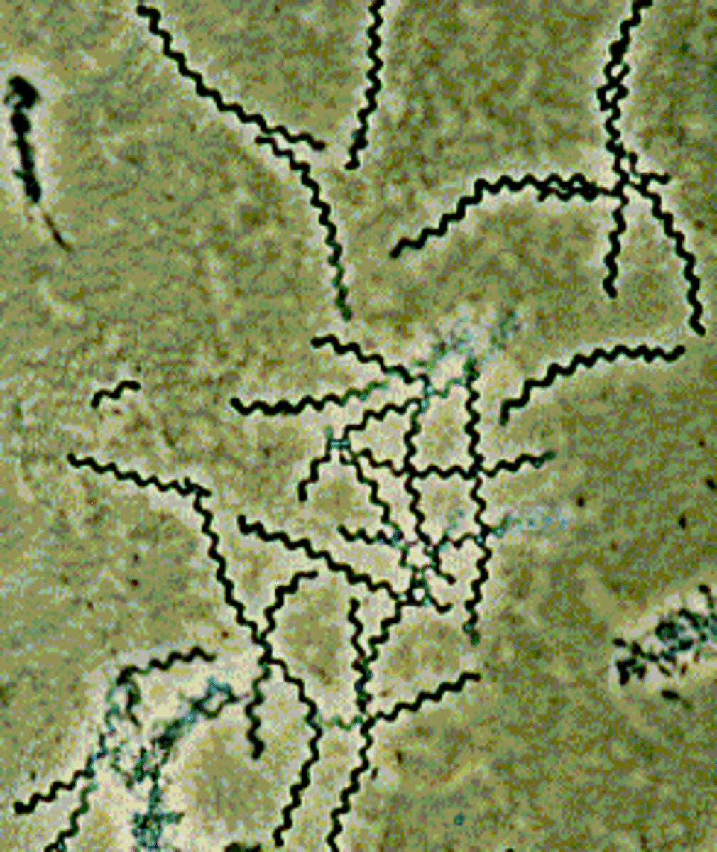
What is the shape of mycelium
Network of long, multinucleate filaments
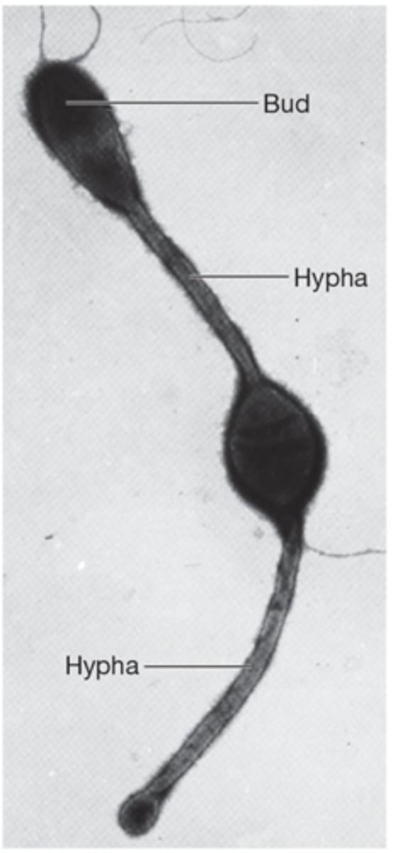
What does pleomorphic mean?
When organisms are variable in shape. Can alter shape/morphology in response to environment
Why haven't bacteria evolved to be bigger?
being small is good for S/V ratio. This is also why rods exist, cause their S/V ratio is higher that cocci
What is the cell envelope composed of?
1) Plasma membrane
2) Cell Wall
3) Capsule or slime layer
What are peripheral proteins and how abundant are they?
Loosely associated proteins with PM, 20-30%
What are integral proteins and how abundant are they?
Amphipathic, which allows them to associate through the whole PM tightly. 70-80%
What is amphipathic?
both hydrophobic and hydrophilic
What is the bacterial version of cholesterol?
hopanoids:
Stabilize membrane
congregate to create microdomains where membrane proteins can sit
What are macro elements/macronutrients?
Minerals that are needed in larger quantities
C,O,H,C,S,P,K,Ca,Mg,Fe
What are micronutrients?
Trace elements
Mc, Zn, Co, Mo, Ni, Cu
Ubiquitous in nature
What are growth factors?
organic compounds that cannot be synthesized by an organism but are essential for growth, survival, and reproductions
What are 4 common types of growth factors?
1) Amino acids: protein synthesis
2) Purines and pyrimidines: Nucleis acid synthesis
3) Vitamins: Function as enzyme cofactors
4) Heme: iron
Name all 5 transport mechanisms for nutrient uptake through PM
1) Passive diffusion
2) Facilitated diffusion: membrane protein requiring
3) Active Transport: ATP requiring
4) Group Translocation: Chemical change while entering
5) Endocytosis: PM forms vesicle
3 types of active transport
1) Primary active transport (ABC transporters)
2) Secondary active transport (cotransport: symport/antiport)
3) Group Translocation
What is special about group translocation?
Chemically modifies a molecule as it is brought into the cell.
PEP: phosphoenolpyruvate
PTS: phosphotransferase system
Phosphorylating things that come in is common, especially sugars
What are siderophores
specialized molecules secreted to bind ferric ion (Fe3+) and transport it into the cell
Gram staining:
Series of stains and washes that help show the nature of the cell wall. Different types of cell walls stain pink or purple.
+ = purple: Big cell wall with peptidoglycan
- = pink: Small cell wall with periplasmic space
What is the structure of peptidoglycan?
A meshlike polymer (sacculus) of identical subunits
Alternating sugars:
NAM: N-acetylmuramic acid
NAG: N-acetylglucosamine
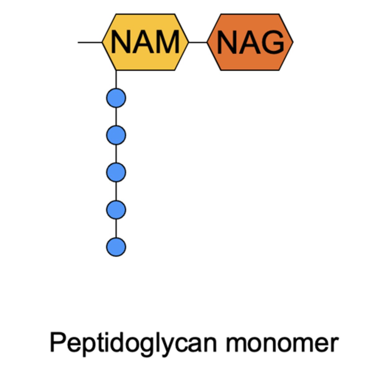
How do peptidoglycan chains interact with one another?
1) Direct crosslinking
2) Gly peptide bridges connect chains
Which sugar is the penta-peptide connected to?
NAM
What is transpeptidation?
Cross-linking of adjacent peptidoglycan strands
3 types of peptidoglycan variation
1) composition of linkage (direct, bridge)
2) Amino acid at #3 position (DAP, L-Lys, L-Hsr)
3) Linkage (3-->4 and 2-->4)
Do bacteria use D or L isomers to bypass human immune response?
D-isomers
What is the peptidoglycan backbone composed of?
Alternating NAM-NAG, via beta-(1,4) glycosidic bonds. (OH's were on opposite sides, thus is beta). Carbons 1 and 4 form bond.
Composition of Gram-Positive Cell Wall
1) primarily peptidoglycan (20-80nm)
2) teichoic acids
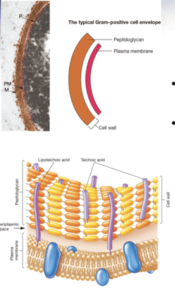
What are teichoic acids?
1) Polymers consisting of phosphate and glycerol groups
2) Have a negative charge
3) covalently bonded to peptidoglycan
What is the function of teichoic acids?
1) helps maintain cell envelope shape
2) protects from environmental substances
3) may bind to host cell
What are lipoteichoic acids (LTA)?
teichoic acids that are connected to lipids in the plasma membrane
Describe the periplasmic space in Gram + bacteria
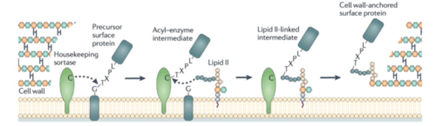
Is in between cell wall and PM, small compared to Gram (-). Has few proteins, example of one is called sortase
How does Sortase work in Gram-positive cell walls?
Proteins that need to be expressed on the outside of the cell wall are clipped by sortase, which places them on a Lipid II which then anchors to peptidoglycan in the cell wall.
Compostion of Gram-Negative Cell Walls
1) More complex than gram +
2) Thin layer of peptidoglycan surrounded by outer membrane (OM)
3) No teichoic acids
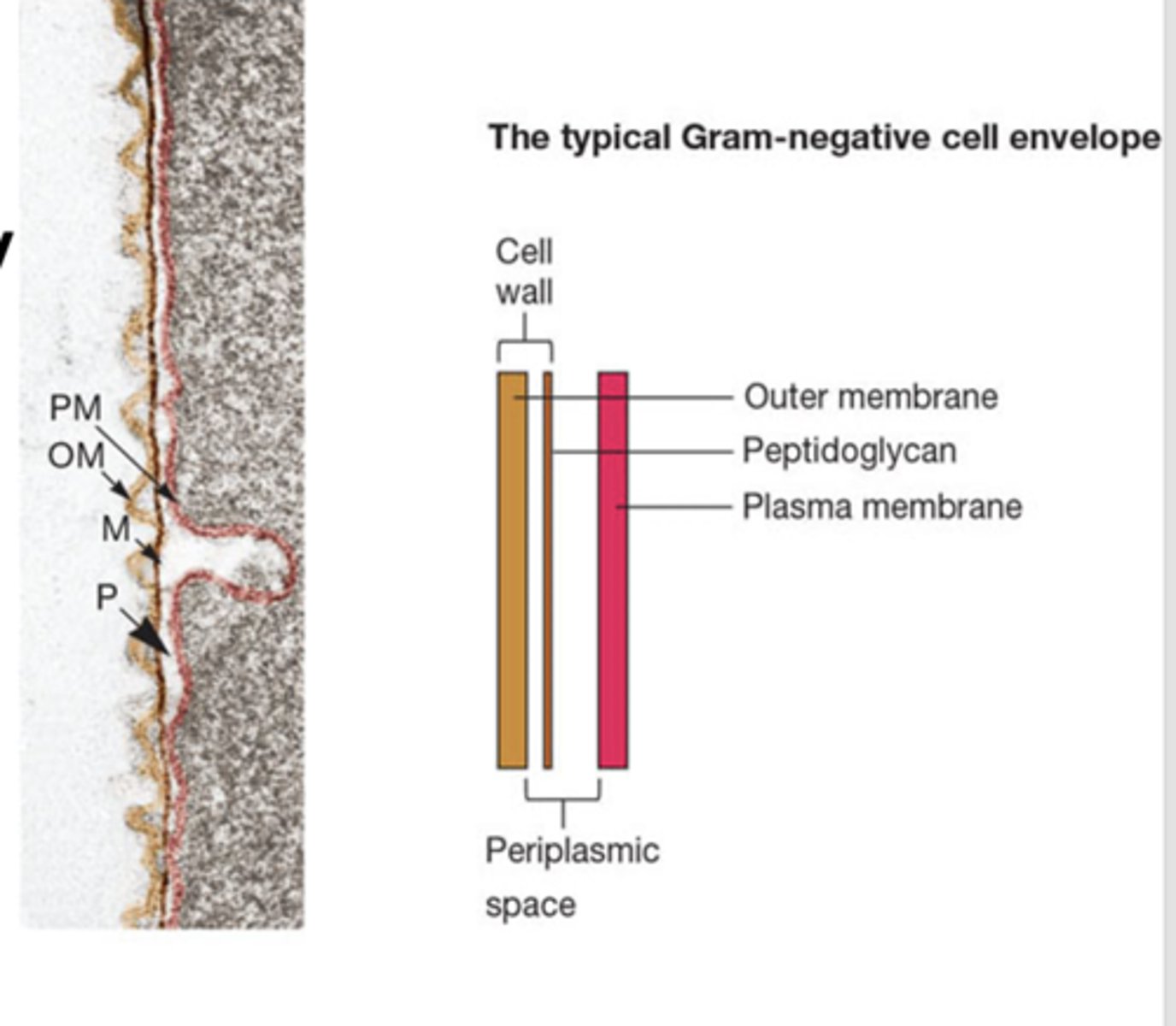
What is the outer membrane composed of? (Gram (-)
1) lipids
2) lipoproteins
3) lipopolysaccharides (LPS)
What is another name for plasma membrane?
inner membrane
How thick is peptidoglycan in Gram-negative bacteria?
2-7 nm (5-10% cell wall weight)
How big is periplasm in gram negative cells?
20-40% of cell volume. Potentially an organelle. Lots of enzymes present
What connects outer membrane to thin peptidoglycan layer in gram negative cells?
Braun's lipoproteins
What are Lipopolysaccharide (LPS)?
1) Found in exterior of of OM in Gram-Negative cells
2) Contributes to negative charge of the cell
3) Creates a permeability layer, and can act as an endotoxin
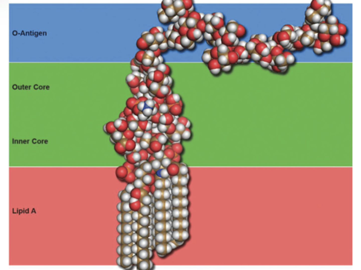
What is the anatomy and function of the lipopolysaccharide (LPS)?
1) Lipid A: Embedded in the outer membrane (hydrophobic). Stabilizes outer membrane surface. Can act as an endotoxin.
2) Core polysaccharide: hydrophilic head area of the membrane (creates negative charge)
3) O-antigen: Hair like protrusions that protect cell
What do Porin proteins do in Gram negative cells?
Increase permeability
What are the 4 steps to the Gram-stain?
1) Crystal violet dye
2) Iodine
3) Destain: ethyl alcohol or acetone
4) safranin (counterstain)
How are Gram postive bacteria stained?
Crystal violet and iodine forms a complex and gets absorbed in peptidoglycan layer. The destain dries out peptidoyglycan, trapping stain in. Safranin stains pink but the purple from initial stain overpowers. PURPLE
How are Gram negative bacteria stained?
Crystal violet and iodine form a complex that attaches to the peptidoglycan layer. The destain squeezes the stain out of the thin peptidoglycan layer (look clear at this point). Safranin stains pink. PINK
What is plasmolysis?
Shrinking of cell due to osmotic pressure
What are Gram-Positive bacteria - peptodyglycan called?
Protoplasts
What are Gram-negative bacteria - peptodyglycan called?
Spheroplasts
3 Types of additional layers of the cell wall
1) Capsules
2) Slime layers
3) S-layers
What are capsules?
Composed of polysaccharides
resistant to phagocytosis and exclude viruses
What are slime layers?
like capsules but unorganized and easily removed. May aid in motility
What are S-layers
Self-assembled layers of protein/glycoprotein
Adheres to peptidoglycan in Gram-Positive, and OM in Gram-negative
What is FtsZ?
A tubulin homolog. Forms ring during septum formation in cell division
What is MreB?
A actin homolog. Foud in rods. Maintains shape by positioning peptidoglycan synthesis machinery
What is CreS?
Intermediate filament homolog. Maintains shape in curved bacteria inhibits peptidoglycan formation, so one side makes more than other.
What are intracytoplasmic membranes?
plasma membrane infoldings
What is the nuceloid?
Where genetic information is stored. Usually not membrane-bound. Nuceloid-associated proteins (NAPs) aid in folding.
What are plasmids?
Pieces of extrachromosomal DNA. Exist and replicated independently. Contains non-essential genes. If they are lost, bacteria is known as cured
What are episomes?
plasmids that can integrate into the genome
What are inclusions?
Granules of nutrients stored for future use. Often known as microcompartments
What are Fimbriae and pili?
Short hair-like protrusions that aid in motility and DNA uptake
What is a sex pilus?
A type of pili that attaches to other bacteria and allows for conjugation (transfer of plasmids)
What is a flagella?
Long thread-like whip structure. Allows for motility. Can be a virulence factor
What is Monotrichous?
single flagellum. (polar, cause only on one end)
What is amphitrichous?
flagella at both ends of cell