chem - amino acids + proteins
 Call Kai
 Learn
 Practice Test
 Spaced Repetition
 Match
 Flashcards
 Knowt Play
1/39

There's no tags or description
Looks like no tags are added yet.
40 Terms
amino acid overview
amino acids = building blocks of proteins
content and arrangement of amino acids in a protein are DETERMINED BY THE GENE THAT ENCODES THE PROTEIN
cellular growth, repair, and maintenance dependent on amino acids
CHEMICAL PROPERTIES OF AMINO ACIDS DETERMINE ITS BIOLOGICAL ACTIVITY
proteins catalyze almost all rxns in living cells - controlling virtually all cellular processes
amino acids basic structure
a single amino acid contains at least 1 amino group and 1 carboxyl functional group
amino acids differ from one another by the chemical composition of their R group (side chain)
amino group from one amino acid linked w/ the carboxyl group of another amino acid by a PEPTIDE BOND
chain of many amino acids linked by many peptide bonds, polypeptide

essential amino acids
humans CANNOT synthesize these; they are needed from diet
arginine (Arg)
histidine (His)
Isoleucine (Ile), leucine (Leu), valine (Val)
lysine (Lys)
methionine (Met)
phenylalanine (Phe)
threonine (Thr)
tryptophan (Trp)
non-essential amino acids
body is able to make these; dietary intake NOT required
alanine (Ala)
asparagine (Asn)
aspartic acid (Asp)
cysteine (Cys)
glutamic acid (Glu)
glutamine (Gln)
glycine (Gly)
proline (Pro)
serine (Ser)
tyrosine (Tyr)
proteins structure
functional proteins have a fundamental structure organized into 4 hierarchal levels
primary
secondary
tertiary
quaternary

primary amino acid structure
amino acids in a linear sequence in a polypeptide chain
determined by DNA sequence that codes for the protein
secondary amino acid structure
patterns of folding that arise from hydrogen bonds between amino acids
alpha helix: rigid coiled structure
beta sheet: folded structure formed by multiple segments of polypeptides lying side by side
adds new properties such as strength and flexibility
tertiary amino acid structure
overall 3D shape of single polypeptide chain
formed by interactions of different R groups
give the protein its functional, physical, and chemical properties
quaternary amino acid structure
multiple folded polypeptide chains (subunits) to form a larger functional protein
example: hemoglobin (4 globin chains + 4 hemes)
chemical properties of proteins
denaturation
protein loses its natural 3D structure due to breaking of bonds
secondary, tertiary, or quaternary structure of protein is disturbed
primary structure remains intact
caused by
heat, strong acid or alkaline environments, enzymes, UV light, other substances

proteins overview
play key roles in every cell throughout the human body
major functions: antibodies, enzymes, messengers, transporters, structural
basic structure: all contain C, H, O, N, S
N sets them apart from carbs and lipids
typical protein contains 200-300 amino acids
some smaller, some much larger
ex: insulin = 6000 dalton

general chemical properties of proteins
the structure of a protein directly affects its chemical properties
proteins contain many ionizable groups on the R groups as well as on the N- and C- terminal ends
overall charge of protein depends on combination of amino acids present and the pH of the solution
proteins are AMPHOTERIC compounds
they can act as both an acid (donates H+) and a base (accepts H+)
function as effective buffers
pH dependent behavior
acid pH: protein can accept H+, acting as a base
alkaline pH: protein donates H+, acting as acid
zwitterion
contains an equal number of positively and negatively charged functional groups

general chemical properties of proteins
isoelectric point (pI): the pH at which a protein has a net electrical charge of ZERO
when pH > pI → protein has a NEGATIVE charge
when pH < pI → protein has a POSITIVE charge
most proteins have pI values in the pH range of 5.5 — 8.0
difference in net charge depending on pH is the basis for several procedures used to separate and quantify proteins

protein metabolism
involves protein catabolism and anabolism
to be utilized as energy source, protein must be broken down into amino acids (catabolism)
amino acids are recycled through catabolism of body proteins to be used for energy or synthesis of new proteins

protein catabolism
digestion
proteolytic enzymes digest proteins (supplied by diet) into constituent amino acids
absorption
amino acids absorbed into the bloodstream from the small intestine
transamination
amino group is transferred to an alpha-keto acid (pyruvate or oxaloacetate)
deamination
the remaining C skeleton of the amino acid is deaminated, releasing ammonia (NH3)
urea cycle
ammonia converted into urea in the liver, excreted in urine
energy production
C skeletons of deaminated amino acids enter citric acid cycle or glycolysis to produce energy (ATP)
protein anabolism
synthesis of NEW proteins from amino acids
absorption
amino acids obtained from protein or synthesized within body (non-essential amino acids)
translation
amino acids transported to ribosomes, assembled into chains according to instructions provided by mRNA (transcription)
folding/modification
polypeptide chains fold into their functional protein structures, undergo post-translational modification
protein assembly
multiple polypeptides chains combine to form functional
nitrogen balance
nitrogen balance
maintaining equal intake and excretion of amino acids
healthy adults
positive nitrogen balance
nitrogen intake exceeds loss
examples: pregnancy, children, recovery from illness, athletic training
negative nitrogen balance
more nitrogen is excreted than incorporated into the body
examples: excessive tissue destruction, burns, wasting diseases, high fevers, starvation

protein classifications
proteins do everything in the living cell
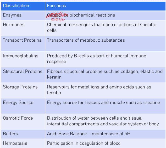
proteins are responsible for many different functions, so they are commonly classified by the function they perform
classifications:
enzymes - catalyze biochemical rxns
hormones - chemical messengers that control actions of specific cells
transport proteins - transporters of metabolic substances
immunoglobulins - produced by B cells as part of humoral immune response
structural proteins - fibrous structural proteins such as collagen, elastic and keratin
storage proteins - reservoirs for metal ions and amino acids such as ferritin
energy source - energy source for tissues and muscle such as creatine
osmotic force - distribution of water between cells and tissue, interstitial compartments and vascular system of body
buffers - acid-base balance; maintenance of pH
hemostasis - participation in coagulation of blood
simple proteins
contain peptide chains of ONLY amino acids
may be globular or fibrous in shape
globular: transporters, enzymes, and messengers
fibrous: structure to cells such as connective tissue, tendons, bone and muscle
conjugated proteins
consist of proteins and non-proteins
non-amino part is referred to as the prosthetic group
prosthetic group defines characteristics
examples: lipid, carbohydrate, porphyrin, metal. etc.
metalloproteins: metal ion attached to protein (ferritin, ceruloplasmin)
glycoprotein: protein w/ 1 or more sugar molecules attached (haptoglobin, erythropoietin, collagen, antibodies)
lipoprotein: protein + lipid (HDL, LDL)
nucleoprotein: protein + nucleic acid (chromatin)
total protein - more common test
fasting NOT required, serum or plasma
lipidemia may interfere
hemolysis unacceptable (RBC intracellular proteins released), false increase
total protein concentrations lower at birth adult levels by age 3
total protein reference range
normal: 6.5 - 8.3 g/dL
recumbent: 6.0 - 7.8 g/dL
refers to bedridden pts
total protein - Kjeldahl method
REFERENCE METHOD for QUANTITATION
more lengthy: involves digestion, distillation, and titration
NOT suitable for CSF or urine
NO LONGER ROUTINELY USED
TIME CONSUMING
RELIES ON ASSUMPTION that average mass of protein is 16% (can actually vary from 15.1 - 16.8%)
measures amount on Nitrogen in specimen
commonly used for food, soil, fertilizer, and wastewater analysis to determine protein content since N is a key component of amino acids
total protein method - biuret method
MOST WIDELY USED and recommended
principle: in ALKALINE medium and the presence of AT LEAST 2 PEPTIDE BONDS (linkages) — cupric (Cu 2+) will complex with groups in peptide bond to form a VIOLET-colored chelate
absorbance measured at 540 nm using a spectrophotometer
COLOR & ABSORBANCE ARE PROPORTIONAL TO NUMBER OF PEPTIDE BONDS
REFLECT THE TOTAL PROTEIN CONCENTRATION
do not use with lipemic samples (messes w/ absorbance → can centrifuge to clear up)
not suitable for CSF or urine
total protein - dye binding
dye binding
methods based on the ability of most proteins to bind to dyes
used to determine protein concentration in sample
methods are simple and fast
unequal dye binding response of individual proteins requires caution
examples: bromophenol blue, ponceau S, amino black 10B
albumin dye binding methods
pH of solution is adjusted so that albumin is positively charged and binds to the dye by electrostatic forces
albumin + dye has different absorption maximum than dye alone
albumin concentration is proportional to the albumin-dye complex
methods: principle for all is that ALBUMIN binds to DYE causing a SHIFT in ABSORPTION MAXIMUM
methyl orange:
non specific for albumin
HABA (hydroxyazobenzene-benzoic acid)
more specific for albumin
many interferences (salicylates, bilirubin)
BCG (bromocresol green)
sensitive, quick readings
overestimates low albumin levels
hemoglobin slightly interferes
BCP (bromocresol purple) - REFERENCE METHOD
SPECIFIC, SENSITIVE, and PRECISE
commonly preferred
bilirubin interferes
underestimates renal insufficiency
abnormal protein levels
hypoproteinemia (< 6.5 g/dl)
decreased protein levels
negative nitrogen balance
liver failure, starvation, glomerular disease, nephrotic syndrome, edema, burns, and catabolic hormones (like increased estrogen)
hyperproteinemia (> 6.5 g/dl)
increased protein levels
dehydration
excessive production, primarily gamma-globulins
multiple myeloma (abnormal proteins)
steroids, excess growth hormones (anabolic)
fractionation, identification, and quantitation of specific proteins
when evaluating total protein, may be useful to determine albumin and globulin fractions
compare them as a ratio (A/G ratio)
measure total protein and albumin
subtract albumin from total protein to get globulin
Globulins = Total Protein - albumin
A/G (albumin/globulin) ratio
reference range: 0.8 - 2
ratio is not a specific marker for disease
decreased A/G ratio
liver disease, kidney disease, autoimmune disease, malnutrition
increased A/G ratio
high albumin or low globulin levels
dehydration (concentrated blood), pregnancy, malnutrition
albumin
a transporter synthesized in the liver
most abundant protein in the plasma
functions
transports thyroid hormones, unconjugated bilirubin, fat-soluble hormones, iron, fatty acids, calcium, magnesium and drugs such as salicylic acid
maintains oncotic pressure (force exerted by proteins in plasma - deals w blood pressure)
binds toxic heavy metals
albumin reference range
normal: 3.5 - 5.0 g/dL
causes of hypoalbuminemia
decreased liver function (liver makes albumin)
kidney disease
severe malnutrition
acute inflammatory response
negative acute phase reactant
causes of hyperalbuminemia
not clinically significant usually
dehydration
excessive albumin infusion
urinary protein - quantitative methods
24 hour urine collection - you collect the entire day not just one excretion
turbidimetric
principle: proteins precipitated as fine particles → turbidity measured spectrophotometrically
rapid, easy to use; unequal sensitivity for individual proteins
biuret
principle: proteins concentrated by precipitation → redissolved in alkali then reacted w Cu2+ → Cu2+ forms color complex w peptide bonds
accurate
folin-lowry
principle: initial biuret rxns + oxidation of tyrosine, tryptophan, and histidine residues by folin phenol reagent → measurement of resultant blue color
very sensitive
dye binding (coomassie blue, ponceau S)
principle: protein binds to dye, causes shift in absorption maximum
limited linearity: unequal sensitivity for individual proteins
CSF protein
CSF formed in choroid plexus of ventricles of the brain by ultrafiltration of blood plasma
INCREASED CSF protein when increased permeability of barriers where ultrafiltration occurs
bacterial, viral, and fungal meningitis, Multiple Sclerosis, obstruction, neoplasms
DECREASED CSF protein
hyperthyroidism, fluid leaking CNS

CSF protein reference range
15 - 45 mg/dl
MILLIGRAMS NOT GRAMS
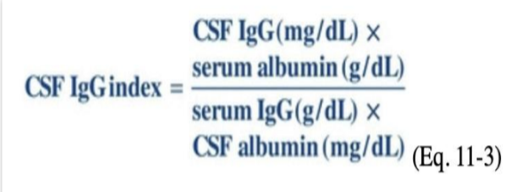
CSF IgG index
differentiate elevated IgG concentrations due to local CNS production from leakage of plasma into CSF
increased local production seen in Multiple Sclerosis
compare IgG concentrations from CSF and serum with albumin
CSF IgG index reference ranges
normal: 0.26 → 0.70
value GREATER than 0.7 → CNS production of IgG (MULTIPLE SCLEROSIS)
value BELOW 0. 25 → Blood-Brain Barrier compromised (tumor, strokes, etc.)
CSF IgG index equation