BCOR 2500 Reading Guide for Exam #3
 Call Kai
 Learn
 Practice Test
 Spaced Repetition
 Match
 Flashcards
 Knowt Play
1/86

There's no tags or description
Looks like no tags are added yet.
87 Terms
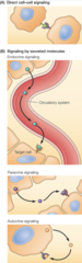
What are the different modes of cell-cell signaling? How is each type of signaling carried out by the cell?
1. Direct Cell-Cell Contact: direction interaction of a cell with its neighbor. Critical in regulating behavior of cells in animal tissues
-Ex: integrins and cadherins (function not only as cell adhesion molecules, but in response to cell-cell and cell-matrix contacts)
2. Action of Secreted Signaling Molecules: cells express a variety of proteins that function as cell surface receptors that interact with signaling molecules on the surface of neighboring cells
-Endocrine signaling
-Paracrine signaling
-Autocrine signaling
-Secreted signaling molecules can travel long distances, act on adjacent cells, or act on the cell that produces them
What characteristics of steroid hormones enable them to carry out their mode of signaling?
-Synthesized from cholesterol, making them hydrophobic
-Freely diffuse across the plasma membrane, facilitated by carrier proteins, and bind to transcription factors inside their hormonally-responsive target cells (contain related domains for ligand binding, DNA binding, and transcriptional activation)
-Ligand binding regulates their function as activators/repressors of target genes, so steroid hormones regulate gene expression
-After entering target cell, bind to intracellular receptors, forming complex that translocates to the nucleus
What are the major steps in glucocorticoid signaling?
1. Glucocorticoids diffuse across the plasma membrane and bind to the glucocorticoid receptor
2. In the absence of ligand, the receptor is bound to Hsp90 chaperones in the cytoplasm
3. Glucocorticoid binding causes conformational changes that displace the receptor from Hsp90 and expose nuclear localization signals, allowing nuclear import as receptor dimers
4. The activated receptors then bind recognition sites in DNA and associate with coactivators with histone acetyltransferase (HAT) activity to stimulate transcription of their target genes

How are genes under the control of the Thyroid Hormone Receptor regulated?
-Thyroid hormone receptor binds its recognition sites in DNA in the presence/absence of thyroid hormone
-In absence: receptor associates with corepressors with histone deacetylase (HDAC) activity, resulting in repression of target gene expression
-In presence: thyroid hormone binds the receptor, inducing conformational changes that displace the coreceptors and cause association with coactivators with histone acetyltransferase (HAT) activity. This results in target gene expression

What role do HDAC and HAT have in switching genes on and off under control of the Thyroid Hormone Receptor?
-HDAC: represses target gene expression (switch genes off) under thyroid hormone receptor
-HAT: enhances target gene expression (switch genes on) under thyroid hormone receptor
What's the role of Nitric Oxide (NO) in stimulating synthesis of cyclic GMP?
-Major paracrine signaling molecule in the nervous, immune, and circulatory systems
-Can diffuse directly across the plasma membrane of its target cells (but has distinct function from that of steroids)
-Alters activity of intracellular target enzyme guanylyl cyclase (sGC), stimulating the synthesis of cyclic GMP inside the cell
-Ex: dilation of blood vessels
What's the mechanism of signaling used by Nitric Oxide (NO) gas molecules?
Alters activity of intracellular target enzyme guanylyl cyclase (sGC), stimulating the synthesis of cyclic GMP inside the cell
What are the main groups of peptide signaling molecules?
1. Peptide Hormones
2. Neuropeptides
3. Polypeptide Growth Factors
Peptide Hormones
-Main group of peptide signaling molecules
-Signaling molecule composed of amino acids
-Ex: insulin and hormones produced by pituitary gland (GH, FSH, prolactin, etc...)
Neuropeptides
-Main group of peptide signaling molecules
-A peptide signaling molecule secreted by neurons
-Ex: enkephalins and endorphins, which act as natural analgestics that decrease pain response in CNS
Growth Factors
-Main group of peptide signaling molecules
-Variety of signaling molecules that regulate animal cell behavior and differentiation
-Ex: nerve growth factor (NGF) which is a polypeptide that regulates the development/survival of neurons
-Ex: epidermal growth factor (EGF) which stimulates cell proliferation
-Ex: platelet-derived growth factor (PDGF) which is stored in blood platelets and released during blood clotting at the site of a wound (stimulates movement of fibroblasts to clot, leading to regrowth of damaged tissue)
-Ex: cytokines regulate blood cells and lymphocytes; include erythropoietin and interleukin-2
-Ex: membrane-anchored growth factors remain associated with membrane, functioning as signaling molecules during direct cell-cell interactions
What is the basic structural unit of a peptide signaling molecule?
An amino acid!
Do peptide signaling molecules act at the surface of the cell or do they cross the plasma membrane?
All can't cross plasma membrane of target cells, so they bind to cell surface receptors
-This is because they are hydrophilic (most)
What are the main steps leading up to the hormonal activation of adenylyl cyclase by the hormone epinephrine?
1. Binding of hormone promotes interaction of receptor with heterotrimeric G proteins composed of α, β, and γ subunits and with GDP bound to its α subunit
2. The receptor catalyzes exchange of GDP for GTP
3. The activated G protein α subunit dissociates from the receptor and stimulates adenylyl cyclase
4. This catalyzes the conversion of ATP to cAMP

What role do GTPase activating proteins (GAPs) have in regulating G-protein signaling?
-Protein that stimulates GTP hydrolysis of bound GTP by the small GTP-binding proteins (heterotrimeric G proteins)
-"Off-switch" of sorts
-Accelerates the conversion of GTP to GDP (which inactivates the G protein)

What are the main steps leading to the regulation of glycogen breakdown?
1. Hormones (like epinephrine), bind to their receptors, stimulating adenylyl cyclase (can convert ATP into cAMP)
2. cAMP activates protein kinase A which consists of 2 subunits
3. Binding of cAMP to the regulatory subunits induces a conformational change that causes dissociation of the catalytic subunits
4. Protein kinase A activates phosphorylase kinase which activates glycogen phosphorylase (from inactive 𝑏 form to its active 𝑎 form)
5. Glycogen phosphorylase catalyzes the breakdown of glycogen to glucose-1-phosphate

What amino acid residue is phosphorylated during glycogen breakdown?
Tyrosine or serine/threonine residues
How is protein kinase A regulated (activated/inactivated)?
Activation: receptor stimulation and binding of cAMP to regulatory subunits leads to G protein-mediated activation of adenylyl cyclase, synthesis of cAMP, and activation of protein kinase A
Inactivation: low cAMP levels, phosphorylation, degradation of activated protein kinases. Protein Kinase A consists of 2 regulatory (R) and 2 catalytic (C) subunits in this form

What are the main steps that lead to expression of cAMP-inducible genes?
1. Receptor stimulation leads to G protein-mediated activation of adenylyl cyclase, synthesis of cAMP, and activation of protein kinase A
2. The free catalytic subunit of protein kinase A translocates to the nucleus and phosphorylates the transcription factor CREB (CRE-binding protein)
3. This leads to the recruitment of coactivators and expression of cAMP-inducible genes

Compare the differences between receptor tyrosine kinases and non-receptor tyrosine kinases
Receptor Tyrosine Kinases
-Transmembrane proteins
-Activated by extracellular ligands
-Bind to signaling molecules on cell surface
-Undergo dimerization and autophosphorylation
-Trigger downstream signaling
-Recruit SH2-domain containing proteins
-Autophosphorylate tyrosine residues on cytoplasmic domain
-Ligand binding induces receptor dimerization
Non-Receptor Tyrosine Kinases
-Located in cytoplasm
-Lack transmembrane domain
-Activated through protein-protein interactions
-Mediate intracellular signals that are initiated in the receptor tyrosine kinases
What are the main steps that lead to activation of receptor tyrosine kinases?
1. Each receptor consists of an N-terminal extracellular ligand-binding domain, a single transmembrane α helix, and a cytosolic C-terminal domain with tyrosine kinase activity
2. Growth factor binding induces ligand receptor dimerization
3. This results in receptor autophosphorylation as the two polypeptide chains cross-phosphorylate one another (First, phosphorylation of tyrosine residues within the catalytic domain increases protein kinase activity. Second, phosphorylation of tyrosine residues outside of the catalytic domain creates specific binding sites for additional proteins that transmit intracellular signals downstream of the activated receptors)
4. The association of these downstream signaling molecules with receptor tyrosine kinases is mediated by protein domains that bind to specific phosphotyrosine-containing peptides, including SH2 domains
5. These domains bind to specific phosphotyrosine-containing sites of activated receptors
Simplified: ligand-binding, receptor dimerization, autophosphorylation, and recruitment/activation of downstream signaling proteins
What is the role of Ras in ERK MAP kinase signaling?
-A family of small GTP binding proteins (encoded by the ras oncogenes) that couple growth factor receptors to intracellular targets, including the ERK MAP kinase pathway
-Act as molecular switch and activates Raf upon binding GTP
-Interference with Ras function blocks growth-factor-induced cell proliferation
-Ras induces abnormal growth characteristic of cancer cells
-Required for response of normal cells to growth factor stimulation
-Recruits and activates RAF, which is the first kinase in a cascade that phosphorylates and activates MEK
-MEK phosphorylates and activates ERK
-The activated ERK then triggers a series of events in the nucleus that control genes involved in cell growth, survival, and differentiation
How is Ras regulated in ERK MAP kinase signaling?
Ras proteins alternate between inactive GDP-bound and active GTP-bound states
-Guanine nucleotide exchange factors (GEFs) that promote the exchange of GDP for GTP
-Deactivated by GTPase-activating proteins (GAPs) that hydrolyze GTP to GDP

Under what circumstances would Ras directly be responsible for causing abnormal cell growth in ERK MAP kinase signaling?
-Mutations in Ras can have the effect of inhibiting GTP hydrolysis by the Ras proteins
-Mutated Ras proteins remain continuously in the active GTP-bound form, driving the unregulated proliferation of cancer cells (even in the absence of growth factor stimulation)
-ERK MAP kinase pathway: constitutively active due to mutations. Activated ERK can translocate to the nucleus and phosphorylate transcription factors, altering gene expression
PI 3-kinase/Akt pathway forms the initial steps in many signaling pathways. What are the steps that lead to AKT activation?
1. PI 3-kinase is recruited to activated receptor tyrosine kinases via its SH2 domain
2. PI 3-kinase phosphorylates the 3 position of inositol, converting PIP2 to PIP3
3. Akt is recruited to the plasma membrane by binding to PIP3 via its pleckstrin homology (PH) domain
4. Activated as a result of phosphorylation by two other protein kinases (PDK1 and mTORC2) that also bind PIP3
5. Akt phosphorylates target proteins, including direct regulators of cell survival (such as Bad), several transcription factors (some activated by phosphorylation, others inhibited), and other protein kinases, including GSK-3 (which is inhibited by Akt phosphorylation)
6. If not inhibited by Akt, GSK-3 phosphorylates metabolic enzymes, the translation initiation factor eIF2B, and transcription factors

What are some signaling pathways regulated by Akt regulation?
1. mTOR: Akt inhibits TSC, leading to activation of GTP-binding protein Rheb and mTORC1 in response to GF stimulation. mTORC1 stimulates translation by phosphorylating S6 kinase
2. GSK-3: inhibited by Akt phosphorylation. If not inhibited, phosphorylates metabolic enzymes & transcription factors
3. Bad: phosphorylated by Akt; target protein that regulates cell survival
4. FOXO: inhibited by Akt phosphorylation. In absence of Akt activity, FOXO stimulates gene transcription to inhibit cell proliferation or induce cell death
What are the main steps leading to activation of transcription for the TGF-β/Smad pathway?
-TGF-β receptors are dimers of type I and II polypeptides
-The type II receptor phosphorylates and activates type I
-Type I phosphorylates a Smad protein
-Phosphorylated Smads form complexes and translocate to the nucleus to activate transcription of target genes

What role does ubiquitination and proteasomes play in the activation of the NF-κB pathway?
-Phosphorylation causes conformational changes that enable a ubiquitin ligase (β-TrCP) to recognize and ubiquitinate IκB for proteasomal degradation, freeing NF-κB to translocate to the nucleus and induce expression of its target gene
-In the inactive state, homo- or heterodimers of NF-κB are bound to IκB in the cytoplasm
-Activation of the TNF receptor leads to the recruitment of adaptor proteins that activate IκB kinase
-Phosphorylation marks IκB for ubiquitination by β-TrCP, causing its degradation by the proteasome
-This allows NF-κB to translocate to the nucleus & activate transcription of its target genes

What is the mode of signaling in the Notch signaling pathway?
-Direct cell-cell interactions during development (embryonic)
-Notch serves as a receptor for direct cell-cell signaling by transmembrane proteins (ex: Delta) on neighboring cells
-The binding of Delta (ligand binding) leads to proteolytic cleavage of Notch by presenilin as part of the γ-secretase complex that also includes nicastrin, Aph-1, and Pen-2
-This releases the Notch intracellular domain, which translocates to the nucleus and interacts with the CSL transcription factor to induce gene expression

What are DNA damage checkpoints?
A cell cycle checkpoint that ensures that damaged DNA is not replicated and passed on to daughter cells
-Monitor progress through the cycle

What are the functions of DNA damage checkpoints during cell cycle progression?
-Ensure that damaged or incompletely replicated DNA is not passed on to daughter cells
-Only complete genomes will be transmitted to daughter cells
-DNA damage checkpoints in G1, S, and G2 lead to cell cycle arrest in response to damaged or unreplicated DNA
-The spindle assembly checkpoint arrests mitosis if the chromosomes are not properly aligned on the mitotic spindle
What are the events that lead to the activation of the maturation promoting factor (MPF)?
-Triggered by the rising levels of M-cyclin which bind to Cdk, forming an inactive complex
-Cdk1 forms complexes with cyclin B during G2
-Cdk1 is phosphorylated on Thr161 by the kinase CAK, which is required for Cdk1 activity, as well as on Tyr15 and Thr14 in vertebrate cells— by the kinase Wee1, which inhibits Cdk1 activity
-Dephosphorylation of Tyr15 and Thr14 by Cdc25 phosphatases activates MPF at the G2-to-M transition

What are the events that lead to the termination of MPF activity?
MPF activity is terminated toward the end of mitosis by proteasomal degradation of cyclin B due to its ubiquitination by APC/C, which is followed by dephosphorylation of Cdk1

What types of enzymes are involved in MPF regulation?
-Kinases: Wee1 phosphorylates and inactivates Cdk1
-Phosphatases: cdc25 phosphatases are primary activators, removing inhibitory phosphate group added by Wee1, allowing Cdk1 to become active
-Ubiquitinases: APC/C is crucial for degradation of cyclin B, which inactives MPF and allows the cell cycle to progress past the M phase
How does Cdk-4,6/cyclin D activity drive G1 progression?
1. In its underphosphorylated form, Rb binds to members of the E2F family, repressing transcription of E2F-regulated genes
2. Phosphorylation of Rb by Cdk4, 6/cyclin D complexes results in its dissociation from E2F (releases E2F transcription factor)
3. E2F then stimulates expression of its target genes, which encode proteins required for cell cycle progression
4. E2F induces cyclin E, and Cdk2/cyclin E complexes inactivate the APC/C ubiquitin ligase and drive progression through the restriction point and into S phase

What are the main targets of Cdk4,6/cyclin D and what are their roles in the regulation of transcription?
-Main targets of Cdk4,6/cyclin D complexes is Rb, which functions with E2F transcription factors to regulate genes required for cell cycle progression from G1 to S phase

Compare the 2 different responses to DNA damage
1. Single-Stranded/Unreplicated DNA: ATR activation. ATR phosphorylates and activates the Chk1 protein kinase to inhibit the cdc25 protein phosphatases. This inhibition leads to arrest at DNA damage checkpoints
2. Double-Stranded Break: ATM activation. ATM phosphorylates and activates the Chk2 protein kinase to inhibit the cdc25 protein phosphatases. This inhibition leads to arrest at DNA damage checkpoints

What role do checkpoint kinases play in cell cycle arrest in response to DNA damage?
Arrest the cell cycle by phosphorylating and inhibiting or degrading cdc25 phosphatases
What are checkpoint kinases?
A protein kinase (chk1 or chk2) that brings about cell cycle arrest in response to damaged DNA
-Ensure cell cycle proceeds correctly and without errors, especially in response to DNA damage
What is MDM2's role in the regulation of p53, both in the absence of DNA damage and when there are high levels of DNA damage?
-In the absence of DNA damage, p53 is degraded by the proteasome due to MDM2-mediated ubiquitination, thus keeping p53 levels low
-In the presence of DNA damage, ATM and Chk2 phosphorylate p53, which blocks MDM2-mediated ubiquitination. In turn, p53 levels rapidly increase and p53 activates transcription of the gene encoding the Cdk inhibitor p21, leading to inhibition of Cdk2/cyclin E or cyclin A complexes and cell cycle arrest
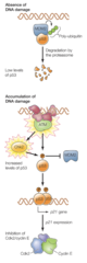
How is p53 modified by MDM2?
-p53 normally arrests the cell cycle in G1 in response to damaged DNA and is required for apoptosis
-Targeted for proteasomal degradation by MDM2 in absence of DNA damage
-Phosphorylated p53 is not recognized by MDM2 so has increased levels during DNA damage
-MDM2 modifies p53 by ubiquitination which targets it for degradation by the proteasome and by inhibiting it's transcriptional activity
What roles do APC/C ubiquitin ligases play in the metaphase-to-anaphase transition?
Targets key regulatory proteins (commonly cyclins) for proteasomal degradation to exit mitosis
-Ubiquitinates securin, leading to activation of separase
What roles do Securins play in the metaphase-to-anaphase transition?
Binds to and inhibits separase
What roles do Separases play in the metaphase-to-anaphase transition?
Degrade cohesin, breaking the link between sister chromatids and initiating anaphase
What role do cohesins and condensins have in chromatin condensation?
-Cohesins bind to DNA during S phase and maintain the linkage between sister chromatids following DNA replication
-As the cell enters M phase, the condensins are phosphorylated by Aurora B kinase and Cdk1, the cohesins by Aurora B and Polo-like kinases
-The cohesins are replaced by condensins along most of the chromosome, remaining only at the centromere due to a kinetochore-associated phosphatase that antagonizes cohesin phosphorylation
-The condensins drive chromosome condensation by packing the chromatin into loops

Which kinases regulate chromatin condensation?
Cdk1, Aurora, and Polo kinases
What structural changes occur between G2, prophase, and metaphase?
G2- cell growth and protein synthesis in preparation for mitosis, chromatin condensation, centrosomes begin to move apart
Prophase- centrosomes separate and move to different sides of the nucleus
Prometaphase- nuclear envelope breaks down, chromosome shuffling
Metaphase- spindles align chromosomes at the metaphase plate

What type of remodeling occurs at the nuclear envelope during mitosis and what are the enzymes and structural proteins involved?
-Breakdown and reformation process
-Cdk1/cyclin B phosphorylates the nuclear lamins as well as proteins of the nuclear pore complex and inner nuclear membrane, allowing envelope to disassemble
-Phosphorylation of the lamins causes the filaments that form the nuclear lamina to dissociate into free lamin dimers

Compare the differences between embryonic and typical eukaryotic cell cycles
Eukaryotic Cell Cycle
-Divided into four phases by the timing of mitosis and DNA synthesis (M, G1, S, and G2)
-During S phase, DNA replication occurs
-Cytokinesis occurs during M phase
-G0 phase where cells only divide when demanded
-Significant growth during G1/G2
-Highly regulated
-Cells are differentiated
Embryonic Cell Cycle
-No G1 or G2 phase (no growth- only cytoplasmal division)
-DNA replication occurs very rapidly
-Very short S phases
-Alternating M phases
-Less regulated- focused on rapid proliferation
What could account for different cell cycle times in embryonic and typical eukaryotic cell cycles?
Rapid embryonic divisions that occur without growth, leading to a decreasing cell size during cleavage = much quicker cell cycle time in embryos versus typical eukaryotic cell cycles
How is the centromeric region maintained during chromatin condensation?
Phosphatase maintains centromere-associated cohesins in an unphosphorylated state; thus, the sister chromatids remain linked at the centromere
-Condensin complexes and chromatin looping
How are cohesins replaced by condensins?
-Condensins are phosphorylated as the cell enters M phase by both Cdk1 and Aurora B kinase, whereupon they replace the cohesins along most of the length of the chromosome
-The condensins then induce chromatin condensation by forming DNA loops, leading to the formation of metaphase chromosomes
What are the differences between endothelial cells and epithelial cells?
Endothelial Cells
-Line vessels of the circulatory and lymphatic systems
-Fully differentiated cell, capable of proliferation
-Proliferation allows them to form new blood vessels for repair of damaged tissue
-Proliferation is triggered by VEGF
-More permeable- allow for transport of water and oxygen
Epithelial Cells
-Able to proliferate to replace damaged tissue
-Commonly found in external surfaces (skin) and internal organs, cavities, and ducts
-Tightly packed
What is the role of Vascular Endothelial Growth Factors (VEGF) in cell proliferation?
VEGF is secreted by cells deprived of oxygen, leading to the outgrowth of new capillaries into tissues lacking adequate blood supply. VEGF stimulates endothelial cells to proliferate by signaling endothelial cells to multiply, migrate, and survive

What are stem cells and how do they proliferate to become fully differentiated cells?
A cell that divides to produce daughter cells that can either differentiate or remain as stem cells
-Divide to produce one daughter cell that remains a stem cell and a second that proliferates and differentiates
-The differentiating cell receives signals that direct it to become a specialized cell
What are the steps leading up to replacing damaged cells that have shed from surface epithelium?
1. Hemostasis
2. Inflammation
3. Proliferation (keratinocytes and fibroblasts release growth factors)
4. Migration (new cells migrate across wound surface to seal the breach)
5. Remodeling (once wound is closed, tissue enters remodeling phase to restore strength/function)
Division of stem cells gives rise to a population of transit-amplifying cells, which undergo three or four divisions before differentiating into the cell types of the surface epithelium. The surface epithelial cells continually undergo programmed cell death and are shed

What is "hematopoietic stem cell transplantation"?
A clinical procedure in which transplantation of hematopoietic stem cells is used in the treatment of cancer and diseases of the hematopoietic system
How is hematopoietic stem cell transplantation applied to the treatment of cancer?
-Hematopoietic stem cells are among the most rapidly dividing cells of the body, so the toxic effects of anticancer drugs on these cells frequently limit the effectiveness of chemotherapy in cancer treatment
-Transplantation provides an approach to bypass this toxicity, allowing the use of higher drug doses to treat the patient's cancer effectively
-The patient is treated with high doses of chemotherapy that would normally not be tolerated because of toxic effects on the hematopoietic system
-The potentially lethal damage is repaired by transferring new hematopoietic stem cells to the patient following completion of chemotherapy
-In some cases, stem cells are obtained from the patient prior to chemotherapy, stored, and then returned to the patient once chemotherapy is completed
-Stem cells to be transplanted can be obtained from a healthy donor whose tissue type closely matches
What are induced pluripotent stem cells?
An adult somatic cell that has been reprogrammed in culture by 4 transcription factors to resemble an embryonic stem cell
How are induced pluripotent stem cells made?
Adult mouse fibroblasts (or any adult somatic cells that can develop into any cell type, such as blood cells) in culture are converted to pluripotent stem cells by infection with retroviral vectors carrying genes to express four transcription factors: Oct4, Sox2, Klf4, and c-Myc

Why was it necessary to make induced pluripotent stem cells?
-Induced pluripotent stem cells generated by transient expression of reprogramming factors thus provide a ready source of pluripotent stem cells for transplantation therapy and are currently being tested in clinical trials for the treatment of many of the same diseases that are in trials with embryonic stems cells
-Avoids ethical concerns (created with patient's own cells)
-Prevent immune rejection
-Enable regenerative medicine
What is transdifferentiation?
Differentiation of one type of somatic cell into another differentiated cell type
What is the medical significance of transdifferentiation?
-Regenerative medicine
-Disease modeling
-Drug discovery

What role do phagocytes play in the removal of apoptotic cells?
The receptors on phagocytes recognize phosphatidylserine on the cell surface and are guided to bind there, triggering them to engulf the cell
What interactions take place between apoptotic cells and phagocytes?
-Apoptotic cells and cell fragments are recognized and engulfed by phagocytic cells
-One of the signals recognized by phagocytes is phosphatidylserine on the cell surface
-In normal cells, phosphatidylserine is restricted to the inner leaflet of the plasma membrane, but it is translocated to the outer leaflet during apoptosis where it is recognized by phosphatidylserine receptors of phagocytic cells
What role do caspases have during the process of apoptosis?
-Inhibitor of DNase ICAD: DNA fragmentation
-Nuclear lamins: fragmentation of nucleus
-Cytoskeletal proteins: cytoskeletal disruption, cell fragmentation, membrane blebbing
-Golgi peripheral membrane proteins: fragmentation of Golgi
-Scramblase: translocation of phosphatidylserine to cell surface
What are the roles of effector proteins in the mitochondrial pathway of apoptosis? (Focus on Bax and Bak)
-Induce caspase activation and promote cell death; downstream effectors that directly induce apoptosis
-Oligomerize to form pores in the mitochondrial outer membrane, leading to the release of cytochrome c from mitochondrial intermembrane space (this then triggers caspase activation since cytochrome c forms a complex with Apaf-1)

What steps lead to cytochrome C release during apoptosis?
1. Apoptotic signal/stimulus
2. BCL-2 family (Bak/Bax) protein regulation- move to outer membrane and form pores
3. Cytochrome c detaches from membrane once it permealizes, and can exit the mitochondria in the cytosol
What role does p53 have in DNA damage-induced apoptosis?
-DNA damage leads to activation of the ATM and Chk2 protein kinases, which phosphorylate and stabilize p53, resulting in rapid increases in p53 levels
-The p53 protein then activates transcription of genes encoding the proapoptotic regulatory proteins PUMA and Noxa, leading to cell death
-p53 activates transcription of proapoptotic Bcl-2 family members

What role does caspase-8 have in the mitochondrial pathway of apoptosis?
-TNF and other cell death receptor ligands consist of three polypeptide chains, so their binding to cell death receptors induces receptor trimerization
-Caspase-8 is recruited to the receptor and activated by adaptor molecules
-Once activated, caspase-8 can directly cleave and activate effector caspases
-Caspase-8 cleaves the proapoptotic regulatory protein Bid, which activates the mitochondrial pathway of apoptosis, leading to caspase-9 activation

What type of enzyme is caspase-8?
Initiator capsase; cysteine protease
Endocrine Signaling
Type of cell signaling in which signaling molecules (hormones) are carried through the circulatory system to act on distant target cells
-Ex: Estrogen is produced by the ovaries and stimulates development and maintenance of the female reproductive system
-Hormones travel through bloodstream to distant targets
Paracrine Signaling
Type of cell signaling in which a molecule released from one cell acts locally to affect nearby target cells/neighboring cells in close proximity
-Ex: neurotransmitters carrying signals between nerve cells at a synapse
Autocrine Signaling
Type of cell signaling in which a cell produces a signaling molecule to which it also responds (acts on same cell that secreted it)
-Ex: response of cells of vertebrate immune system to foreign antigens. Certain T-lymphocytes respond to antigenic stimulation by increasing the number of responsive T-cells and amplifying the immune response
Non-Receptor Tyrosine Kinases
-Lack extracellular ligand-binding domains and transmembrane helixes
-Mostly cytoplasmic and often activated by protein-protein interactions often initiated from ECM signals
-Amplifies signals from other receptors
-Intracellular
-Include cytokine receptor superfamily
-Ligand binding induces receptor dimerization which leads to activation of associated nonreceptor tyrosine kinases as a result of cross-phosphorylation
-Activated kinases phosphorylate tyrosine residues of the receptor, creating phosphotyrosine-binding sites for downstream signaling molecules

Receptor Tyrosine Kinases
-A membrane-spanning tyrosine kinase that is a receptor for extracellular ligands
-Each receptor consists of an N-terminal extracellular ligand-binding domain, a single transmembrane α helix, and a cytosolic C-terminal domain with tyrosine kinase activity
-Growth factor binding induces receptor dimerization, which results in receptor autophosphorylation as the two polypeptide chains cross-phosphorylate one another
-On cell surface and receives signals there to be transmitted to cell
-Ligand binding induces receptor dimerization and autophosphorylation
-Receptor phosphorylation creates binding sites for downstream signaling molecules (SH2 domains)

Activation of Ras, Raf, MEK, and ERK downstream of receptor tyrosine kinases
1. Growth factor binding to a receptor tyrosine kinase leads to autophosphorylation and formation of binding sites for the SH2 domain of a guanine nucleotide exchange factor (GEF)
2. This recruits the GEF to the plasma membrane, where it can stimulate Ras GDP/GTP exchange
3. Activated Ras-GTP complex then activates the Raf protein kinase
4. Raf phosphorylates and activates MEK, a dual-specificity protein kinase that activates ERK by phosphorylation on both threonine and tyrosine residues (Thr-183 and Tyr-185)
5. ERK phosphorylates a variety of nuclear and cytoplasmic target proteins
Explain the regulation of FOXO
-In the absence of growth factor stimulation, the FOXO transcription factor translocates to the nucleus and induces target gene expression
-Growth factor stimulation leads to activation of Akt, which phosphorylates FOXO
-This creates binding sites for the cytosolic chaperone 14-3-3, which sequesters FOXO in an inactive form in the cytoplasm
What is the effector enzyme for cAMP?
Protein Kinase A
What is the effector enzyme for cGMP?
Protein Kinase G (PKG)
What is the effector enzyme for IP3?
IP₃ receptors on the ER, causing Ca²⁺ release
During G-Protein Signaling, what terminates the active state of the Gα subunit?
Hydrolysis of GTP to GDP
The PI 3-kinase/Akt pathway promotes cell survival primarily by what?
Phosphorylating and inactivating Bad
Role of cyclin-dependent kinases (cdks)
Phosphorylate key target proteins to drive cell cycle transitions
What event triggers the metaphase-to-anaphase transition?
Activation of separase following degradation of securin
What does caspase activation during apoptosis cause?
Cleavage of cytoskeletal and nuclear proteins
Which event marks the mitochondrial pathway of apoptosis?
Release of cytochrome c and formation of apoptosome