622 11/10 GLA Type 2-4 Hypersensitivities
 Call Kai
 Learn
 Practice Test
 Spaced Repetition
 Match
 Flashcards
 Knowt Play
1/33

There's no tags or description
Looks like no tags are added yet.
34 Terms
Summary of Hypersensitivities Chart

Anaphylaxis
a systemic type 1 hypersensitivity reaction mediated by widespread mast cell activation throughout the body
Type II Hypersensitivity
antibody-mediated process in which the body’s cells are attacked by the cells of the immune system causing cell death, inflammation, and changes in the cell’s function
can be cytotoxic or non-cytotoxic
antigens are on a cell membrane
When do Type II Hypersensitivity reactions occur?
when our antibodies attack our own cells or tissues
Opsonization
the process by which antibodies coat the surface of a cell after antibodies formed by B cells respond to the cell-surface antigen
the presence of antibodies on the cell surface will cause these cells to be targeted and killed by the immune system
3 Mechanisms of Cell Death in Type II Hypersensitivity
phagocytosis
complement activation
ADCC
Phagocytosis
mechanism of cell death in type ii hypersensitivies, opsonized cells are targeted by phagocytic cells such as macrophages, which recognize the Fc portion of the coating antibodies
ex: autoimmune hemolytic anemia
Example of Type II Hypersensitivity that uses Phagocytosis
autoimmune hemolytic anemia
antibodies bind to the antigens of rbcs, macrophages recognize the Fc receptors, and these rbcs are targeted for destruction via phagocytosis, resulting in anemia
Complement Activation
mechanism of cell death in type ii hypersensitivies, can be activated by cell surface antibodies, recognizes complement on cell surface like C3b, this can seperately stimulate phagocytosis and cell death
alternatively, complement forms MAC and cell dies
Antibody-Dependent Cell-Mediated Cytotoxicity (ADCC)
mechanism of cell death in type ii hypersensitivies, the antibodies on the opsonized cell are detected by NK cells, the NK cells bind the Fc portion of the autoantibodies using their Fc receptors which causes the NK cell to release perforin and granzymes
Phagocytosis Diagram

Inflammatory Responses of Type II Hypersensitivity
stimulated by abnormal cell-surface antibodies, can occur via activation of complement or by activation of cells witih Fc receptors
when these cells (macrophages, neutrophils) detect bound antibody, they are activated and amplify the inflammation
2 Examples of Type II Hypersensitivities with Inflammatory Response
rheumatic fever which can cause joint pain, redness and swelling
transplant rejection which causes fever, pain, and swelling over the rejected organ transplant
Loss or Change in Cell Function of Type II Hypersensitivity
occurs when antibodies that bind cells change their normal function, rather than cause death or stimulate inflammation
this process can result in either inhibition or stimulation of the cell
Example of Type II Hypersensitivity Loss of Function
myasthenia gravis, where antibodies to ACh receptors on the muscle cell surface block neurologic transmission
Example of Type II Hypersensitivity Stimulation/Change in Cell Function
graves disease, where continuous stimulation of the thyroid-stimulating hormone receptor leads to hyperthryroidism
MS and Graves’ Disease Diagram

Type III Hypersensitivity
result of antigen-antibody complex formation, which sets off an inflammatory reaction
mediated by circulating immune complex deposition → complement activation → inflammation leading to tissue damage
Immune Complex
a circulating complex of an antibody (usually IgG) bound to an antigen
mediates type iii hypersensitivities
3 Examples of Type III Hypersensitivity
systemic lupus erythematosus (diffuse inflammation in many organs)
polyarteritis nodosa (blood vessel inflammation)
postinfectious glomerulonephritis (kidney inflammation)
Type III Hypersensitivity Mechanism
antigen can be foreign like microbe protein or native
initial immune complex formation (phase i)
immune complex is deposited
complement binds to immune compex, attracting neutrophils, which bind to the complex using a complement receptor
binding activates the neutrophils and revs up inflammation, leading to tissue damage
Type III Hypersensitivity Mechanism Diagram
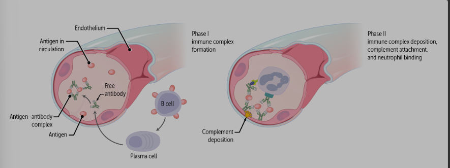
How is the action of immune-complexes different for Type III Hypersensitivity?
it is normal for our immune system to form immune complexes, but these are usually phagocytosed and cleared
however, if plasma cells produce antibodies at an accelerated rate, more immune complexes are formed than can be cleared
Characteristics of Type III Hypersensitivity Clinical Disease
can by systemic or localized depending on where deposition occurs
site of damage does not reflect the original site of infection, because these immune complexes can circulate to sites around the body
2 Examples of Type III Hypersensitivity
serum sickness
systemic lupus erythematosus
Serum Sickness
type iii hypersensitivity, used to occur when injections of large amts of horse serum were used for protection against diptheria
antibodies would develop against these foreign horse proteins and immune complexes would circualte throughout the body, leading to inflammation wherever they deposited, such as capillary beds of blood vessels and joint spaces
not a problem anymore because we no longer use this therapy
Systemc Lupus Erythematosus (SLE)
type iii hypersensitivity, autoantibodies, immune complexes circulate and deposit within tissue
sites can include skin (rash), kidney glomeruli (renal failure, proteinuria), bone marrow (low blood cell counts), and joints (arthritis)
Type IV Hypersensitivity (Delayed Type)
cell-mediated (CD4+ and CD8+ T cells), uses only immune cells (NO antibodies), delayed in that it takes 24-74 hours for one reaction to appear after exposure to an antigen
2 Type IV Hypersensitivity Mechanisms
first, CD8+ cytotoxic t cells recognize foreign or infected cells, which have foreign antigen complex with the MHC class I protein compex on their surface, and directly kill the cells using perforin and granzymes for apoptosis
alternatively, CD4+ hleper t cells can recognive an antigen present on a dendritic cell, which activates the helper t cells and causes them to secrete cytokines, thse cytokines activate circulating CD8+ cytotoxic T cells and macrophages, which then kill the abnormal cell
Example of Type IV Hypersensitivity
contact dermatitis (such as poison ivy)
Poison Ivy Mechanism (Type IV Hypersensitivity)
plant antigens on skin are haptens (too small to be noticed alone), which bind to proteins that form a hapten-protein complex that penetrates the epidermis
phagocytized by APCs and presented to CD8+ cytotoxic t cells at local lymph nodes
the now-sensitized cytotoxic T cells are primed and ready for actvation on secondary exposure
the sensitization phase can last 10-15 days or more
with secondary exposure, the primed CD8+ cells and macrophages induce skin inflammation, leading to the familiar symptoms of itching, redness, and tissue damage
TB Test
PPD skin test for TB exposure takes advantage of type iv hypersensitivity
a noninfectious TB agent is injected in the skin and sets off a reaction that is similar to but less severe that what is seen in contact dermatitis
the test is read 48-72 hours later, and a hard raised skin lesion suggest that the pt has previously been exposed to TB
Hypersensitivities Mnemonic
ACID
allergy/anaphylactic/atopy = type i
cytotoxic or cell dysfunction = type ii
immune complex deposition = type iii
delayed = type iv
Hypersensitivity Reaction
an excessive immune response to an antigen in a sensitized individual