BIO - Unit 2
 Call Kai
 Learn
 Practice Test
 Spaced Repetition
 Match
 Flashcards
 Knowt Play
1/86

There's no tags or description
Looks like no tags are added yet.
87 Terms
Proteins - building block
amino acids
↪ NH2 - CH(R) - COOH
Proteins - primary structure
amino acid chain
Proteins - primary structure - terminus
N-terminus: amino group (NH2) → start of protein
C-terminus: carboxyl group (COOH) → end of protein
Proteins - primary structure - bonds
Peptide bonds between amino acids
Proteins - secondary structure
interactions of nearby amino acids
Proteins - secondary structure - types
α-helix: SAME polypeptide chain
β-pleated sheets: TWO polypeptide chains
β-sheets types
Antiparallel: opposite ways (horizontal lines)
Parallel: same ways (zigzag lines)
Mixed: combination of both

Proteins - secondary structure - bonds
H-bonds between H of NH2 and O of COOH
Proteins - tertiary structure
interactions between R-groups of the same polypeptide chain
Most proteins only have ___ structures
tertiary
Proteins - tertiary structure - bonds
H-bonds
ionic bonds
hydrophobic interactions
disulfide bonds (S-S)
Proteins - quaternary structure
interactions between polypeptide chains
Common examples of quaternary structure
hemoglobin
DNA polymerase
Denaturation
change in temp or pH of environment → primary structure is only intact one
Colloid behavior
diffuse slowly & scatter light when in water
Buffering capacity of proteins
can resist changes in pH → because of their groups that can accept/donate H+
Amphoteric behavior
something can act as both an acid and a base depending on the situation
Different types of protein shapes
Fibrous: support tissues
Globular: transport, enzymes, and regulation
Different types of protein composition
Simple: just amino acids
Conjugated
Conjugated protein terminology
Apoprotein: protein part of conjugated protein
Holoprotein: apoprotein & prosthetic group
Prosthetic group: non-protein component → permanently attached → necessary for protein function
Common eg of conjugated protein
Hemoglobin:
heme: prosthetic group
globin: apoprotein
Different types of protein functions
enzymatic
structural
transport
storage
defense
hormonal/signaling
receptors
regulator (control cellular processes)
Solubility of different protein shapes
Globular: more soluble
Fibrous: less soluble
Allostery
when a molecule binds to a protein at a site that’s NOT the active site
Allosteric enzymes
enzymes that are able to change between T and R states
T and R states of allosteric enzymes
T: tense, active site is not super open → binding is difficult
R: relaxed, configuration of enzyme makes easy binding
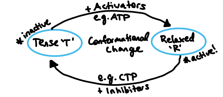
Different types of models for allosteric enzymes
Concerted model: specific ratio (ie 90T:10R) that must be continued so enzymes change from T to R and then at the end R to T to keep the ratio the same
Sequential model: 100% T at the beginning → substrate builds up until forcibly binds to T protein → enzyme actually has 3 subunits! As one substrate binds, second subunit becomes R, then third subunit becomes R → entire protein is R
Enzymes
Biological catalysts → lower activation energy requried
Categories of enzymes
Intracellular: inside cells
Extracellular: outside cells (ie in mouth or small intestine)
Structure of enzymes
Globular protein
Models of enzymes
Lock and key: substrate fits perfectly to active site
Induced fit: substrate doesn’t fit perfectly but active site conforms (more evidence)
Types of inhibitors
Competitive: compete with substrate for active site
Noncompetitive/allosteric: bind to enzyme at an allosteric site and change enzyme shape so substrate can’t bind to active site
Strengths of inhibitors
Reversible: weak bonds, can detach and allow enzyme to work again
Irreversible: strong bonds, permanently stop enzyme from working
Covalent modification
regulation of enzymes by covalently adding/removing chemical groups
Types of covalent modification
Phosphorylation: adding PO4 → using enzyme kinase → usually turns enzyme ON → phosphorylated enzyme
Dephosphorylation: removing PO4 → using enzyme phosphatase → usually turns enzymes OFF
Common examples of monosaccharides
glucose
fructose
galactose
mannose
ribose
deoxyribose
Monosaccharides
Cn(H2O)n → n = num from 3-7
Types of glucose
α-glucose: OH on C1’ on bottom
β-glucose: OH on C1’ on top
Anomeric carbon
only carbon where if OH is on top/bottom it changes whether carbohydrate is α or β
Glucose
bonds store lots of energy → major cellular fuel
blood sugar → glucose traveling through blood
building block of starch, glycogen & cellulose
Fructose
sugar in fruits and honey
component of sucrose
Types of fructose
D-fructose: found in nature
L-fructose: exists in laboratories
Difference between D- and L- carbohydrates
Two different forms that are mirrored images of one another (flipped horizontally)
Galactose
component of lactose
provides cellular energy
building block for tissues, brain, and immune system
Difference between glucose and galactose
Galactose has OH of C4’ on top instead of on the bottom (where glucose OH is)
Glucose molecule
C₆H₁₂O₆

Galactose molecule
C₆H₁₂O₆

Fructose molecule
C₆H₁₂O₆

Mannose molecule
C₆H₁₂O₆
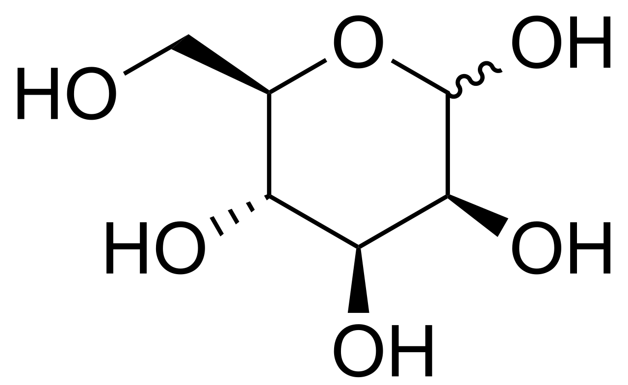
Ribose molecule
C5H10C5

Deoxyribose molecule
C5H10O4

Mannose
helps build proteins → glycosylation
stops bacteria (mainly E. coli) from sticking to urinary tract → offered as a supplement to prevent urinary infections
Types of mannose
α-mannose: OH on C1’ is on the bottom
β-mannose: OH on C1’ is on top
Ribose
present in RNA, ATP and NAD+
important for cellular energy, genetic material and metabolic cofactors
Deoxyribose
forms backbone of DNA
without the hydroxyl group on C2’, it’s more stable than RNA → better for long-term storage
Oligosaccharides
3-10 sugar units → most are disaccharides (2 sugar units)
Succrose/sacchrase
table sugar
energy storage/transport in plants
energy source in humans
Succrose/sacchrase make-up
glucose + fructose

Lactose
milk sugar
helps body absorb important minerals
supports healthy gut bacteria
Lactose make-up
galactose + glucose
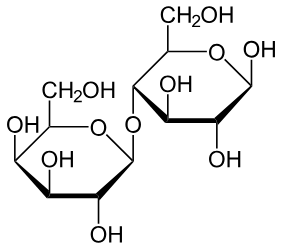
Maltose
produced during starch digestion
Maltose make-up
glucose + glucose

Common examples of oligosaccharides
succrose/sacchrase
lactose
maltose
Polysaccharides
more than 10 sugar units
Common examples of polysaccharides
starch
glycogen
cellulose
chitin
Amylopectin branch frequency
every 20 subunits
Bond formation in amylopectin
α(1→4) in chains & α(1→6) in branch points
Types of starch
amylopectin: branched
amylose: not branched
Bond formation in amylose
α(1→4)
Starch
energy storage in plants
dietary energy in animals
Bond formation in glycogen
α(1→6) at branch points
Glycogen
short-term energy storage in animals and funghi
stored in liver and muscle cells
Glycogen branch frequency
every 10 subunits
Cellulose
gives support to plants
insoluble fibers in humans → adds bulk to stool
primary nutrient for cows and horses
Bond formation in cellulose
β(1→4)
Chitin
forms exoskeletons
tight hydrogen make it very strong
Chitin make-up
N-acetylglucosamine
Chemically modified carbohydrates change
Phosphorylation: addition of a phosphate group - PO4
Glycosidation: replacement of hydroxyl group with an alcohol or amine
Phosphorylation function
increases reactivity and traps molecule inside cell membrane
Glycosidation function
removes reducing properties and forms stable glycosides or nucleotides
Glycoproteins
carbohydrate + protein (main component)
Glycoprotein function
cell recognition
cell receptors
immune functions
Glycosylation
covalent attachment of carbohydrate chains to proteins
Types of glycoproteins
O-linked glycoproteins: carbohydrate attached to O of hydroxyl group of serine (Ser) or threonine (Thr).
N-linked glycoproteins: carbohydrate attached to N of amino acid → usually asparagine (Asn)
Proteoglycans
carbohydrates + protein
Proteoglycan functions
structure
hydration
joint cushioning
cell signaling
extra-cellular matrix
Glycosaminoglycan (GAG) chains
unbranched complex carbohydrates made of repeating disaccharide units