Nursing Health Assessment: Skin Lesions Types
 Call Kai
 Learn
Learn Practice Test
 Spaced Repetition
 Match
Match Flashcards
 Knowt Play
1/50

There's no tags or description
Looks like no tags are added yet.
Name | Mastery | Learn | Test | Matching | Spaced | Call with Kai | Chat |
|---|
No analytics yet
Send a link to your students to track their progress
51 Terms
Macule
Flat, circumscribed area that is a change in color of the skin, less than 1 cm in diameter. Examples: Freckles, flat moles (nevi), petechiae, measles, scarlet fever. (Primary Lesion)

Papule
Elevated, firm, circumscribed area less than 1 cm in diameter. Examples: Wart (verruca), elevated moles, lichen planus, cherry angioma, neurofibroma, skin tags. (Primary Lesion)

Plaque
Elevated, firm, and rough lesion with a flat surface greater than 1 cm in diameter. Examples: Psoriasis, seborrheic and actinic keratoses, eczema. (Primary Lesion)

Wheal
Elevated irregular-shaped area of cutaneous edema; solid, transient, with variable diameter. Examples: Insect bites, urticaria, allergic reaction, lupus erythematosus.

Nodule
Elevated, firm, circumscribed lesion, deeper in dermis than a papule, 1 to 2 cm in diameter. Examples: Dermatofibroma, erythema nodosum, lipomas, melanoma, hemangioma. (Primary Lesion)

Vesicle
Elevated, circumscribed, superficial lesion not extending into the dermis, filled with serous fluid, less than 1 cm in diameter. Examples: Varicella (chickenpox), herpes zoster (shingles), impetigo. (Primary Lesion)

Bulla
Vesicle greater than 1 cm in diameter. Examples: Blister, pemphigus vulgaris, lupus erythematosus. (Primary Lesion)

Pustule
Elevated, superficial lesion similar to a vesicle but filled with purulent fluid. Examples: Impetigo, acne, folliculitis, herpes simplex. (Primary Lesion)

Cyst
Elevated, circumscribed, encapsulated lesion in dermis or subcutaneous layer filled with liquid or semisolid material. Examples: Sebaceous cyst, cystic acne. (Primary Lesion)

Excoriation
Loss of the epidermis; linear, hollowed-out, crusted area. Examples: Abrasion, scratch, scabies. Secondary Lesion.

Fissure
Linear crack or break from the epidermis to the dermis, may be moist or dry. Examples: Athlete's foot, cracks at mouth corners, chapped hands. Secondary Lesion.

Crust
Dried drainage or blood, slightly elevated, variable size and color. Secondary Lesion.

Scale
Thin or thick flake of skin varying in color; usually secondary to desquamated, dead epithelium
Ex. Dandruff
Secondary Lesion
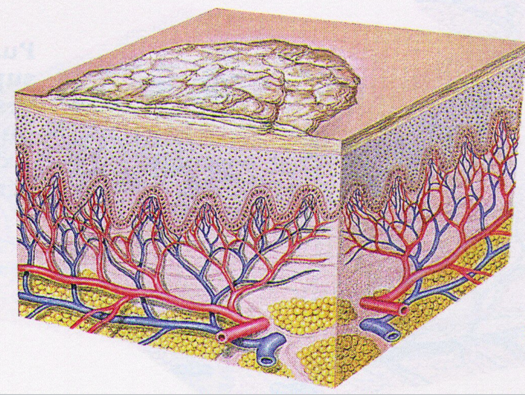
Erosion
Loss of part of the epidermis, depressed, moist, glistening, following rupture of a vesicle or bulla. Secondary Lesion.
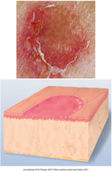
Ulcer
Loss of epidermis and dermis, concave, varies in size. Examples: Pressure ulcer, stasis ulcers, syphilis chancre. Secondary Lesion.

Atrophy
Thinning of the skin surface and loss of skin markings; skin appears translucent and paperlike. Secondary Lesion.

Petechiae
Tiny, flat, reddish-purple, nonblanchable spots in the skin, pinpoint to pinhead size, less than 0.5 cm in diameter. Vascular Lesion.

Purpura
Flat, reddish-purple, nonblanchable discoloration in the skin greater than 0.5 cm in diameter resulting from hemorrhage of blood into skin. Vascular Lesion.

Ecchymosis (Bruise)
Reddish-purple, nonblanchable spot of variable size caused by trauma to blood vessels resulting in bleeding under tissue. Vascular Lesion.

Angioma
Benign tumor consisting of a mass of small blood vessels, varying from small to large. Examples: Cherry angioma, hemangioma. Vascular Lesion.

Annular Lesion
Ring-shaped skin lesion configuration. Example: Ringworm.

Arcuate Lesion
Partial ring skin lesion configuration. Example: Syphilis.

Bizarre Lesion
Irregular or geographic pattern not related to any underlying anatomic structure. Example: Factitial dermatitis.

Circinate Lesion
Circular skin lesion configuration.
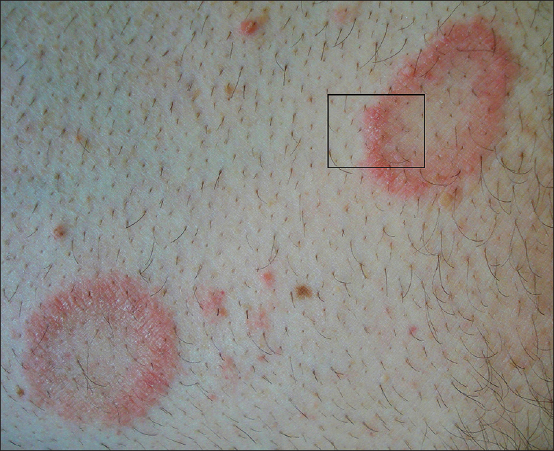
Confluent Lesion
Skin lesions that run together. Example: Childhood exanthems.

Discoid Lesion
Disc-shaped skin lesion without central clearing. Example: Lupus erythematosus.

Discrete Eczematoid Lesion
Lesions remain separate; inflammation with tendency to vesiculate and crust.

Generalized Grouped Lesion
Skin lesions clustered together. Example: Herpes simplex.

Iris Lesion
Circle within circle; bull's-eye skin lesion pattern. Example: Erythema multiforme.

Eczematous Dermatitis
Most common inflammatory skin disorder, including irritant contact, allergic contact, and atopic forms.

Folliculitis
Inflammation and infection of the hair follicle and surrounding dermis.
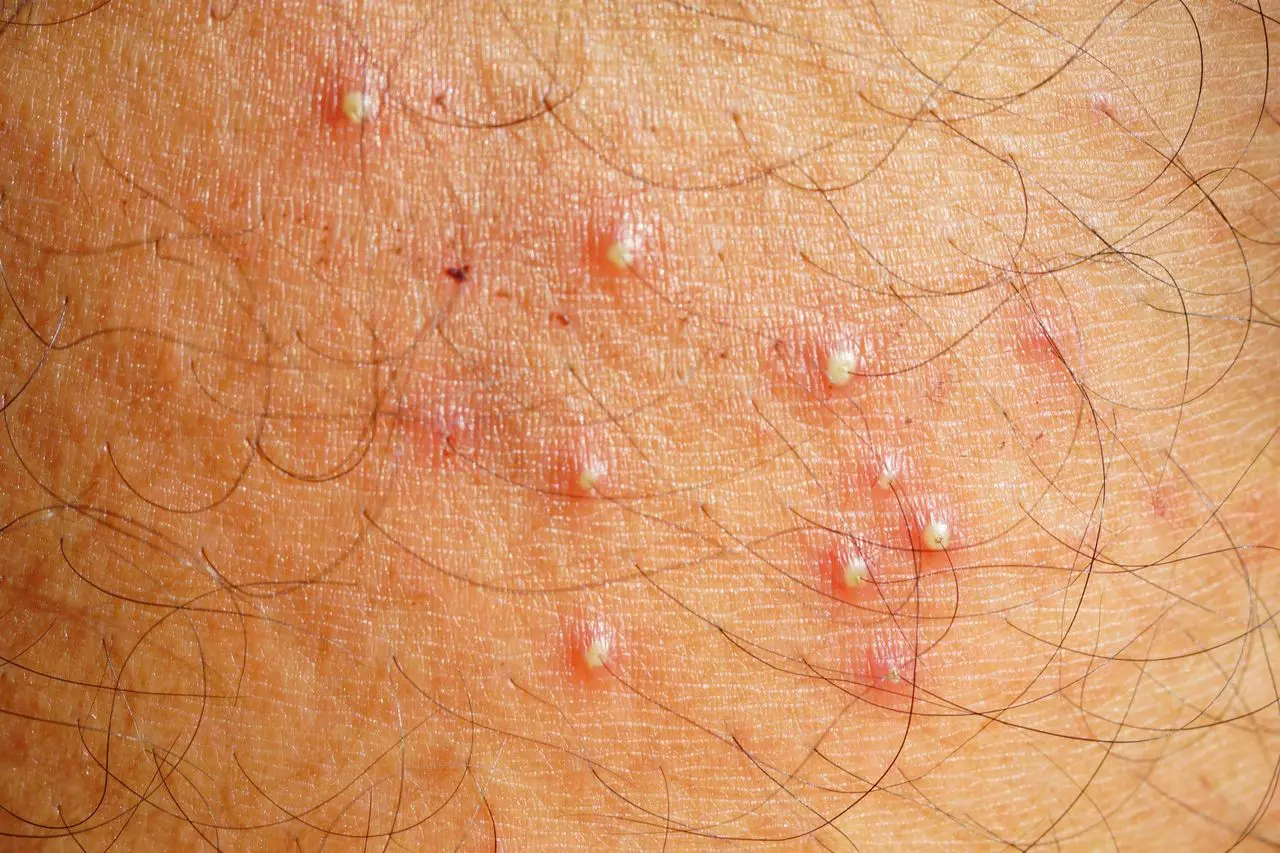
Furuncle (Boil)
Deep-seated infection of the pilosebaceous unit.

Cellulitis
Diffuse, acute infection of the skin and subcutaneous tissue.

Tinea (Dermatophytosis)
Group of noncandidal fungal infections involving the stratum corneum, nails, or hair.

Psoriasis
Chronic and recurrent disease characterized by keratinocyte proliferation.

Rosacea
Chronic inflammatory skin disorder.

Herpes Zoster (Shingles)
Viral skin infection caused by reactivation of the varicella-zoster virus.

Herpes Simplex
Viral infection occurring as Type 1 (associated with oral infection) or Type 2 (associated with genital infection).

Tick-Borne Diseases
Infections such as Lyme disease, Rocky Mountain Spotted Fever, and Colorado Tick Fever that can lead to multisystemic failure.

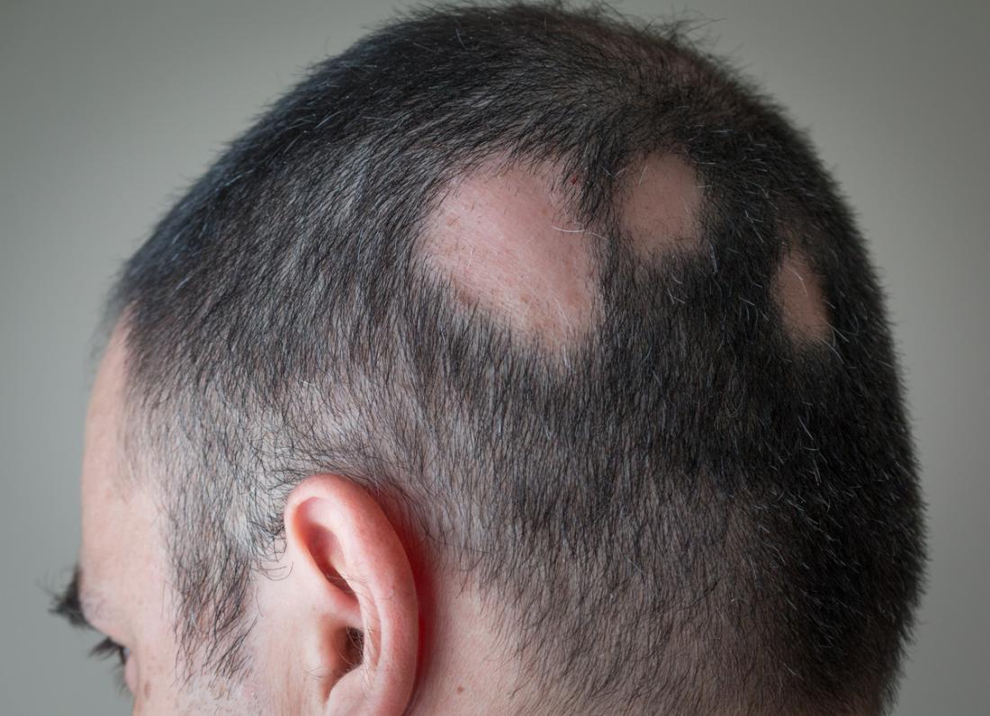
Alopecia Areata
Sudden, rapid, coin-shaped loss of hair, usually from the scalp or face.
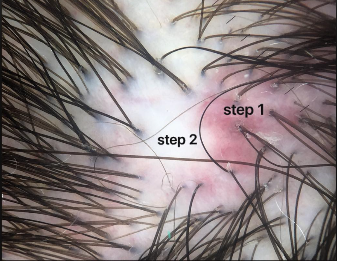
Scarring Alopecia
Hair loss caused by the replacement of hair follicles with scar tissue.
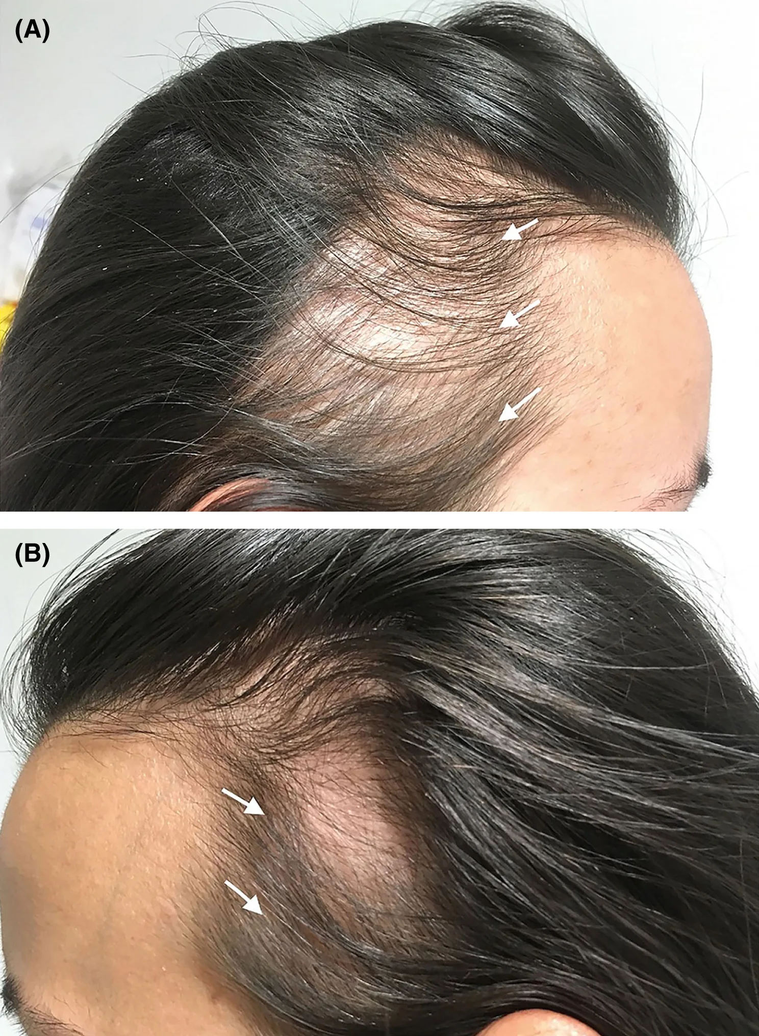
Traction Alopecia
Hair loss resulting from prolonged, tightly pulled hairstyles.
Hirsutism
Growth of terminal hair in women following a male distribution pattern on the face, body, and pubic areas.

Paronychia
Infection and inflammation of the paronychium surrounding the nail.

Onychomycosis
Fungal infection of the nail.

Ingrown Nails
Condition where the nail pierces the lateral nail fold and grows into the dermis.

Nail Avulsion
Injury where part or all of a nail is torn away or removed from the nail bed.

Subungual Hematoma
Trauma to the nail plate severe enough to cause immediate bleeding and pain under the nail.

Onycholysis
Loosening of the nail plate from the nail bed beginning at the distal groove.

Nail Clubbing
Structural change where fingertips enlarge and nails curve around fingertips like an upside-down spoon, becoming red and sponge-like; often associated with low blood oxygen and lung disease.

Koilonychia
Spoon-shaped, abnormally thin nails that lose convexity and become flat or concave; sign of hypochromic or iron-deficiency anemia.
