SB 15: Ch 13.1-13.3 The Eukaryotic Chromosome Pt 1
 Call Kai
 Learn
Learn Practice Test
 Spaced Repetition
 Match
Match Flashcards
 Knowt Play
1/26

There's no tags or description
Looks like no tags are added yet.
Name | Mastery | Learn | Test | Matching | Spaced | Call with Kai | Chat |
|---|
No analytics yet
Send a link to your students to track their progress
27 Terms
What is a chromosome composed of?
Each chromosome (or chromatid, in terms of replication) contains one long linear molecule of double stranded DNA
Shorter molecules recoil faster than longer ones
Drosophila: longest DNA molecule = largest chromosome
How does DNA compact itself to fit in the cell nucleus?
Interactions between two protein categories: histones and nonhistone chromosomal proteins
What are chromatin? What is a chromosome in terms of chromatin, what does chromatin have to do with RNA?
Complexes of DNA, protein, and RNA found in a cell’s nucleus
Chromosome = a piece of chromatin that contains a single DNA molecule
Chromatin = 1/3 DNA, 1/3 histones, 1/3 nonhistone proteins + significant amounts of RNA
Histone proteins and core histones
Small proteins with a preponderance of the basic, positively charged amino acids lysine and arginine
Strong positive charge = bind to and neutralize negatively charged DNA
Basic histone proteins are attracted to phosphate groups along DNA backbone
5 types of molecules: H1, H2A, H2B, H3, and H4
H2A, H2B, H3, and H4 = core of nucleosome (DNA packaging unit), core histones
All 5 appear in chromatin of all euk cells, very similar, changed little throughout evolution
Nonhistone (chromosomal) proteins
Many different kinds
Structural role, help package DNA > proteins that form scaffold of chromosome
Active in replication, DNA Polymerase
Chromosome segregation, motor proteins of kinetochores move cz along spindle and expedite transport of cz from parent to daughter
Largest class = facilitate transcription during gene expression, influence when/where/how frequently genes are transcribed
Nucleosomes
Winding of DNA around histones
Compaction
Basic unit of chromatin, exist in interphase cells and cells undergoing mitosis
Condensins
Protein complexes that form rings around DNA, organize chromosomes into loops
Condensin loops = greatest form of chromosome condensation in mitosis
Compaction
Nucleosome structure
DNA wraps twice around a core made of two copies each of histones H2A, H2B, H3, and H4 (8 total)
Histone H1 associates with the DNA as it enters and leaves the nucleosome
X-ray diffraction: DNA bends sharply as it wraps around core histone octamer
Only some DNA base sequences can bend

Linker DNA
Connects one nucleosome with the next
40 or so base pairs
Histone chaperones?
What is the scaffold, how does chromosomal DNA appear relative to it?
The scaffold is the structural backbone of chromosomes. Chromosomal DNA appears as long loops anchored in the scaffold.
Loops = chromosomal compaction
What are condensins, and what is their role in the scaffold?
Protein complexes that form one of the major components of the scaffold. They make the chromatin loops, which is the next step of chromosomal compaction.
Ring-shaped
Sit on chromatin by forming around nucleosome-studded DNA so the chromosome fiber passes twice through the ring's interior, making a small loop
How do condensins enlarge a loop?
Some subunits are molecular motors that use ATP hydrolysis to move the condensin relative to the chromatin. This extrudes the loop (makes it larger).
How fast is loop extrusion? How big?
A single condensin complex can extrude about 1500 bp of chromatin per second.
Roughly 30 to 400 kb of DNA.
We don’t know when extrusion stops or what determines where condensin rings first form
Why does loop extrusion create a problem? What enzyme solves this and how?
Condensin movement relative to chromatin twists the DNA and causes supercoiling. The supercoils would prevent further extrusion.
DNA topoisomerase II relaxes the supercoils and prevents DNA from tangling in compacted chromosomes.
What are the two major components of chromosomal scaffolds?
Condensin complexes and DNA topoisomerase II.
How did biochemists study the requirements for metaphase chromosome assembly?
They reconstituted chromosome structure in vitro using protein extracts from the large eggs of Xenopus (African frogs).
Sperm chromatin incubated with the egg extract disassembles and then reassembles into metaphase chromosome structures.
Which four purified proteins were enough to mimic most steps of metaphase chromosome assembly?
Histones, histone chaperones, condensins, and topoisomerase II.
What do the reconstitution results suggest about mitotic chromosome condensation?
Researchers could mimic most steps of metaphase chromosome assembly
The major features are nucleosome assembly (histones and histone chaperones) and chromatin loop extrusion (condensins, with help from topoisomerase II).
Why isn't loop formation alone the full story of condensation? What are two proposed explanations for additional compaction, and what is known about them?
Large, loosely packed loops anchored by condensin don't reach the level of condensation seen in mitotic chromosomes.
Nucleosomes within loops interact to form a 300 Å superhelix. This has been seen under the electron microscope, but scientists disagree about whether much chromatin is actually organized this way in cells.
Condensin rings interact with each other in the scaffold, bringing loops closer together.
What is Giesma staining, what does it reveal?
Chromosomes are gently heated, then exposed to Giemsa stain.
The dye preferentially darkens certain regions, producing alternating dark and light
The biochemical basis of G bands isn't understood.
Most think the bands reflect uneven packaging of loops, determined by the spacing and density of short repetitive DNA sequences along the chromosome.
Why do geneticists think banding patterns are an intrinsic property of each chromosome? What do the patterns reproduceability offer?
They are highly reproducible from one generation to the next, so they must be determined by the base-pair sequence itself.
Geneticist designate a gene’s location by its position relative to the bands on the p (short) or q (long) arm of a particular chromosome.
Diagram of banding pattern = idiogram
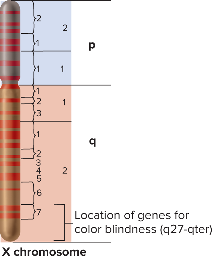
What is FISH, what does it allow us to do?
Fluorescence in situ hybridization
Solves resolution gap. Karyotypes show the whole genome at low resolution, and whole-genome sequencing is ultra-high resolution
Allows us to find the locations of specific DNA sequences with respect to the chromosomes in a karyotype.
Steps/basis of FISH
It uses nucleotide complementarity to locate specific DNA sequences on chromosomes
Spread and fix metaphase chromosomes on a slide, then gently denature the DNA. Add a heat-denatured fluorescent DNA probe. It hybridizes only to complementary regions, which are seen under a fluorescence microscope.
What does a FISH result with a single-gene probe look like?
Fluorescent spots marking where that gene sits. Sister chromatids are so close together that they show one spot per homolog.
What is SKY, and what are FISH and SKY used for?
SKY is a FISH variation with probes from DNAs scattered along each chromosome, labeled with a unique mix of fluors so all 24 human chromosomes show up as different colors. Both techniques detect chromosomal rearrangements (deletions, duplications, inversions, translocations).
x