Pulp Endodontics MAIN Dr. Leibow
 Call Kai
 Learn
Learn Practice Test
 Spaced Repetition
 Match
Match Flashcards
 Knowt Play
1/36

Earn XP
Description and Tags
Operative Exam 1
Name | Mastery | Learn | Test | Matching | Spaced | Call with Kai | Chat |
|---|
No analytics yet
Send a link to your students to track their progress
37 Terms
Pathogenesis of Apical Periodontitis
Irreversible Pulpitis: Progressive degeneration of the pulp.
Symptoms: Usually accompanied by symptoms.
Apical Periodontitis: Can initiate early apical periodontitis.
Pulpal Necrosis: Eventually results in complete pulpal necrosis.
Infection: The necrotic pulp becomes infected.
Purpose of a Root Canal
To prevent and cure apical periodontitis

Apical Periodontitis
Definition: An inflammatory process in the periradicular tissues caused by microorganisms in an infected root canal.
Apical Periodontitis Cause
Microorganisms
Apical Periodontitis Initial Infection
Mostly gram negative and
anaerobic
(e.g. Fusobacterium)
Apical Periodontitis Post-Treatment Infection
Gram positive facultative
anaerobes
(e.g. Enterococcus faecalis)
Goals of Endodontic Therapy
Goal: Prevent or resolve apical periodontitis.
Remove Infection Source: Remove all organic substrate from the root canal system.
Prevent Reinfection: Prevent re-infection of the root canal system.
Removal of Organic Substrate
Complete access
Careful instrumentation
Thorough irrigation
Disinfection
Dissolution and removal of bioburden
Removal of smear layer
Prevention of Re-Infection (3)
3-D Obturation: Complete three-dimensional obturation of the pulp space.
Coronal Seal: Create an impervious coronal seal.
Final Restoration: Perform ongoing inspection and maintenance of the final restoration.
Diagnostic Objectives (3)
Reproduce the Chief Complaint: Reproduce and confirm the patient’s chief complaint.
Determine the Cause: Identify the underlying cause of the complaint.
Eliminate the Cause: Remove or treat the cause of the complaint.
.
o Not Treat Unless You Know The Cause Of The Chief Complaint (Do no harm)
Normal Pulp
Normal Pulp: A clinical diagnostic category in which the pulp is symptom-free and responds normally to pulp testing.
Reversible Pulpitis
Reversible Pulpitis: A clinical diagnosis based on subjective and objective findings indicating that the inflammation should resolve and the pulp return to normal.
Reversible Pulpitis Findings
Percussion: Test performed.
Bite: Test performed.
Cold Test: Positive (++), with a non-lingering response.
Radiograph: Unremarkable.

Symptomatic Irreversible Pulpitis
Symptomatic Irreversible Pulpitis: A clinical diagnosis indicating that the vital, inflamed pulp is incapable of healing.
Lingering Thermal Pain: Pain persists after the thermal stimulus is removed.
Spontaneous Pain: Pain may occur without any stimulus.
Referred Pain: Pain may be felt in another area, making the source difficult to identify.
Symptomatic Irreversible Pulpitis Findings
Percussion: Positive (+) or negative (−).
Bite Test: Positive (+) or negative (−).
Cold Test: Strongly positive (+++) with lingering pain lasting more than 30 seconds.
Heat Test: Strongly positive (+++) or negative (−).
Radiograph: Usually unremarkable.
Asymptomatic Irreversible Pulpitis
Asymptomatic Irreversible Pulpitis: A clinical diagnosis indicating that the vital, inflamed pulp is incapable of healing.
No Clinical Symptoms: The patient may have no noticeable symptoms.
Causes: Inflammation may be caused by caries, caries excavation, or trauma.
Asymptomatic Irreversible Pulpitis Findings
Percussion: Negative (−).
Bite Test: Negative (−).
Cold Test: Positive (+) or negative (−), and the response may linger.
Heat Test: Positive (+) or negative (−).
Radiograph: Caries approaching the pulp.

Pulp Necrosis
A clinical diagnostic category indicating death of the dental pulp.
The pulp is usually nonresponsive to pulp testing.
Pulp Necrosis Findings
Percussion: + to +++ → indicates inflammation of the periodontal ligament, causing pain when the tooth is tapped.
Bite: + to +++ → suggests inflammation around the root/apical tissues; biting pressure aggravates the inflamed periodontal ligament.
Cold: − → no response to cold, which strongly suggests pulp necrosis (assuming the test was performed correctly and compared with control teeth).
Heat: − → also supports loss of pulpal vitality.
Radiographic evidence of apical periodontitis: frequent → inflammation at the root apex may be visible radiographically.
Periapical radiolucency: may or may not be present → an early apical inflammatory lesion can exist before a radiolucency becomes detectable on a conventional radiograph.
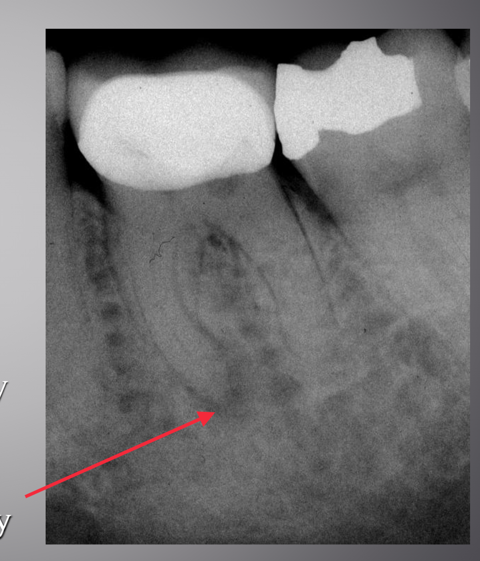
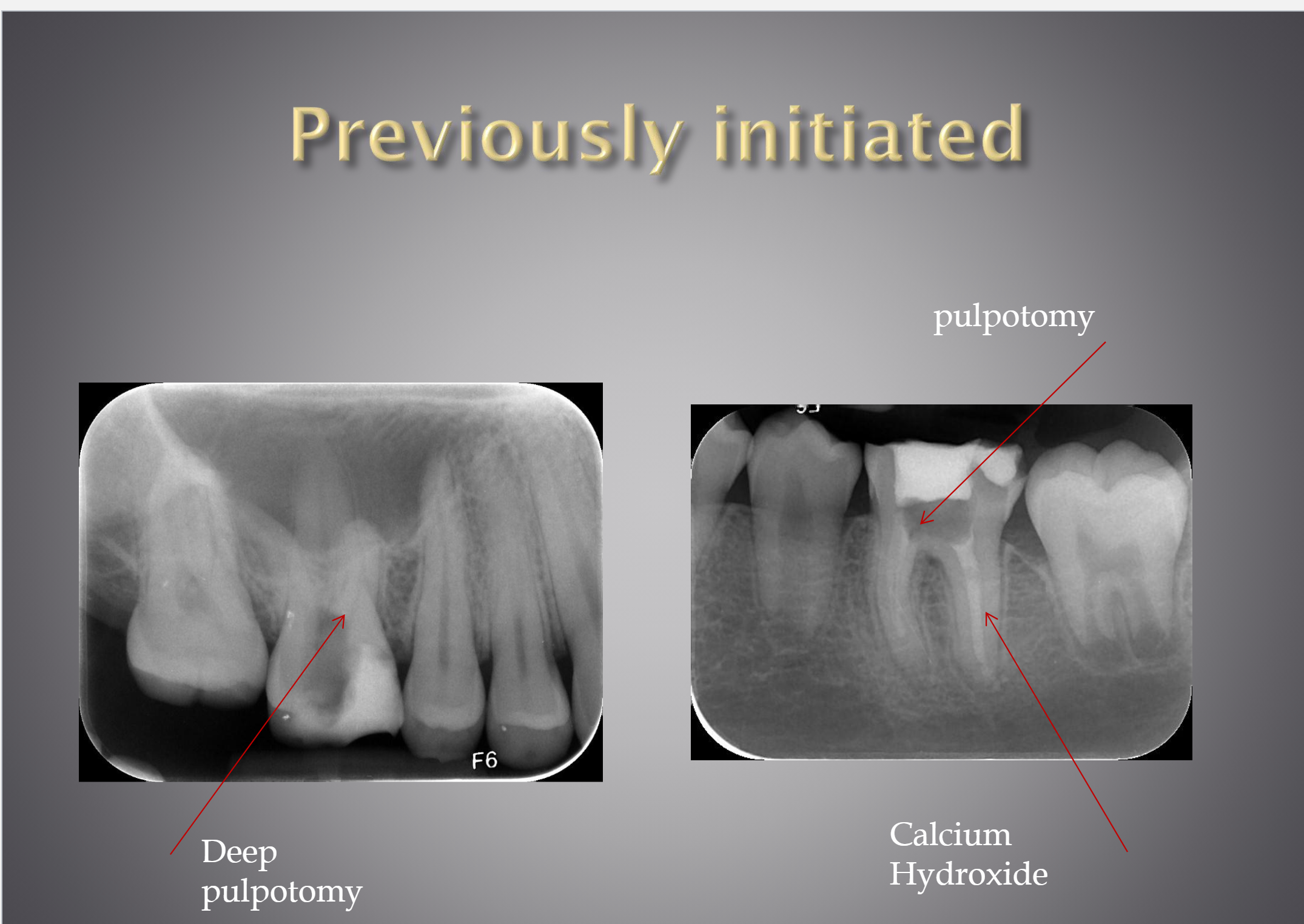
Previously Initiated Therapy
A clinical diagnostic category indicating that the tooth has been previously treated by partial endodontic therapy (eg, pulpotomy, pulpectomy).
Previously Treated
A clinical diagnostic category indicating that the tooth has been endodontically treated and the canals are obturated with various filling materials other than intracanal medicaments.
Previously Treated Findings
Percussion: − to +++ → may be tender because of inflammation of the periodontal ligament.
Bite: − to +++ → biting pain can occur when the apical/periodontal tissues are inflamed.
Cold: − → expected because the pulp has been removed or the tooth has already undergone root canal treatment.
Heat: − → also expected in a root-filled tooth.
Radiographic appearance: variable → depends on the quality/extent of the previous treatment and the status of the periapical tissues.
Root filling evident → indicates the tooth has previously received root canal treatment.
Periapical radiolucency → may be present when there is apical inflammation/bone loss.

Pulpal Diagnosis Summary (6)
1) Pulp Necrosis
No Response to thermal or electrical stimuli
.
2) Previous Root Canal Therapy
Canals are obturated
.
3) Previously Initiated Therapy
Partial endodontic therapy
(e.g. pulpotomy, pulpectomy)
.
4) Reversible Pulpitis
Non-lingering (Thermal tests)
Not spontaneous
.
5) Symptomatic Irreversible Pulpitis
Spontaneous
Pain lingers after stimuli (Thermal tests)
Usually severe
.
6) Asymptomatic Irreversible Pulpitis
No clinical symptoms
Inflammation produced by:
Caries, caries excavation, trauma, etc
Normal Apical Tissue
Teeth with normal periradicular tissues that are not sensitive to percussion or palpation testing.
The lamina dura surrounding the root is intact, and the periodontal ligament space is uniform.
Normal Apical Tissue Photo

Asymptomatic Apical Periodontitis
Inflammation and destruction of apical periodontium that is of pulpal origin, appears as an apical radiolucent area, and does not produce clinical symptoms.
Asymptomatic Apical Periodontitis Photo

Symptomatic Apical Periodontitis
Inflammation, usually of the apical periodontium, producing clinical symptoms including a painful response to biting and/or percussion or palpation.
It might or might not be associated with an apical radiolucent area
Symptomatic Apical Periodontitis Photo
Acute Apical Abscess
An inflammatory reaction to pulpal infection and necrosis characterized by rapid onset, spontaneous pain, tenderness of the tooth to pressure, pus formation, and swelling of associated tissues.
Acute Apical Abscess Photo

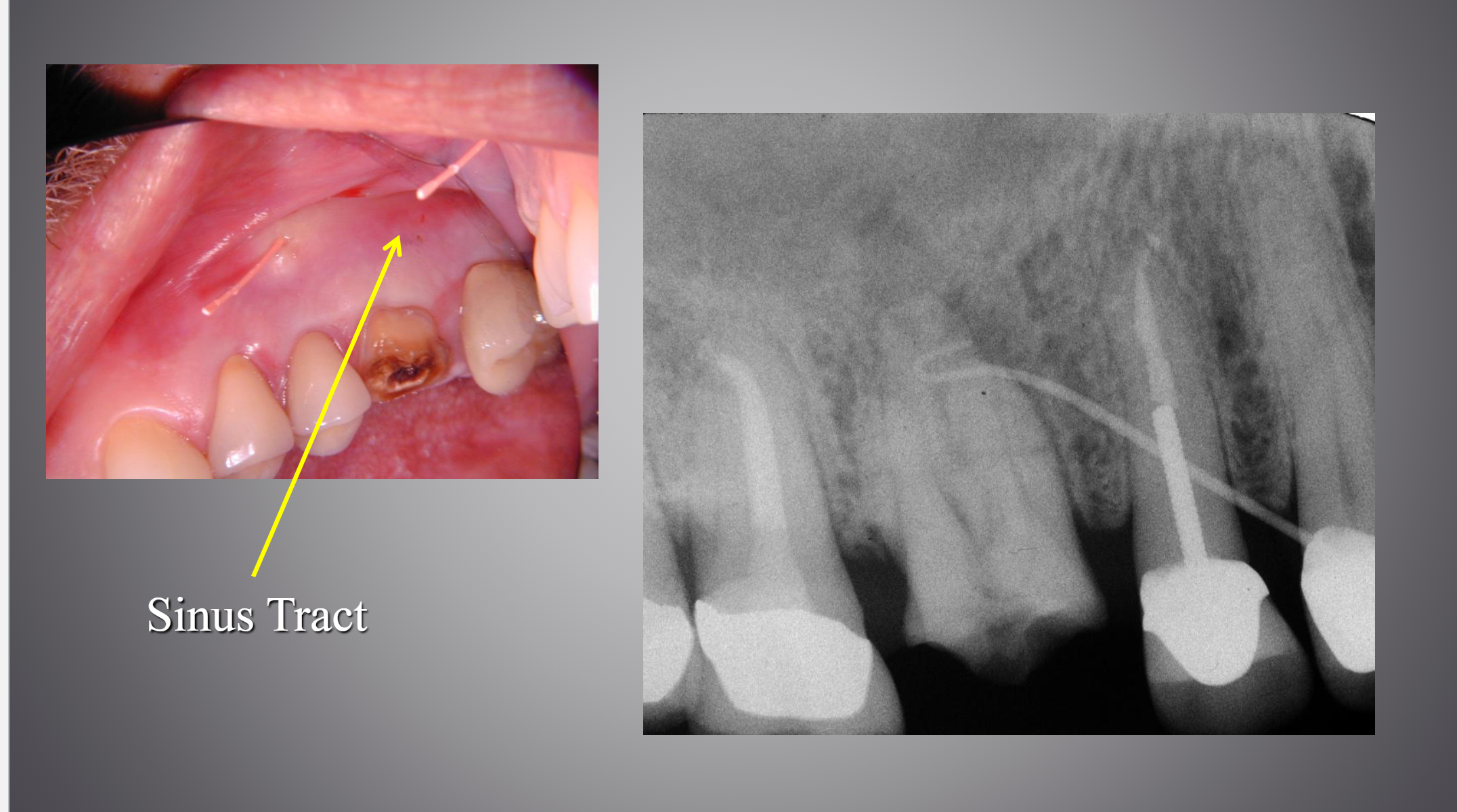
Chronic Apical Abscess
An inflammatory reaction to pulpal infection and necrosis characterized by gradual onset, little or no discomfort, and the intermittent discharge of pus through an associated sinus tract.
Chronic Apical Abscess Photo
Facial Cellulitis
Extraoral/Facial Swelling

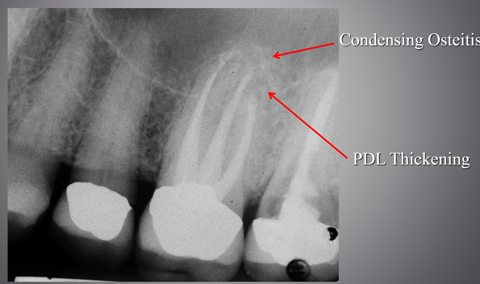
Condensing Osteitis
Diffuse radiopaque lesion representing a localized bony reaction to a low-grade inflammatory stimulus, usually seen at apex of tooth.
Condensing Osteitis Photo

Periapical Diagnosis Summary
1) Normal apical tissues
Asymptomatic, intact lamina dura
.
2) Symptomatic apical periodontitis
pain to biting and percussion
May or may not have associated PA radiolucency
.
3) Asymptomatic apical periodontitis
Cannot elicit pain or altered sensation
Apical radiolucent area
.
4) Acute Apical Abscess
Localized swelling, pain, pus formation
Tender to pressure
Fever? Lymphadenopathy?
PA Radiolucency?
.
5) Chronic Apical Abscess
Minimal or no pain
Pus drains from a sinus tract
.
6) Condensing Osteitis
May be totally asymptomatic
Usually result of irritation from inflamed pulp