A&P Exam 2
 Call Kai
 Learn
 Practice Test
 Spaced Repetition
 Match
 Flashcards
 Knowt Play
1/133

There's no tags or description
Looks like no tags are added yet.
134 Terms
Integumentary System
two or more kinds of tissues grouped together & performing specialized functions constitutes an organ
vital in maintaining homeostasis
15-20% of body mass, - 2m2
Skin
protection from drying out, foreign organism, friction w/ environment
maintain body temperature
excretion (NaCl, urea)
sensory
vitamin D production (necessary for calcium absorption by gut)
lipid soluble substances can be absorbed through skin
Corticosteroids
produced in adrenal cortex → stress response, immune, inflammation
hydrocortisone - immune suppressive drug, allergic reactions or rashes
impaired wound healing, high BP
biomarkers for psychological stress
Urishiol-Induced
poison-ivy
induced immune response, binds & changes shape of protein in cell membrane, body doesn’t recognize as self & attacks
15-30% people no response, 25% strong response
stronger reactions after repeated exposure
Takes weeks to go
Thick Skin
thick on palms & soles of feet
upper back has thickest dermis
Thin skin
located everywhere except palms, soles of feet, & upper back
eyelids = thinnest
Epidermis
stratified squamous epithelium
4 distinct layers
Stratum Basale
Stratum Spinosum
Stratum Granulosum
Stratum Corneum
Stratum Basale
single layer, the stem cells, cuboidal, big nuclei → dividing cells
E) Stratum Basale
Label E

Stratum Spinosum
“prickle layer” larger cells becoming flatter, desmosomes hanging on with cell shrinkage = looks psiny
D) Stratum Spinosum
Label D

Stratum Granulosum
1-3 cell layers thick, keratin granules = stain darkly
C) Stratum Granulosum
Label C

Stratum Lucidum
a single layer of dead skin cells found only in thick skin (palms, soles of feet, upper back)
B) Stratum Lucidum
Label B

Stratum Corneum
abrupt transition, flattened, desiccated, a nucleate cells, loose organelles just keratin filaments, layer of lipids-water barrier, thickest where friction (calluses) = dead layer
A) Stratum Corneum
Label A

Psoriasis
autoimmune disease
skin divides too rapidly (3vs.30 days)
triggers = stress & medications
3% - 7.5 million Americans;
Dermis
uneven boundary helps attachment
dermal papillae meet epidermal ridges
more ridges in thick skin - more surface area
2 Layers -
Papillary Layers - top layer
Reticular Layer - random collagen fibers
Langers Lines = collagen & elastic fibers (follow fiber direction in surgery0
potential to form bone
Blisters
shearing force, fluid in derma-epidermal junctions
Hypodermis
adipose tissue - varies in thickness
muscle for facial muscles, more well developed in other mammals
Melanocytes
produce melanin, replicate slower
scattered among basal cells
goes to tip of processes, “injected” into skin cells
UV radiation stimulates melanin production
2 Factors of Melanocytes
Dark to protect from UV radiation
Light to synthesize Vitamin D ‘
Humans have a lot of sweat glands, less hair to deal with heat but more UV damage
Eumelanin
brownish/black
genetically determined
Pheomelanin
reddish/yellow
genetically determined
Light Skin
melanin degraded in stratum spinosum, lasts longer in darker skin, larger granules
Jaundice
yellow skin
carotenes in fat, blood vessels near skin
Cyanosis
bluish skin (slow moving blood)
Albanism
genetic mutation = less melanin = white skin
Nerve Supply of Skin
lots of sensory receptors
free nerve endings = heat/touch/cold/pain
surround hair follicles = tactile (ex. whiskers)
Lamellated Corpuscles (encapsulated nerve endings)
deep pressure, large
unmyelinated nerve w/ concentric lamellae with fluid between
Tactile Corpuscles (fine touch)
in dermal papillae - touch receptors
fingers, toes, & lips
tapered cylinders just beneath epidermis
nerve takes a spiral path, Schwann cells surround
Hair
humans are covered all over in hair, except on palms, soles, lips, nipples, & part of external reproductive organs
on average each person’s head carries about 100,000 hair follicles
each hair grows for several years, will fall out & be replaced
Hair Scent
hair carries an individual’s scent, by which he or she may be recognized → attract mates
Temperature Regulation - Hair
hair = traps air = insulator
sweat glands = cool off
hair follicles produce hair
distribution of hair influenced by sex hormones
Sweat Gland
coiled & release to surface
Sebaceous Gland
secrete sebum - fatty/cell debris, coats hair & skin surface
holocrine gland - release products by disintegrating the whole cell
directly on skin - labia minora, areola, eyelids, lips
Hair Follicle
hair/sebaceous gland canal to surface
bulb w/ dermal papilla
sheaths surround
starts at surface → moves down into skin
arrector pili muscle = goose bumps
Proteins in Hair
hair is extremely complex & sophisticated composite, where proteins are combined with lipids & minerals to form a unique structure w/ extraordinary properties
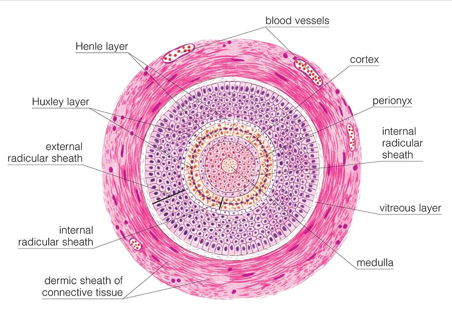
Human Follicle Cross Section
hard keratin
medulla - central part-large open cells (only in thick hair)
cortex - cuboidal cells to keratin filled
cuticle = squamous cells to outside
Human Hair Color
no biological function of human hair color
melanin found in two forms - eumelanin & phaeomelanin (mixture of both in most people’s hair)
decreased melanocyte activity = grey hair
Eccrine Sweat Glands
simple coiled glands
not associated w/ hair follicles
down growth from epidermis
secretory & duct segments
temp regulation
NaCl, urea, ammonia excreted
Apocrine Sweat Glands
coiled tubular/branched (sometimes)
large lumen
same down growth as hair = retain connections
store products in lumen, unlike eccrine
protein rich secretion, lipids - odor when bacteria digest
functional at puberty
pheromones in many mammals
Ceruminous Glands
glands of external auditor meatus = secretes ear wax
Mammary Glands
secrete milk, milk lines from axilla to groin, like sebaceous/seat = secrete lipid
number of glands = number of offspring
Nails
tightly compacted cornified epithelial cells
plates at keratinzied cells - hand keratine
nail plates resting on nail bed
nail bed = stratum basale/spinosum
nail - modified stratum corneum
pale luma - partially keratinized cells, become transparent after
Tattoos
pigment in dermis layer
phagocytes engulf pigment, epidermis flakes off, stable in upper dermis, does migrate deeper over decades → fading
Skin Repair
basal cell proliferation under injury
if all epidermis gone; hair, sweat gland cells have stem cells
3rd degree etc. → need skin graft
Healing of Wounds & Burns
inflammation is a normal response to injury or stress
blood vessels in affected tissues dilate & become more permeable → fluids leak into damage tissue
inflammed skin - reddened, swollen, warm, painful
Cuts
a shallow cut results in epidermal cells long its margin to divde more rapidly
deep cuts result sin blood vessels breaking which causes a clot to form:
Clot & dried tissue fluid form scab
Growth factor stimulate new tissue formation
phagocytic cells remove dead cells & debris
excess collagenous fibers may form scars
First Degree Burns
superficial, partial-thickness
Second Degree Burns
deep, partial thickness
Third Degree Burns
full-thickness, autograft, allograft (from another cadaver), skin substitutes
Regulation of Body Temperature (skin)
vitally important because even slight shifts can disrupt rates of metabolic reactions
skin plays a role in homeostatic mechanism that regulates body temperature
Heat Production & Loss
heat is a product of cellular metabolism
most active body cells are the heat producers: skeletal muscle cells, cardiac muscle cells, cells of certain glands such as the liver
primary means of heat loss is radiation, there is also conduction, convection, & evaporation
Hyperthermia
abnormally high body temperature
humid day = evaporation doesn’t happen, core> 106 degrees F
skin dry, weak, dizzy, nauseous , headache, rapid pulse
Hypothermia
abnormally low body temperature
shivering & coldness to confusion, lethargy
Age Changes in Skin
epidermis & hypodermis thins
melanocytes decrease
sweat/sebaceous glands = less activity
healing = 4x slower
age spots - sun damage & weakened vessels in dermis = bruise
hair = grey/white, nails = weak
Skin Cancer
most common human cancer - half Americans >65 develop at least once
Basal Cell Carcinoma (90% in US) = rarely spread
Squamous cell carcinoma - squamous cells elsewhere in body also
Light skin & sun exposure
Can metastasize (lower lip)
Adenocarcinoma (glands) - colon, lung
Melanoma
clusters of melanocytes
melanoma = when cells become malignant
spread in lymph nodes, to liver & lungs, brain, but STILL melanoma cells
Asymmetry, Border (ragged), Color (uneven), Diameter (>5 mm usually)
biopsy to check for cancer cells - staged as to how thick it is if spread
Skeletal System
206 bones
80 bones in axial skeleton
126 bones in appendicular
20% of body weight
Associated Connective Tissue - cartilage, tendons ligaments, discs
Bones are made up of many tissues = one tissues, cartilage, dense connective, blood & nervous tissue
Functions of Bone
maintain body shape
protect internal organs (brain, spinal cord)
system of levers for the muscle system to act upon
mineral storage = Ca2+ & phosphorus
Hold blood forming cells
surface feature for muscle (insertions & origins)
Long Bone Anatomy
Diaphysis - main tubular shaft of long bone, midsection
Metaphysis - wide, flared neck portion, between epiphysis & diaphysis
Epiphysis (proximal/distal) - rounded end/tip of long bone
Epiphyseal Line - remnant of epiphyseal plate, after bone longitudinal growth stops
Periosteum - covers outer surface of bone
Compact Bone - forms outer protective shell of bones
Spongy Bone - (cancellous bone) porous, makes up interior of bones
Articular Cartilage (hyaline) - covers & protects the end of bones
Medullary Cavity - hallow central space inside the diaphysis
Marrow - soft, spongy tissue found inside the center cavities of bone → produce blood cells
Nutrient Artery - main blood vessels for long bones

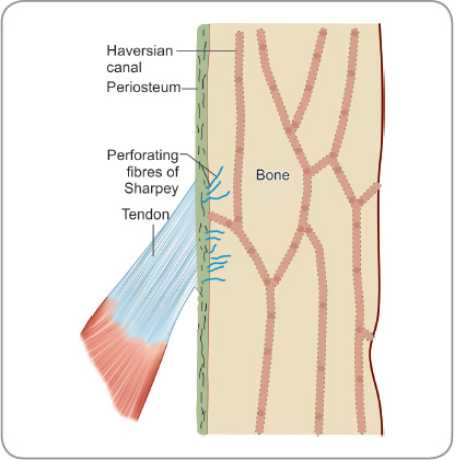
Periosteum
Covers outer surface of bone (except for articulations - cartilage)
outer fibrous layer
collagen fibers from ligaments + tendons go into the bone tissue at an angle
Endosteum
lines bone cavities
1 cell layer thick lining
bone stem cells line marrow cavity & trabeucle of spongy bone
Bone Marrow
red bone marrow = where blood cells are made
yellow bone marrow = fat storage
as you grow, amount of red bone marrow doesn’t increase in proportion to bone growth
yellow marrow forms
lot of red bone marrow in sternum, pelvis
What type of tissue is bone?
Bone is a connective tissue = cells + extracellular matrix
Matrix is mineralized → support & protection
Calcium phosphate in form of hydroxyapatite crystals
30: 70% organic/inorganic salts
without organic minerals = fragile
Osteocytes
maintain bone tissue
mature bone cells
transport nutrients & waste
extracellular matrix of bone is largely collagen & inorganic salts
Collagen = resilience
Inorganic salts = bone hard
Mature Bone
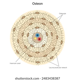
osteons (haversion systems) 4-20 lamellae w/ central nerve, artery, vein
osteocytes trapped in matrix, connected to each other by canaliculi (processes of osteocytes)
sometimes chain of 15
Osteon
cylinder shaped unit containing bone cells & matrix lamella that surround a central canal; havarain system
many come together to form the compact bone
osteons run longitudinally with the axis of bone, functioning as weight-bearing pillars, resisting compression
Central Canal
contains blood vessels & nerves surrounded by loose connective tissue
runs down the middle of osteons
Blood in these vessels nourishes bone cells associated with the central canal via gap junctions between osteocytes

Perforating Canal
connects central canals to one another
contain larger blood vessels & nerves by which the smaller blood vessels & nerve fibers in central canals communicate with the surface of bone & medullary cavity
Lamellae
layers of extracellular matrix concentrically clustered around a central canal
Lacunae
small fluid-filled cavities within the bone matrix that house the osteocytes
Canaliculi
channels/tunnels that connect lucanae to each other and to blood vessels
Nutrient Foramina
blood supply to bone (very important = healing/nutrients)
Spongy Bone
rarely contains osteons
Trabeculae (braching bony plate that separates irregulater spaces within spongy bones) surrounding red bone marrow spaces
composed of osteocytes & extracellular matrix, but the bone cells do not aggregate around central canals
get nutrients from substances diffusing into the canaliculi
Immature Bone
develops in fetus
does not have organized lamella = “woven bone”
relatively more cells/area
adult: tooth sockets, remodeling
Bone Development & Growth
parts of skeletal system begin to develop during first few weeks of prenatal development
human skeleton is initially hyaline cartilage + fibrous membrane (300 bones at birth)
by 25 bone the skeleton is almost completely hardened
Osteogenic Cells
develops into osteoblasts
Osteoblast
forms bone tissue
synthesize organic components of matrix (collagen, bone matrix proteins)
hormones stimulate activity
Osteoclast
function in resorption of bone (destruction of bone matrix)
derived from WBCs = bone resorption
large, motile cells
secrete enzymes that digest matrix
Intramembranous Ossification
(between membranes) flat bones of the skull, clavicles, sternum, & some facial bones (mandible, maxillae, & zygomatic bones)
during their development (osteogenesis) membranelike layers of embryonic connective tissue appear at the sites of future bone
Mesenchymal cells that are part of the connective tissues enlarge & differentiate into osteoblasts
Osteoblasts form bony matrix around themselves
Dense networks of blood vessels supply these connective tissues
Spongy bone forms in all direction along the blood vessels

Endochondral Bones/Ossification
bone begins as hyaline cartilage
Form models for future bones
Most bones of skeleton
cuff of bone forms in mid-region - BONY COLLAR
cartilage cells in center swell, secrete calcium
calcified cartilage inhibits diffusion = kills cells
blood vessels grow in, bone stem cells move in
secondary center of ossification - leaves cartilage epiphyseal plate
when that cartilage stops growing bone has reached its max length

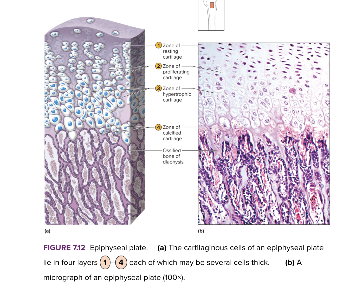
Zone of Resting Cartilage (growth at epiphyseal plate)
first layer of cells
closet to end of epiphysis
resting cells
anchors epiphysis plate to epiphysis
Zone of Proliferating Cartilage (growth at the epiphyseal plate)
2nd layer of cells
many rows of young cells
undergoing mitosis

Zone of Hypertrophic Cartilage (growth at epiphyseal plate)
older cells left behind when new cells appear
cells enlarging and becoming calcified
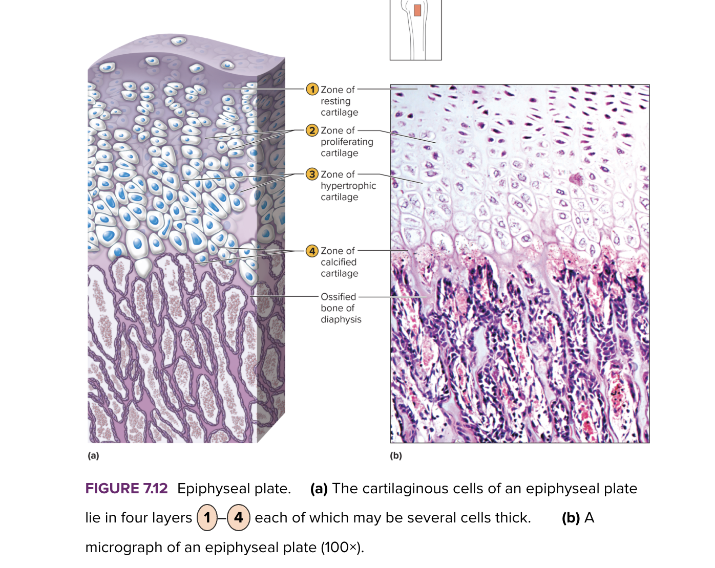
Zone of Calcified Cartilage (growth at epiphyseal plate)
4th layer of skin
thin, dead cells, calcified extracellular matrix

Homeostasis of Bone Tissue
bone remodeling occurs throughout life
bone resorption - action of osteoclasts
bone deposition - action of osteoblasts
10-20% of skeleton is replaced each year
Factors Affecting Bone Development & Repair
Deficiency of Vitamin A - disrupts normal growth & remodeling of bone
Deficiency of Vitamin C - results in fragile bones
Deficiency of Vitamin - rickets, osteomalacia = weak
Insufficient Growth Hormone = dwarfism
Excessive Growth Hormone = gigantism, acromegaly
Insufficiency Thyroid Hormone = delays bone growth
Physical Stress - stimulates bone growth
Sex Hormones
promote bone formation: stimulate ossification of epiphyseal plates
Fractures
break in the bone
simple/compound (open) - infection
single: horizontal, oblique, spiral
comminuted → 2 bone fragments
Healing in Bone
1 Day = hematoma formation (fibrin mesh)
3 Days = inflammation, macrophages remove debris, periosteum & endosteum response w/ proliferation of bone stem cells
1 Week = soft callus → granulation, matrix
3-6 Weeks = callus → ossification, woven bone
8+ Weeks = remodeling → absorb/deposit, strength, lamellate
Osteoporosis
imbalance: bone resorption + bone formation
no symptoms, but increase in fractures
bone mineral density is reduced & microarchitectures deteriorates
spongy bone = more active (more surface area) - ends of long bones + vertebrae problems
30-35 loss begins
age = decrease in androgens, especially estrogen, smoking, malnutrition, genetics
exercise, nutrition, & hormones help
Bone Functions: Support, Movement, & Protection
gives shape to head, etc.
support body weight
protect vital organs (ex. lungs)
bones & muscles interact when move
Bone Functions: Blood Cell Formation
hematopoiesis
red bone marrow
Bone Functions: Inorganic Salt Storage
most abundant salt is calcium phosphate
others = magnesium, sodium, potassium, carbonate ions
Skeleton Organization
206 bones
actual number of bones varies person to person
Axial Skeleton
skull, hyoid bone, thoracic cage, vertebrae
Appendicular skeleton
shoulder girdle, upper limbs, pelvic girdle, lower limbs