8. Nervous System: Neuron Physiology
 Call Kai
 Learn
 Practice Test
 Spaced Repetition
 Match
 Flashcards
 Knowt Play
1/27

There's no tags or description
Looks like no tags are added yet.
28 Terms
neuron plasma membrane
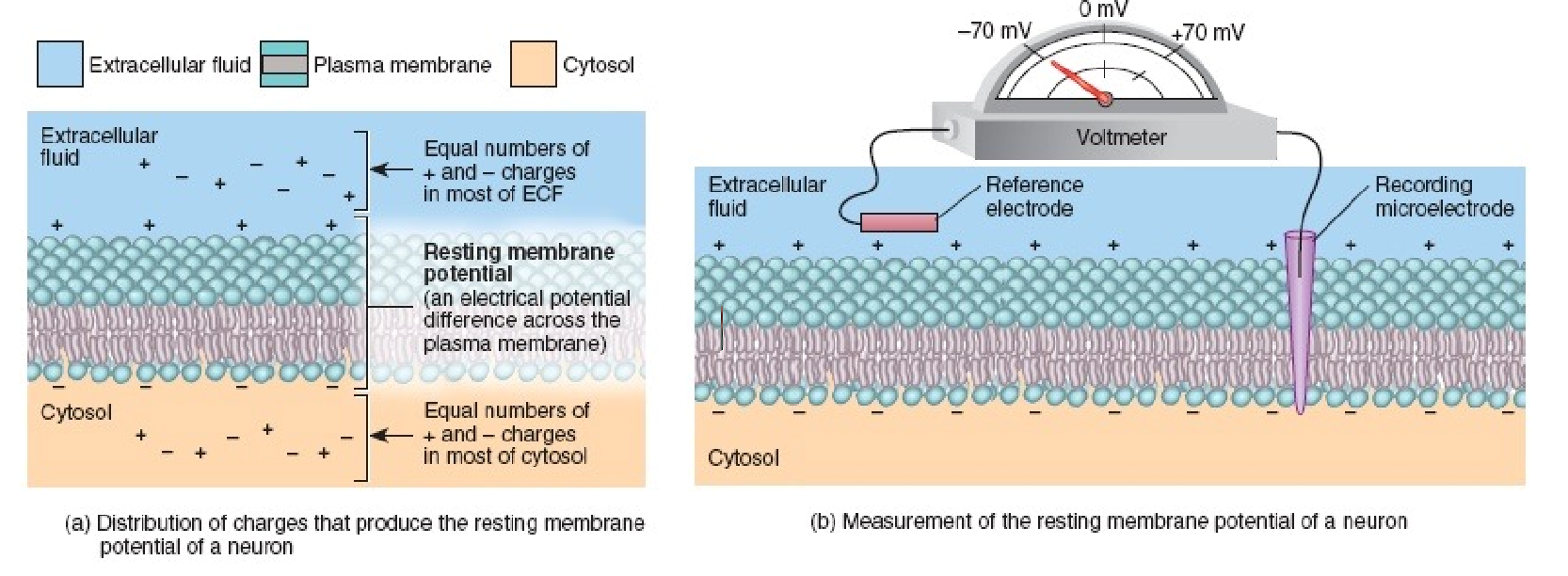
creates barrier separating ICF from ECF
ICF + ECF contain (+) and (-) ions/proteins
both overall electrically neutral
unequal distribution of specific ions build up close to PM due to ion channels + ion pumps
establishes resting membrane potential
establishes electrochemical gradients
polarized → RMP = -70mV
neuron integral proteins
ion channels
leak channels
voltage-gated channels (VGCs)
ligand-gated channels (LGCs)
membrane pumps
Na+/K+ pump

resting membrane potential (RMP)
measure of electrical charge difference across PM (in volts)
neurons → -70mV
inside of cell negative compared to outside
established by unequal distribution of specific ions that build close to PM due to ion channels + ion pumps
K+ leak channels → numerous + always open
K+ exit cell down electrochemical gradient
contributes greatly
(-) proteins → cannot leave
Na+ leak channels → few
Na+ ions enter cell
balanced by redistribution via Na+/K+ pump
resting membrane potential (RMP)

graded potential
temporary, local change in RMP
caused by LGCs opening on PM of dendrite
select ions move in/out of cell down electrochemical gradient
can be
depolarizing = more +
hyperpolarizing = more -
vary in amplitude → depend on stimulus strength
strength influenced by
# of open ion channels
duration that channel is open

depolarizing graded potential
occurs when membrane potential becomes more positive = less polarized
influx of (+) charged ions
inhibition of efflux of (+) charged ions
increase likelihood threshold value will be reached = -55mV
AP can occur

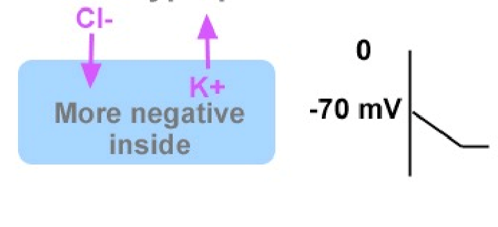
hyperpolarizing graded potentials
occurs when membrane potential becomes more negative = more polarized
influx of (-) charged ions
efflux of (+) charged ions
summation
multiple graded potentials adding together
can have depolarizing OR hyperpolarizing effect

threshold
-55mV in neurons
summated graded potentials depolarize enough to reach this level → VGNCs open
Na+ influx = AP initiated → propagated along axon
subthreshold stimulus
stimulus too weak to bring membrane potential to required level
action potential (AP)
sequence of rapidly occurring events in which membrane potential reverses, then returns to resting state
electrical signal propagated along axon length
shape is always same
amplitude of graph determined by VG ion channels
frequency can altered

resting state
MP = -70mV
all VGNCs + VGKCs closed

depolarization
MP = -55mV
(+) from graded potential depolarized MP
VGNCs open in response to depolarization @ -55mV = Na+ influx

repolarization
MP = +30mV
VGNC close
VGKC open = K+ efflux
hyperpolarization
MP = -80 to -90mV
VGKCs still open
take longer to close → responsible for prolonged hyperpolarization

return to resting state
MP = -70mV
VGKCs close
K+ leak channels + Na+/K+ pump working in background constantly → redistributes ions = restores RMP
VGNCs need time to rest to original configuration

refractory period
period of time after AP is generated when threshold stimulus CANNOT generate second AP
divided into:
absolute
relative

absolute refractory period
period of time where second AP cannot be fired no matter strength of duration of stimulus
during depolarization + early repolarization
VGNCs need time to return to original configuration
no subsequent APs can fire

relative refractory period
period of time where second AP can only be fired w/ specific stimulus
during late repolarization
VGNCs have returned to original configuration
some VGKCs still open
requires SUPRATHRESHOLD stimulus to fire subsequent AP

voltage-gated Na+ channels (VGNCs)
-70mV (RMP) = closed + can open
-50mV to +30mV = open (activated)
+30mV to -70mV = closed + cannot open (inactivated)

all-or-nothing principle
initiation of AP only occurs if threshold stimulus is reached
increasing stimulus past amount required to attain threshold increases AP frequency
BUT frequency limited by absolute refractory principle

continuous conduction
occurs in unmyelinated axons
AP initiation → VGNCs open = local current w/ intracellular ion flow
local current depolarizes adjacent PM region to threshold → more VGNCs in new region open
refractory period prevents backward conduction
K+ efflux repolarizes membrane

saltatory conduction
occurs in myelinated axons
VG ion channels only @ nodes of Ranvier
intracellular local current swiftly moves through insulated areas of axon
electrical activity “leaps” from node to node
greatly increases speed of AP propagation

neural synapse action
AP depolarizes axon terminal
voltage-gated Ca2+ channels = Ca2+ enters cell
calcium entry = exocytosis of synaptic vesicle contents
NTs diffuse across synaptic cleft → bind w/ receptors on postsynaptic cell
NT binding = response in postsynaptic cell
opening of ligand-gated channels = influx of ions → intracellular electrical environment changed
can be excitatory or inhibitory

excitatory post-synaptic potential (EPSP)
ligand binding to post-synaptic receptor causes depolarization
eg. acetylcholine (ACh)
released by pre-synaptic neuron
binds to post-synaptic ligand-gated Na+ channels
Na+ influx
depolarizing graded potential → closer to threshold

inhibitory post-synaptic potential (IPSP)
ligand binding to post-synaptic receptor causes hyperpolarization
eg. gamma-aminobutyric acid (GABA)
released by pre-synaptic neuron
binds to post-synaptic ligand-gated Cl- channels
Cl- influx
hyperpolarizing graded potential → further from threshold

neurotransmitter regulation
NTs removed from synaptic cleft to regulate its action (effect)
reuptake by pre-synaptic neuron
NT components actively transported back into pre-synaptic neuron
enzymatic degradation
enzymes in synaptic cleft break down NTs
diffusion
NTs diffuse away from synaptic cleft

depression treatment
depression = serotonin deficiency
drugs prevent serotonin reuptake = prolong serotonin action

botox (botulinum toxin)
inhibits release of NTs from somatic motor neurons, ex. ACh
no NTs to bind onto myocyte = no post-synaptic AP initiated in myocyte = temporary muscle paralysis
neuromuscular junction → where motor neurons + myocytes contact
myocytes electrically excitable
APs in myocytes = contraction