Patient Year 2

0.0(0)
Studied by 1 person
call kaiCall Kai
learnLearn
examPractice Test
spaced repetitionSpaced Repetition
heart puzzleMatch
flashcardsFlashcards
GameKnowt Play
Card Sorting

1/78

flashcard set

Earn XP

Description and Tags

dermatology

79 Terms

1
New cards

LEARN SKIN structure

knowt flashcard image
2
New cards

epidermis structure includes

as you go up epidermis cells get squashed and nuclei get lost

stratum corneum cells lose nuclei and become dead = get bits of hair can look like dust

<p>as you go up epidermis cells get squashed and nuclei get lost</p><p>stratum corneum cells lose nuclei and become dead = get bits of hair can look like dust </p>
3
New cards
<p>learn for dermatology workshop</p>

learn for dermatology workshop


4
New cards
term image
5
New cards
term image

A = papule

B= vesicles

c= hives

d= fissure
6
New cards
term image

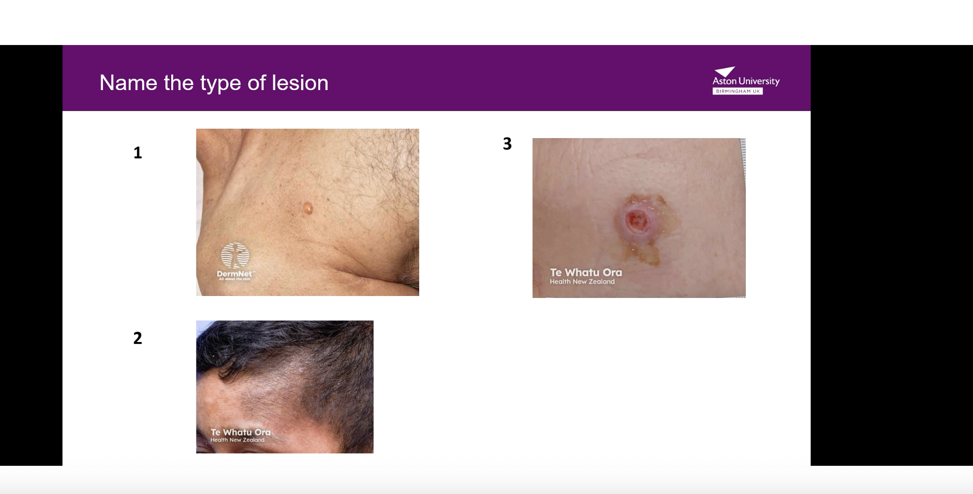
1 = bulla

B = patch

C = ulcer

7
New cards
term image

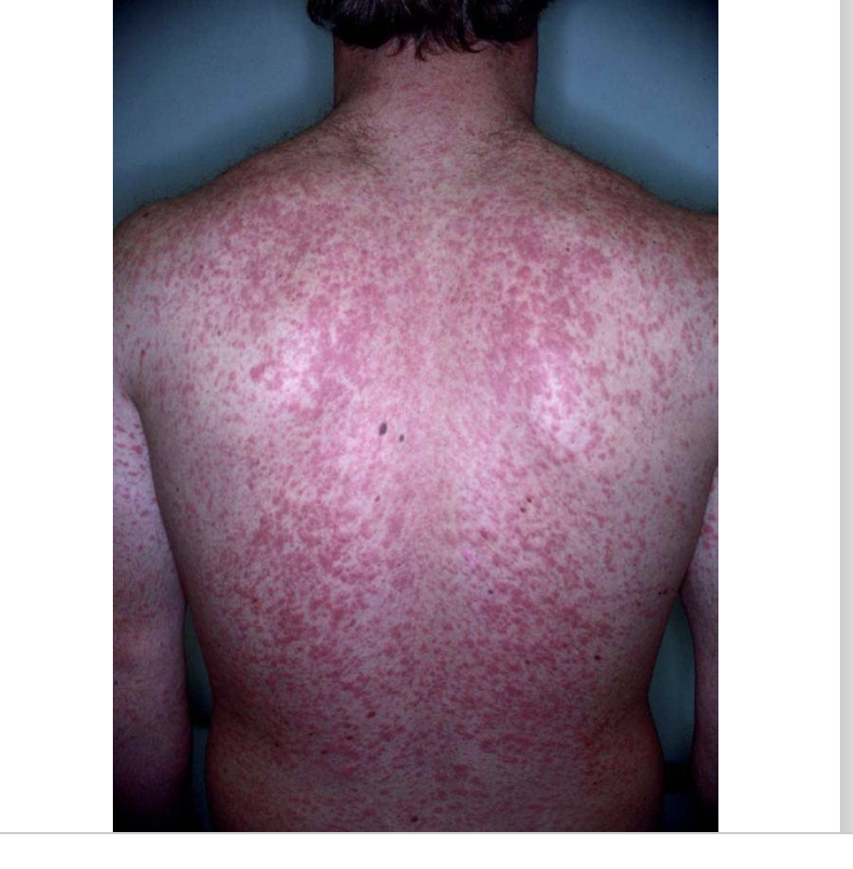
rash is Morbilliform means it looks exactly like measles—blotchy, red, and widespread.

8
New cards
term image
9
New cards
term image
10
New cards
<p>bulls blistering </p>

bulls blistering

11
New cards
<p>lichenoid plus</p>

lichenoid plus

12
New cards
<p>lupus erythematous</p>

lupus erythematous

13
New cards
<p>pustular adrs</p>

pustular adrs

14
New cards
<p><br><span>Purpura/cutaneous vasculitis</span></p>


Purpura/cutaneous vasculitis

15
New cards
<p>red man syndrome </p>

red man syndrome

16
New cards
<p>phototoxic picture </p>

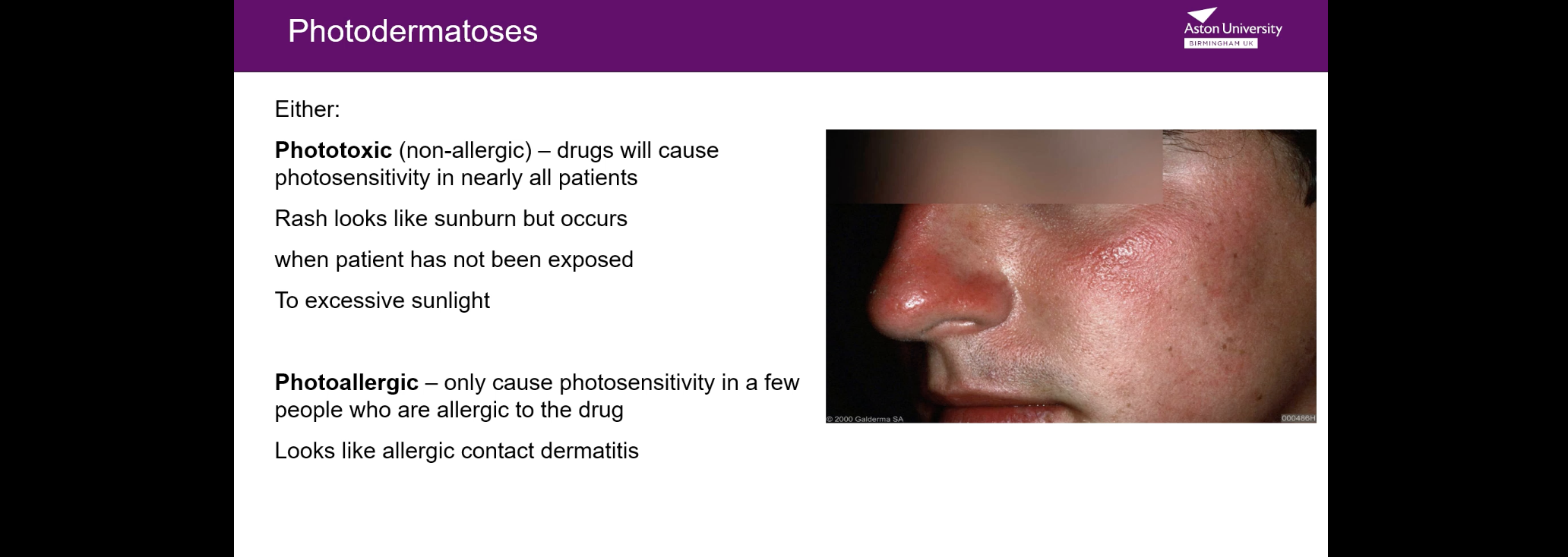
phototoxic picture

17
New cards
<p>superficial basal cell carcinoma </p>

superficial basal cell carcinoma

18
New cards
<p>nodular bcc</p>

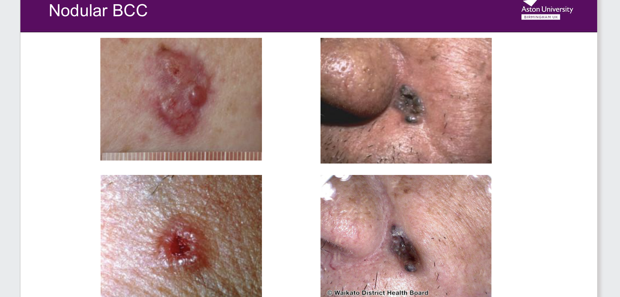
nodular bcc

19
New cards
term image
20
New cards
term image
21
New cards
term image
22
New cards
<p><span>It begins in the pigment-producing cells called melanocytes in the top layer of the skin and grows horizontally before potentially moving into deeper layers. [1, 2, 3]</span></p>

It begins in the pigment-producing cells called melanocytes in the top layer of the skin and grows horizontally before potentially moving into deeper layers. [1, 2, 3]

23
New cards
<p><mark>an aggressive type of skin cancer that grows vertically downward into the deep layers of the skin rather than spreading across the surface</mark></p>

an aggressive type of skin cancer that grows vertically downward into the deep layers of the skin rather than spreading across the surface

24
New cards
<p><mark>a rare type of skin cancer that develops on the palms of the hands, the soles of the feet, or under the fingernails and toenails</mark></p>

a rare type of skin cancer that develops on the palms of the hands, the soles of the feet, or under the fingernails and toenails

25
New cards
<p>hsdhsh</p>

hsdhsh

26
New cards
term image
27
New cards
term image
28
New cards
term image

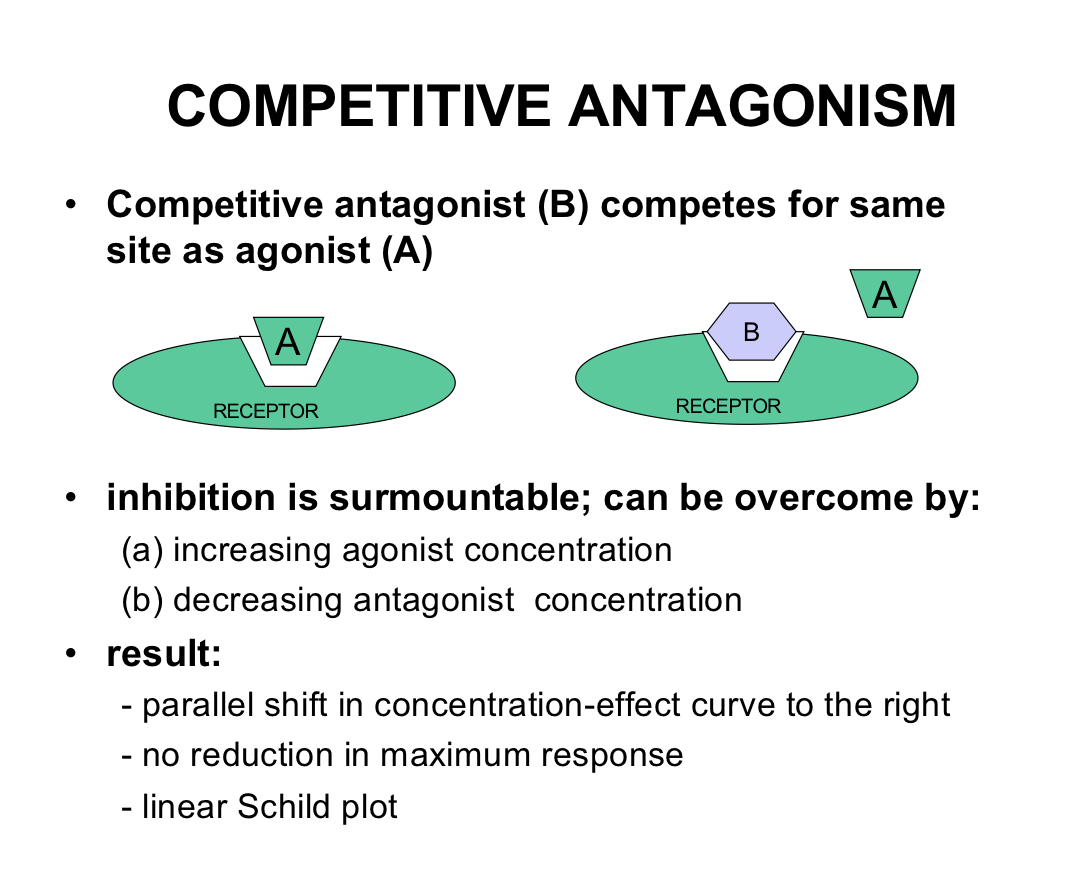
same image

what that anxiolytic effect but not sedative effect

<p>same image </p><p>what that anxiolytic effect but not sedative effect </p>
29
New cards
term image
30
New cards
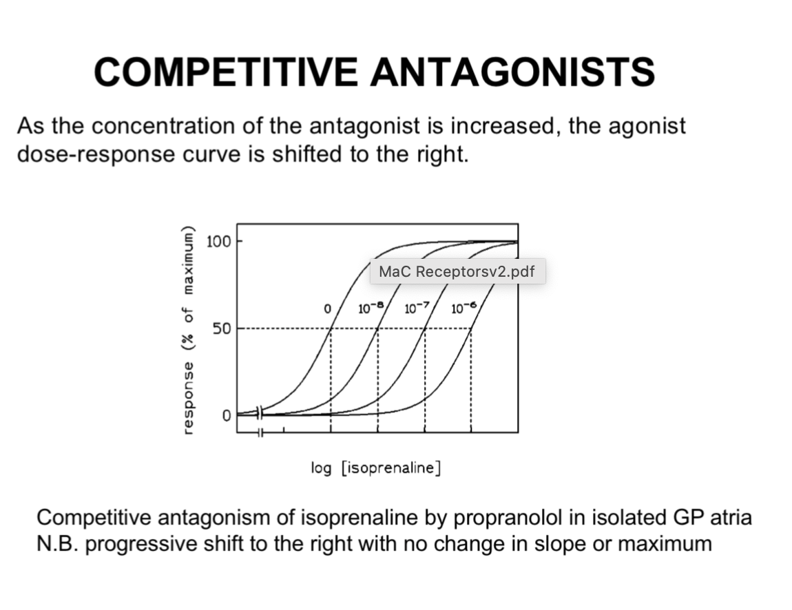
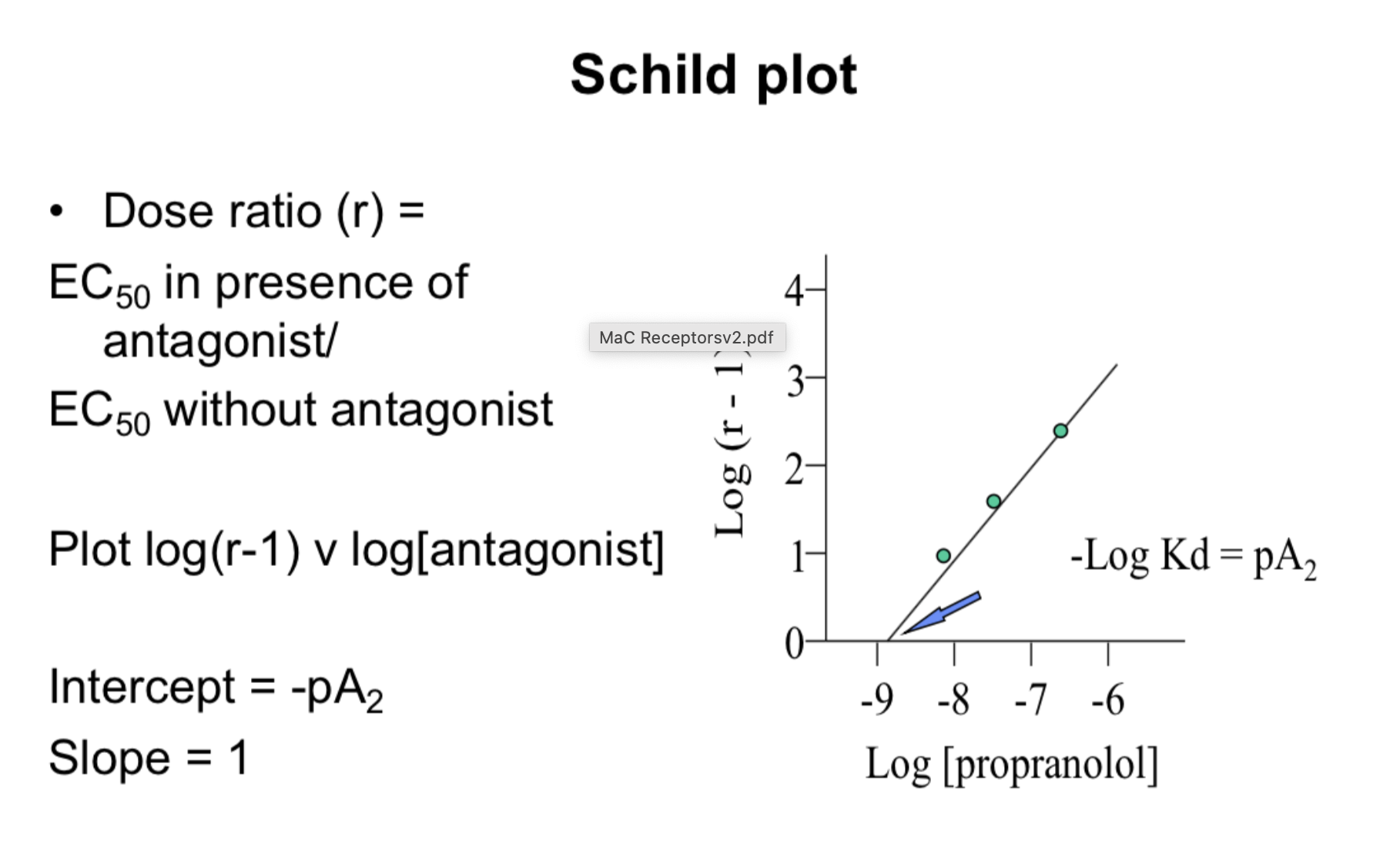
<p>look at ec50 extrapolate to left hand side you cna see down there</p><p>the concentration goes to the right</p>

look at ec50 extrapolate to left hand side you cna see down there

the concentration goes to the right

she this to plot a schlid plot to work out affinity of atagonist to the recepto
31
New cards
term image
32
New cards
term image
33
New cards
<p>lifting of bottom of curve telling us theres a partial agonist </p>
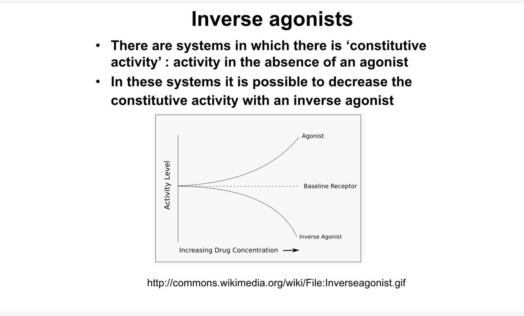
lifting of bottom of curve telling us theres a partial agonist

34
New cards
term image
35
New cards
term image
36
New cards
<p>measuring ligand activity </p>

measuring ligand activity

37
New cards
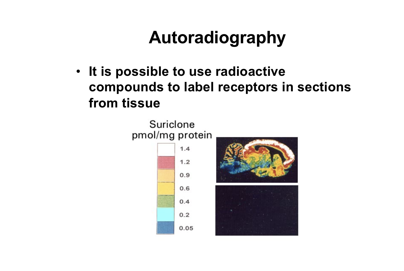
<ul><li><p><span><strong>Slicing the Tissue:</strong>&nbsp;Researchers take an organ (like a brain or a kidney) and cut it into incredibly thin slices.</span></p></li><li><p><span><strong>Applying the "Hot" Ligand:</strong>&nbsp;They bathe these slices in a solution containing a radiolabeled (radioactive) ligand. This ligand binds specifically to its target receptors on the tissue slice.</span></p></li><li><p><span><strong>Washing Away Excess:</strong>&nbsp;The slice is washed so that only the ligand tightly bound to a receptor remains.</span></p></li><li><p><span><strong>Exposing to Film or Digital Sensors:</strong>&nbsp;The tissue slice is placed directly against a piece of photographic film or a high-sensitivity digital radiation detector.</span></p></li><li><p><span><strong>Creating the Map:</strong>&nbsp;Over time, the radioactivity emitted by the bound ligand exposes the film (much like light exposes traditional camera film). When developed, it produces a clear, high-resolution image where dark spots or hot spots correspond exactly to areas with a high density of receptors.</span></p></li></ul><p></p>
  • Slicing the Tissue: Researchers take an organ (like a brain or a kidney) and cut it into incredibly thin slices.

  • Applying the "Hot" Ligand: They bathe these slices in a solution containing a radiolabeled (radioactive) ligand. This ligand binds specifically to its target receptors on the tissue slice.

  • Washing Away Excess: The slice is washed so that only the ligand tightly bound to a receptor remains.

  • Exposing to Film or Digital Sensors: The tissue slice is placed directly against a piece of photographic film or a high-sensitivity digital radiation detector.

  • Creating the Map: Over time, the radioactivity emitted by the bound ligand exposes the film (much like light exposes traditional camera film). When developed, it produces a clear, high-resolution image where dark spots or hot spots correspond exactly to areas with a high density of receptors.


38
New cards
<p><span>Ligand binds → GPCR activates G protein → receptor phosphorylation → reduced G-protein coupling → desensitisation.</span></p>

Ligand binds → GPCR activates G protein → receptor phosphorylation → reduced G-protein coupling → desensitisation.

39
New cards
<p>check Google; docs </p>

check Google; docs

40
New cards
<p> thyroid blood test</p>

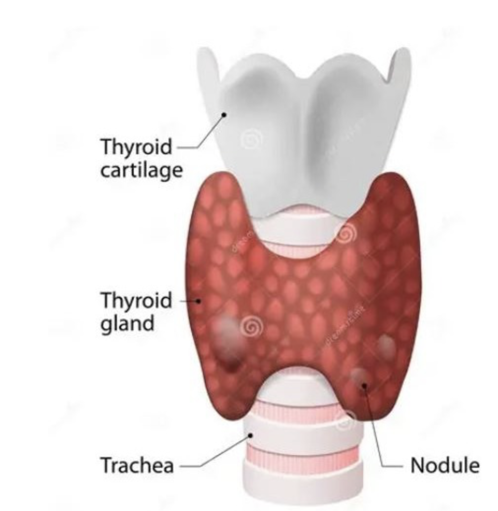
thyroid blood test

41
New cards
term image
42
New cards
term image
43
New cards
term image
44
New cards
<p><span style="font-size: calc(var(--scale-factor)*12.03px);"> distributed portal triads (seen above).</span><br><span style="font-size: calc(var(--scale-factor)*12.00px);">Examination of a tract in cross section should reveal a bile duct and branches</span><br><span style="font-size: calc(var(--scale-factor)*12.03px);">of the hepatic artery and hepatic portal vein. Lymphatic vessels are also</span><br><span style="font-size: calc(var(--scale-factor)*12.00px);">present, but are difficult to see in standard paraffin sections</span></p>

distributed portal triads (seen above).
Examination of a tract in cross section should reveal a bile duct and branches
of the hepatic artery and hepatic portal vein. Lymphatic vessels are also
present, but are difficult to see in standard paraffin sections

45
New cards
term image
46
New cards
term image
47
New cards
term image
48
New cards
term image
49
New cards
<p>cardiac = minimum of 4</p><p>use pre prepared intravenous fluids that has k+ already not get a bag and adding k+</p><p>as not mixing properly the infusing its can get concentrated bit of KCL</p><p></p>

cardiac = minimum of 4

use pre prepared intravenous fluids that has k+ already not get a bag and adding k+

as not mixing properly the infusing its can get concentrated bit of KCL


effervescent stand K better modified release of k if not washed down with water

50
New cards
term image
51
New cards
<p>SEVERE LIVER DISEASE cant transfer the calcidofl into </p>
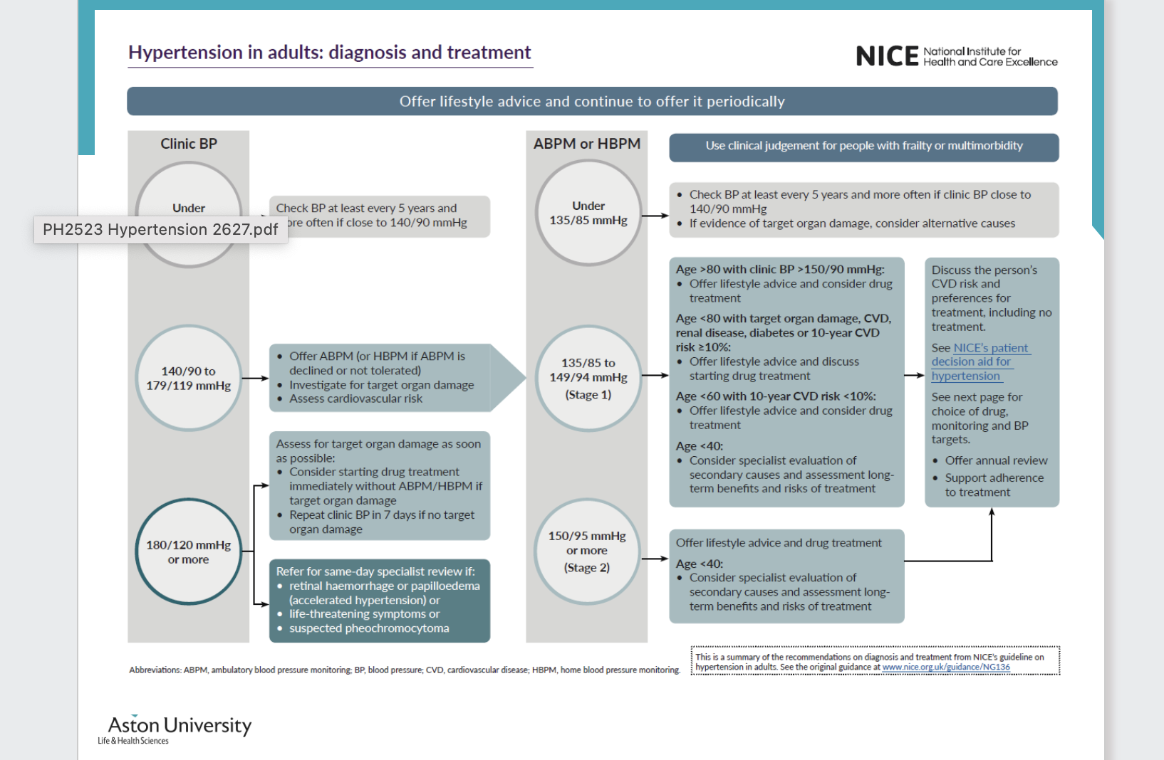
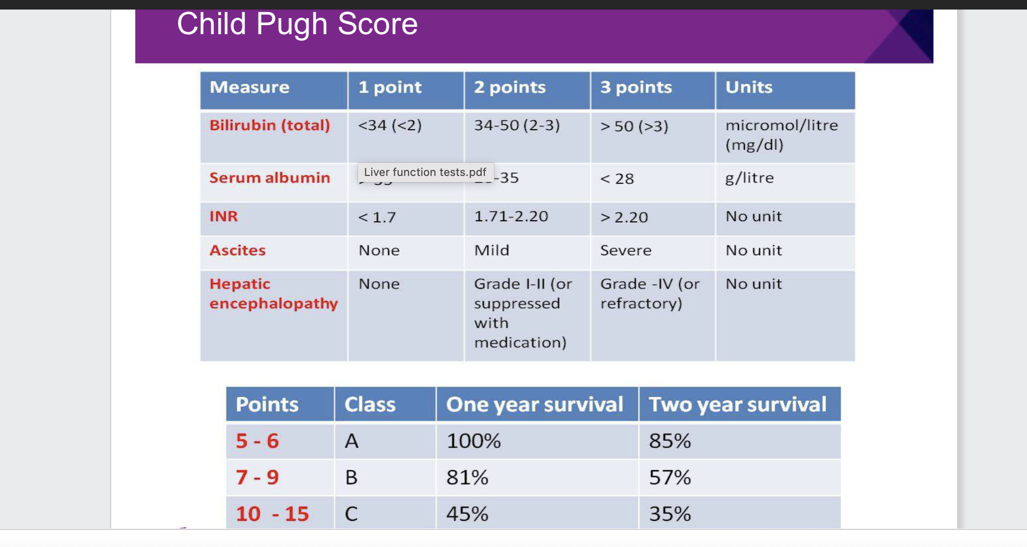
SEVERE LIVER DISEASE cant transfer the calcidofl into

52
New cards
term image
53
New cards
term image
54
New cards
term image
55
New cards
term image
56
New cards
term image
57
New cards
term image
58
New cards
term image
59
New cards
term image
60
New cards
term image
61
New cards
term image
62
New cards
term image
63
New cards
term image
64
New cards
term image
65
New cards
term image
66
New cards
term image
67
New cards
term image
68
New cards
<ul><li><p>Erythroblast = the whole immature red blood cell.</p></li><li><p>Iron granules = the little dark blue deposits surrounding its nucleus.</p></li><li><p>Ring sideroblast = the entire erythroblast when it contains iron granules arranged in a ring around its nucleus.</p></li></ul><p></p>
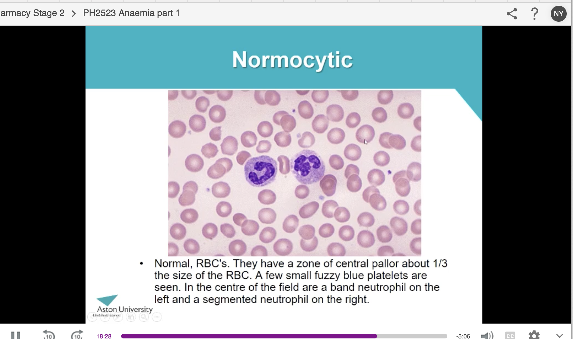
  • Erythroblast = the whole immature red blood cell.

  • Iron granules = the little dark blue deposits surrounding its nucleus.

  • Ring sideroblast = the entire erythroblast when it contains iron granules arranged in a ring around its nucleus.


69
New cards
term image
70
New cards
term image
71
New cards
term image
72
New cards
term image
73
New cards
term image
74
New cards
term image
75
New cards
term image
76
New cards
term image
77
New cards
term image
78
New cards
79
New cards
term image