AHS3263: Fluids Support & Supportive Devices
 Call Kai
 Learn
 Practice Test
 Spaced Repetition
 Match
 Flashcards
 Knowt Play
1/42

There's no tags or description
Looks like no tags are added yet.
43 Terms
PCV:
Hemoconcentration (INCREASED concentration of RBCs)
Hemodilution (DECREASED concentration of RBCs)


The SAME amount of RBCS & LOW plasma is due to DEHYDRATION & HEMOCONCENTRATION = LOW plasma & NORMAL RBCs
What you lose & gain is PLASMA
Anemia = LOW RBCs
Polycythemia = HIGH RBCs
Hemoconcentration/Hemodilution:
INCREASE in CONCENTRATION of BLOOD CELLS resulting from the LOSS of PLASMA or water
Example:
Dehydration
Fluid overload
Dehydration vs. Renal Disease:
USG
Urine specific gravity
Dog = 1.015-1.045 (adequate 1.030)
Cat = 1.035-1.060 (adequate 1.040)
USG can INCREASE when DEHYDRATED
Look at the blood chemistry panels of BUN & Creatinine to determine RENAL DISEASE
Clinical Example:
7-year-old dog
PCV = 58 (36-60)
TP = 8.0 (7-7.4)
BUN – 55 (6-25) (HIGH)
Creatinine 3.0 (0.5-1.6) (HIGH)
USG = 1.058 (1.015-1.045) (HIGH)
Dog is DEHYDRATED b/c USG is HIGH - dehydration can cause BUN & CREATININE to INCREASE
Clinical Example:
13 year old cat
PCV – 28 (29-48)
TP = 6.2 (3.4-5.6)
BUN – 69 (6-33) (HIGH)
eCreatinine – 3.5 (0.6-2.4) (HIGH)
USG = 1.008 (1.035-1.060) (LOW)
Cat has RENAL DISEASE b/c the BUN & CREATININE are HIGH & USG is LOW
Fluid Therapy:
Indications:
Replacement of lost fluids
Maintain basic water needs
Can be used for:
Dehydration
Low blood pressure
Hypovolemia (LOW blood volume)
INCREASE circulating BLOOD VOLUME
INCREASE CARDIAC OUTPUT
Body Composition:
60% of body weight is WATER
Physiologic Changes Under Anesthesia:
Blood pressure
Respiration
Insensible fluid loss
Circulation
Decreased inotropy, cardiac output, vasodilation
Routes of Administration for Fluids:
Intravenous (IV)
FAST & versatile
Subcutaneous (SC)
SLOW over time
Intraosseous (IO)
FAST, alternative to IV
Enteral
LARGE animals (large colon impactions)
Intravenous (IV) Route:
Multiple sites
Consider reason for IV when choosing site
Aseptic technique
Central lines sutured in place (done sterility)
Monitor daily
If fever of unknown origin, REMOVE IV & replace elsewhere
Can keep IV catheter for 3 days
Administering IV Fluids:
Gravity-fed system
Fluids elevated
Flow by gravity
Buretrol placed between fluid bag & administration set
Helps DECREASE the risk of fluid overload
Smaller animals

Fluid pumps
Ensure accuracy

Subcutaneous Route:
USED to administer LARGE VOLUMES of fluid for SLOW ABSORPTION over time
GRADUAL fluid replacement
Subcutaneously administered fluids should be ISOTONIC (a fluid that has the same salt & particle concentration as an animal's blood & body tissues)
Owners should be instructed to use a new needle for each fluid administration
Intraosseous Route:
Offers an ALTERNATIVE for patients in which IV ACCESS is NOT possible (i.e., neonatal patients or small/young patients with cardiovascular collapse)
Provides RAPID dispersion of fluids through the BONE MARROW & MEDULLARY VENOUS CHANNELS
Various sites can be used
Insertion site should be aseptically clipped & prepared
Periosteum anesthetized
INVASIVE procedure

Enteral Route:
Often USED for LARGE animal patients
Typically USED when LARGE COLON impaction is TREATED
May be used simultaneously with IV fluids
When enteral fluids are being administered at scheduled intervals, boluses of no more than 8 L at a time should be given to average adult 500-kg horse
HOME administration to SMALL animal patients
Esophagostomy or gastrostomy tube
Types of Intravenous Fluids:
Crystalloids
Electrolytes in water
USED for shock, surgery, maintenance of fluids
Osmolality
Hypotonic
Isotonic
Hypertonic
Contain WATER & SMALL-molecular-weight solutes such as ELECTROLYTES
PASS FREELY through vascular endothelium (from the blood vessels)
Colloids
Natural
Synthetic
Contain LARGE-molecular-weight solutes
DO NOT FREELY diffuse across vascular endothelium
STAY in the INTRAVASCULAR SPACE
USED to support expansion of BLOOD VOLUME & PRESSURE
GIVEN much SLOWER than crystalloids

Phases of Fluid Therapy:
Resuscitation phase
RESTORE vascular volume
REVERSE hypovolemic shock
“Shock dose”
Mixed fluid resuscitation
Restricted approach “slowlus”
Replacement phase
CORRECT DEHYDRATION
REPLACE & MAINTAIN fluids
Isotonic crystalloids
Monitor escalation of fluid rate with losses
Replacement fluid rate = Dehydration + Ongoing losses + Maintenance
Maintenance phase
After dehydration is corrected
Hypotonic & isotonic crystalloids
IV Fluids:
Typical surgical fluid rate:
Dogs: 5mL/kg/hr
Cats: 3mL/kg/hr
Can INCREASE or DECREASE depending on status of animal – age, condition, disease, etc
Crystalloids:
ONE-THIRD of IV fluids administered will STAY in the INTRAVASCULAR SPACE
TWO-THIRDS will DIFFUSE into the INTERSTITIAL SPACE
Related to ONCOTIC PRESSURE = pulls fluid INTO blood vessels
Colloids:
Colloids DON’T PASS FREELY through the VASCULAR ENDOTHELIUM
The presence of colloids in the INTRAVASCULAR SPACE DRAWS WATER into the space creating OSMOTIC or ONCOTIC PRESSURE
Some solute concentrations (Ca2+, K+) MUST be KEPT within a NARROW RANGE to MAINTAIN NORMAL HEART & MUSCLE FUNCTION
Colloid Solutions:
Contain LARGE-molecular-weight solutes
USED to support BLOOD VOLUMES & BLOOD PRESSURE
STAYS primarily in INTRAVASCULAR SPACE
What is the main COLLOID in the body?
ALBUMIN
Colloids - Two Basic Types:
Synthetic colloids
Dextran
Hetastarch
SHOULD NOT be BOLUSED too QUICKLY or can result in COAGULOPATHY
Blood products
Plasma
Whole blood
Hemoglobin based oxygen carriers – no longer used
Monitoring Fluid Therapy:
Resuscitation phase
Assess cardiovascular stability, blood volume, perfusion parameters in conjunction with BP
Replacement phase
Assess DEHYDRATION
Fluid Therapy Considerations:
Cardiac patients – FLUID OVERFLOW
Hypoproteinemic (LOW protein - albumin) patients
Young patients
Burn patients (Ooze out protein)
Hemorrhage
Volume Overload (Overhydration):
Pulmonary edema (fluid inside lungs)
Auscult lungs
Cerebral edema (fluid inside brain tissue)
Subcutaneous edema - skin feels “jelly-like”
Chemosis (swelling of the conjunctiva)

Nasal discharge
Dyspnea (difficulty breathing)
Coughing
Pitting edema (swelling under the skin - skin leaves a temporary dent when you press your finger into it)

Fluid Therapy Assessment:
WEIGH the animal DALIY
Overhydration is determined by WEIGHTING the animal as 1 L = 1 kg
Important Considerations:
Potassium (K+)
CAN’T GIVE QUICKLY, can cause HEART STOPPAGE
Careful spiking a bag, DO NOT BOLUS
Calcium (Ca2+)
Will cause transfused blood to CLOT
DO NOT use LRS or PLASMALYTE with transfusions
Hetastarch
Given QUICKLY can cause COAGULAPATHIES
SQ Administration:
DO NOT GIVE Normosol
Why?
b/c it is formulated fo formulated for IV administration NOT SQ administration
Contains MAGNESIUM
Supportive Care:
Positioning
Heat
Fluids
Pain
Patient Positioning:
DEPENDS on surgery
Supportive devices for positioning:
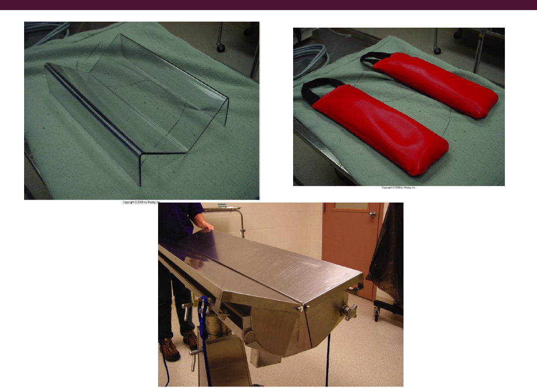
Slip “NOOSE” portion of tie OVER PAW to area PROXIMAL to CARPUS/TARSUS
Make a LOOP in the tie & place it DISTAL to CARPUS/TARSUS over paw
Attach to table
Securing Patient to Table - Abominal Surgery:
CROSS FRONT limbs to INCREASE stability when patient in dorsal recumbency
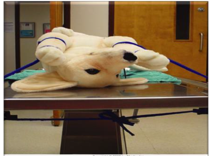
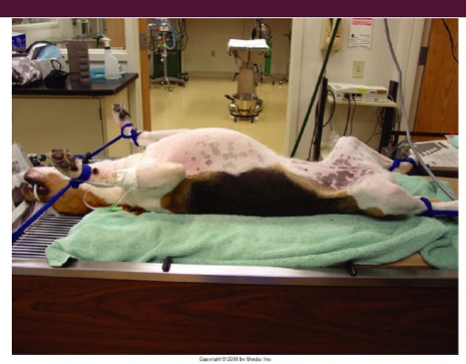
Trendelenburg Position:
Trendelenburg Position = placing the animal on its back with the HEAD LOWER than the rest of the BODY
Used for a spay
Puts PRESSURE on the DIAPHRAGM - making it harder for the animal to breath

Patient Positioning with Devices:
WARMING devices need to be ACTIVATED
Heated table, heating pads, hot water bottles, circulating air warmers, etc

Body Temperature:
Almost ALL animals will experience DECREASE in body temperature UNDER ANESTHESIA
Risk of HYPOTHERMIA especially in SMALL animals
What factors contribute to the DECREASE in body temperature?
NOT a lot of FAT
DRUGS
SURGERY
What devices & techniques should be used as available & appropriate to PREVENT hypothermia/MAINTAIN body temperature?
Heated circulating water blankets
Insulated blankets
Warm air blankets
Plastic wrap
Warmed cages & tables
What are the consequences to the hypothermic patient?
DECREASED HEART RATE
DECREASED RESPIRATORY RATE
DECREASED BLOOD PRESSURE
Temperature SHOULD BE 100 F to 102.5 F (for dog)
Catch-Up vs. Prevention:
It is much more DIFFICULT to WARM UP a COLD BODY
Post Surgery Hypothermia:
Body temperature SHOULD STEADILY RISE
100º F, DISCONTINUE heat sources
Severe hypothermia
Warm-air blanket /warm- water bath
Monitor closely
Heat Sources:
Water-circulating heating pads
Forced-air warming blankets: BAIR HUGGER
Preventing Hypothermia:
Circulating warm water pad

Warm air convection blankets

Heated surgical tables
Warmed: towels, intravenous fluids
IV fluid warmer - placed RIGHT NEXT (CLOSED) to the patient

Insulation: Plastic bubble wrap, towels, Infant socks over distal limbs
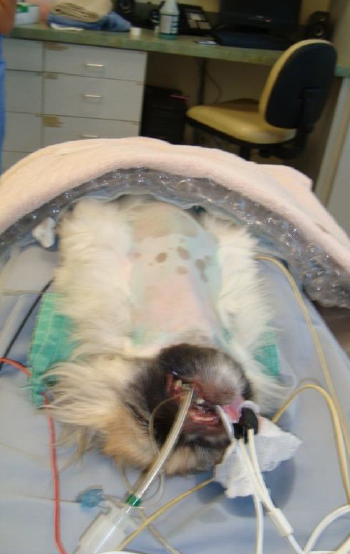
Warming continues AFTER SURGERY
Burns:
You ALWAYS NEED a BARRIER between a DIRECT HEATING ELEMENT & the PATIENT
Heated Blanket, towels, etc can cause BURNS, PRESSURE SORES
NO HOT WATER BOTTLES - if it’s going to be used, shake it to get rid of the hot spots

NO ELECTRIC BLANKETS
Additional Surgical Equipment:
Suction
Used for abdominal exploratory surgery to remove blood & other fluids from the surgical area

Surgical tips

Poole suction tip
All of the holes around the poole suction ALLOW it to suck up LARGE AMOUNTS of fluid WITHOUT getting CLOGGED
Perfect AFTER irrigating the abdominal cavity
Used during abdominal surgery

Yankauer
Used to suction fluids from larger surgical areas - can be used for joints

Frazier
Used for neurosurgery

Cautery
Used to coagulate (seal) blood vessels - uses HEAT to heat tissue & seal blood vessels

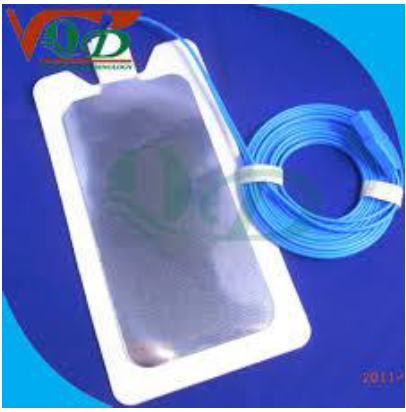
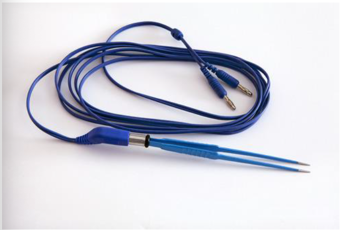
Bipolar & Monopolar:
Monopolar - MOST COMMON
Current GOES THROUGH patient
NEEDS to be GROUNDED
Ground has to be somewhere CLOSE to patient

Bipolar
BETTER CONTROL
Current DOES NOT go through patient
Current GOES BETWEEN TIPS
Used for neurosurgery
Disposable - Heat tip

Which is the true statement?
Colloids have LARGER molecules than crystalloids
Volume overload can include all of the following:
Cough
Chemosis
Pulmonary edema
EXCEPT: skin tenting
Choose the correct statement:
Hemoconcentration is normally seen with DEHYDARTION
Hot water bottles are an appropriate source of heat:
FALSE
Bipolar cautery requires a ground:
FALSE