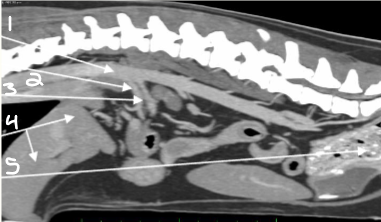
Radiology
 Call Kai
 Learn
Learn Practice Test
 Spaced Repetition
 Match
Match Flashcards
 Knowt Play
1/110

There's no tags or description
Looks like no tags are added yet.
Name | Mastery | Learn | Test | Matching | Spaced | Call with Kai | Chat |
|---|
No analytics yet
Send a link to your students to track their progress
111 Terms
density
On a radiograph, a structure’s borders are visible only when it contacts a structure of a different subject _________
falciform, retroperitoneal, subcutaneous
In what three locations do dogs typically put on fat?
falciform, retroperitoneal, inguinal
In what three locations do cats typically put on fat?
liver
What organ is highlighted yellow?

stomach
What organ is highlighted green?

spleen
What organ is highlighted pink?

small intestines
What organ is highlighted purple?

colon
What organ is highlighted blue?
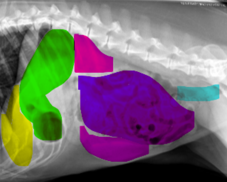
liver
What organ is highlighted yellow?

stomach
What organ is highlighted green?

kidneys
What organ is highlighted purple?

colon
What organ is highlighted blue?

superimposition
The advantage to using CTs to view abdominal organs is that you can view structures without ________________ because you can look at 3 individual planes: transverse, sagittal, dorsal
liver
What organ is highlighted?

liver
What organ is highlighted?
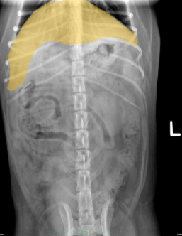
spleen
What organ is highlighted?
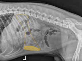
spleen
What organ is highlighted?
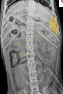
kidneys
What organ is shown?

kidneys
Mineral opacities in what organ are shown?

ureters, urinary bladder, urethra
Injected contrast is necessary to view what parts of the urinary tract on a radiograph?
right
The _______ kidney is more cranial and difficult to see (less surrounding fat)
left
The _______ kidney is more caudal and easier to see (more surrounding fat)
fundus
The ___________ of the stomach lies dorsal and to the left
pylorus
The ____________ of the stomach lies ventral and to the right
ventral
In _________ recumbency, gas rises into the dorsal fundus
dorsal
In ___________ recumbency, gas rises to the ventral stomach
left, midline
In felines, the stomach lies predominantly in the ________ cranial quadrant of the body with the pylorus on the _________
right
In canines, the stomach extends from the left body wall to the right body wall with the pylorus on the _________ side
DV (gas in dorsal fundus)
What view is shown?
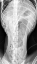
VD (gas in ventral stomach)
What view is shown?

left lateral (gas in pylorus on right)
What view is shown?

right lateral (gas in fundus on left)
What view is shown?

duodenum
What organ is highlighted?

duodenum
What organ is shown?

colon
What organ is shown?

colon
What organ is shown?

colon
What organ is highlighted?

colon
What organ is highlighted?
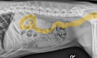
cecum
What organ is highlighted?

cecum
What organ is highlighted?

left
The colon descends along the _________ side of the body
right kidney
What does 1 label?

fundus of stomach
What does 2 label?
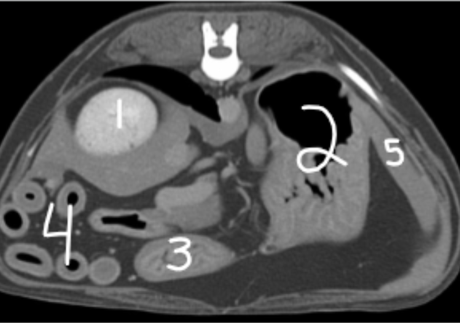
pylorus of stomach
What does 3 label?

small intestines
What does 4 label?
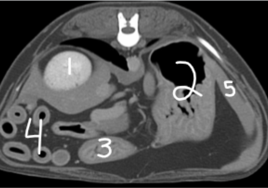
spleen
What does 5 label?

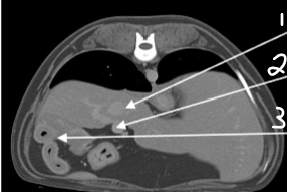
aorta
What does 1 label?

lung
What does 2 label?

liver
What does 3 label?

gall bladder
What does 4 label?

falciform fat
What does 5 label?

caudal vena cava
What does 1 label?
portal vein
What does 2 label?

duodenum
What does 3 label?

right kidney
What does 1 label?

right adrenal gland
What does 2 label?
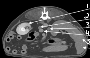
left adrenal gland
What does 3 label?

caudal vena cava
What does 4 label?

spleen
What does 5 label?
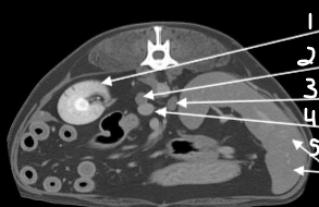
aorta
What does 1 label?

caudal vena cava
What does 2 label?

left kidney
What does 3 label?
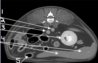
spleen
What does 4 label?

small intestine
What does 5 label?

small intestine
What does 1 label?

colon
What does 2 label?
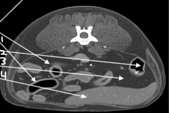
mesenteric fat
What does 3 label?

spleen
What does 4 label?

colon
What does 1 label?
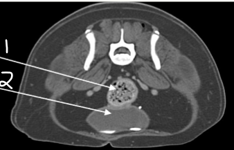
bladder
What does 2 label?

gall bladder
What does 1 label?

heart
What does 2 label?

lung
What does 3 label?

liver
What does 4 label?

small intestine
What does 5 label?
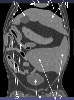
bladder
What does 6 label?
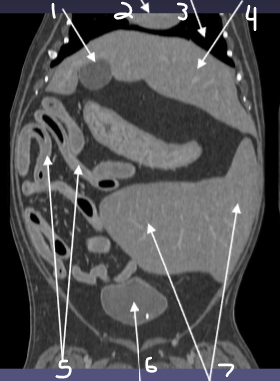
spleen
What does 7 label?

duodenum
What does 1 label?
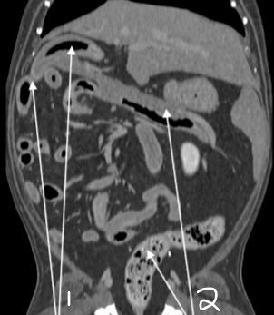
colon
What does 2 label?

fundus of stomach
What does 1 label?

left kidney
What does 2 label?

fundus of stomach
What does 1 label?

caudal vena cava
What does 2 label?

esophagus
What does 3 label?

spleen
What does 4 label?
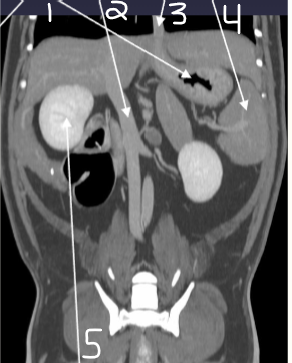
right kidney
What does 5 label?
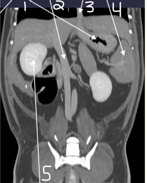
right kidney
What does 1 label?

liver
What does 2 label?

duodenum
What does 3 label?
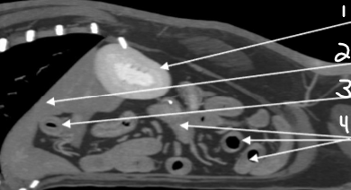
small intestine
What does 4 label?

pylorus of stomach
What does 1 label?

bladder
What does 2 label?

spleen
What does 3 label?

gall bladder
What does 4 label?

caudal vena cava
What does 1 label?

colon
What does 2 label (blue)?
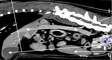
gall bladder
What does 3 label?
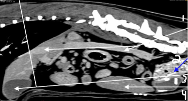
spleen
What does 4 label?

aorta
What does 1 label?

celiac artery
What does 2 label?