Oxygenation & Perfusion Flashcards
 Call Kai
 Learn
 Practice Test
 Spaced Repetition
 Match
 Flashcards
 Knowt Play
1/167

Earn XP
Description and Tags
Comprehensive vocabulary flashcards covering the anatomy, physiology, assessment, diagnostic testing, pharmacology, and clinical nursing interventions for oxygenation and perfusion.
168 Terms
Oxygenation:
The process of providing life-sustaining oxygen to the body's cells.
Factors essential to normal functioning of the respiratory system:
integrity of the airway system to transport air to and from the lungs
properly functioning alveolar system in the lungs
to oxygenate venous blood
to remove carbon dioxide from the blood
properly functioning cardiovascular system and blood supply
to carry nutrients and waste to and from body cells
Airway:
pathway for the transport and exchange of oxygen and carbon dioxide
lined with mucus, which traps cells, particles, and infectious debris
also helps to protect the underlying tissues from irritation and infection
begins at the nose and ends at the terminal bronchioles
divides into upper and lower airways
Functions of the Upper Airway:
warm, filter, and humidify inspired air
Components of the upper airways:
nose
pharynx
larynx
epiglottis
Function of the Lower Airway (Tracheobronchial Tree):
conduction of air, mucociliary clearance, and production of pulmonary surfactant
Components of the lower airway (tracheobronchial tree):
trachea
right and left main stem bronchi
segmental bronchi
terminal bronchioles
Cilia:
microscopic hair-like projections that propel trapped material and accompanying mucus toward the upper airway so they can be removed by coughing
removal is facilitated when mucus is watery in consistency
an adequate fluid intake is necessary for ciliary action and to produce watery mucus
Lungs:
main organs of respiration
located within the thoracic cavity on the right and left sides
extend from the base of the diaphragm to the apex above the first rib
How many lobes does each lung have?
the right lung has 3 lobes
the left lung has 2 lobes
How is each lobe divided?
each lobe is subdivided into segments and lobules
the main bronchus branches to each lung from the trachea
the bronchus subdivides into bronchioles
What are the lungs composed of?
the lungs are composed of elastic tissue
What is the function of surfactant?
it reduces surface tension between alveoli, preventing their collapse
when surfactant production is reduced, the lung becomes stiff and the alveoli collapse
Pleura:
serous membrane lining the lungs and thoracic cavity
visceral pleura-covers the lungs
parietal-covers the thoracic activity
Pleural Space:
lies between the two layers
pleural fluid between the membranes acts as a lubricant and as an adhesive agent to hold the lungs in an expanded position
What is intrapleural pressure, and what is its funtion?
it’s the negative (subatmospheric) pressure within the pleural space that helps keep the lungs expanded and prevents collapse
Alveoli
Small clusters of air sacs situated at the end of the terminal bronchioles where pulmonary gas exchange occurs.
Surfactant
A substance produced in the lungs that reduces the surface tension of the fluid lining the alveoli, preventing their collapse.
Intrapleural Pressure
The subatmospheric (negative) pressure present within the pleural space that acts adhesively to keep the lungs expanded.
Gas Exchange:
the intake of oxygen and the release of carbon dioxide
this is made possible by pulmonary ventilation, respiration, and perfusion
Pulmonary Ventilation
The mechanical movement of air into and out of the lungs, comprising active inspiration and passive expiration.
Inspiration:
the active phase, involves movement of muscles and the thorax to bring air into the lungs
Expiration:
the passive phase, is the movement of air out of the lungs
Respiration
The process of gas exchange between atmospheric air in the alveoli and blood in the pulmonary capillaries via diffusion.
Perfusion
The process by which oxygenated capillary blood passes through body tissues.
Two phases of pulmonary ventilation:
inspiration (inhalation) and expiration (exhalation)
During Inspiration:
the diaphragm contracts and descends (lengthening the thoracic cavity)
the external intercostal muscle contract (lifting the ribs upward and outward)
the sternum is pushed forward, enlarging the chest from front to back
the combination of an increased lung volume and decreased intrapulmonic pressure allows atmospheric air to move from an area of greater pressure (outside air) into an area of lesser pressure (within the lungs)
During Expiration:
the diaphragm relaxes and moves up
the ribs move down
the sternum drops back into position
there is a decreased volume in the lungs and an increase in intrapulmonic pressure
as a result, air in the lungs moves from an area of lesser pressure to one of lesser pressure and is expired
Factors Affecting Pulmonary Ventilation:
condition of the respiratory muscles
lung compliance
airway resistance
How the condition of the respiratory muscles affects pulmonary ventilation:
weak muscles lead to less effective inhalation and exhalation
the accessory muscles of the abdomen, neck, and back are used to maintain respiratory movements at times when breathing is difficult
these muscles are used to facilitate breathing; the movement is called retraction
Retractions:
retractions are inward pulling of the chest tissues during breathing, often indicating increased work of breathing
How lung compliance affects pulmonary ventilation:
low compliance means the lungs are stiff and require more effort to inhale
the ability of the lungs to adequately fill with air during inhalation is achieved by the normal elasticity of lung tissue, aided by the presence of surfactant
emphysema, a chronic lung condition, and the normal changes associated with aging are examples of conditions that result in decreased elasticity of lung tissue, which, in turn, decreases compliance
How does surfactant affect lung compliance?
surfactant reduces surface tension in the alveoli, helping them remain open and making lung expansion easier
What can cause airway resistance to increase?
foreign substances (food, coin, toys, or by liquids)
secretions (excessive or thickened secretions)
tissues (tumors or edema of the respiratory tract)
a decrease in the size of air passages resulting from constriction or poor neck positioning can also impede respiration
bronchial constriction in asthma is an example of airway resistance related to a decrease in the size of air passages
e.g., bronchial constriction in asthma
Lung Compliance
The ease with which the lungs can be inflated, determined by lung tissue elasticity and surfactant presence.
Airway Resistance
Any impediment or obstruction met by air as it travels through the respiratory tract, caused by foreign objects, secretions, tumors, edema, or bronchoconstriction.
Which respiratory organ is the site of gas exchange?
alveoli
Where does gas exchange occur in the respiratory system?
at the alveolar-capillary membrane, where the alveoli meet the surrounding blood capillaries
Diffusion
The passive movement of gas molecules from an area of higher concentration or pressure to an area of lower concentration or pressure.
How do oxygen and carbon dioxide move during gas exchange?
Oxygen (O2) move from the alveoli into the blood
carbon dioxide (CO2) moves from the blood into the alveoli to be exhaled
Factors Affecting Diffusion:
changes in the surface area available
thickening of the alveolar-capillary membrane
partial pressure, or pressure resulting from any gas in a mixture depending on its concentration
How do the changes in the surface area available affect diffusion?
removal of a lung or the presence of a disease that destroys lung tissue can decrease the surface area available (e.g., lung disease)
incomplete lung expansion or the collapse of alveoli (atelectasis) prevents pressure changes and the exchange of gas by diffusion in the lungs
conditions that predispose a patient to atelectasis are obstructions of the airway by foreign bodies, mucus, airway constriction, external compression by tumors or enlarged blood vessels, and immobility
Atelectasis
Incomplete expansion or collapse of lung alveoli, which hinders normal pressure changes and respiratory gas exchange.
How does the thickening of the alveolar-capillary membrane affect diffusion?
from diseases such as pneumonia or pulmonary edema, makes diffusion more difficult
How does partial pressure, or pressure resulting from any gas in a mixture depending on its concentration affect diffusion?
when oxygen is administered, an increased amount of oxygen is available, resulting in greater diffusion across capillary membranes
for example, when a person is at higher altitudes or in the presence of toxic fumes, less oxygen is available for diffusion
Perfusion:
the flow of blood through body tissues to deliver oxygen
How does blood flow affect gas exchange?
blood flow through the lungs affects how much oxygen and carbon dioxide can be exchanged
What does oxygenated blood deliver to body tissues?
oxygen for cells to use
Factors affecting perfusion:
position-sitting, standing, or lying down
perfusion is greater in dependent areas
person’s activity level
greater activity results in an increased need for cellular oxygen by the body’s tissues and a subsequent increase in cardiac output (CO) and consequently in increased blood return to the lungs
adequate blood supply and proper cardiovascular functioning-to carry oxygen and carbon dioxide to and from the lungs
Where is the respiratory center located?
in the medulla in the brainstem, immediately above the spinal cord
What stimulates the respiratory center?
an increased concentration of carbon dioxide and hydrogen ions
decreased amount of oxygen in the arterial blood
chemoreceptors in the aortic arch and carotid bodies are sensitive to the same arterial blood gas levels and blood pressure
Stimulation of the medulla does what?
increases the rate and depth of ventilation (both inspiration and expiration) to blow off carbon dioxide and hydrogen and increase oxygen levels
Hypoxia
A condition in which an inadequate amount of oxygen is available to cellular tissues, commonly caused by hypoventilation.
Hypoventilation:
decreased rate or depth of air movement into the lungs
If a problem exist in ventilation, respiration or perfusion what may occur?
hypoxia
Symptoms of hypoxia:
dyspnea
elevated blood pressure
increased respiratory and pulse rates
pallor
cyanosis
anxiety, restlessness, confusion, and drowsiness
Dyspnea:
difficultly breathing
Chronic hypoxia:
altered though processes
headaches
chest pain
enlarged heart
clubbing of the fingers and toes
anorexia, constipation, decreased urinary output, decreased libido, weakness of extremity muscles, and muscle pain
What is vital for exchanges of gases?
an adequately functioning cardiovascular system
The cardiovascular system is composed of:
the heart and the blood vessels
Three components of ciculation:
the pump (the heart)
the pipes (the blood vessels)
the fluid (the blood)
Atria:
upper chambers that receive blood returning to the heart
Ventricles:
lower chambers that pump blood out of the heart
Arteries and arterioles:
carry blood away from the heart
venules and veins:
return blood toward the heart
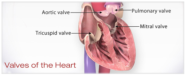
heart valves:
maintain one-way blood flow and prevent backflow
four heart valves:
tricuspid
mitral
pulmonary
aortic
capillaries:
exchange oxygen, carbon dioxide, nutrients, and wastes between blood and body tissues
blood vessel pathway:
arteries → arterioles → capillaries → venules → veins
Oxygen Transport:
blood is squeezed through the heart and out into the body by contractions starting in the atria, followed by contraction of the ventricles, with a subsequent resting of the heart
deoxygenated blood is carried from the right side of the heart to the lungs, where oxygen is picked up and carbon dioxide is released, and then returned to the left side of the heart
this oxygenated blood is pumped out to all other parts of the body and back again
oxygen is carried predominantly via red blood cells due to binding between the hemoglobin in RBCs and oxygen
once the blood cells reach the tissues, internal respiration must occur
Deoxygenated blood:
low in oxygen, high in carbon dioxide
Oxygenated blood:
high in oxygen, low in carbon dioxide
Internal Respiration
The exchange of oxygen and carbon dioxide between circulating systemic blood and metabolizing tissue cells.
Sinoatrial (SA) Node
A specialized mass of cardiac tissue in the upper right atrium acting as the heart's natural pacemaker by initiating electrical impulses at regular intervals.
the heart’s natural pacemaker: starts the electrical impulse
Atrioventricular (AV) node:
it delays the electrical impulse so the atria can finish pushing blood into te ventricles before the ventricles contract
Electrical conduction pathway:
SA node → AV node → Bundle of His → bundle branches → Purkinje fibers
Dysrhythmia (Arrhythmia)
A disturbance in the rate, rhythm, or conduction of electrical impulses through the heart.
What can cause Dysrhythmia (Arrhythmia)
caused by an abnormal rate of electrical impulse generation from the SA node, or from impulses originating from a site or sites other than the SA node
abnormal conduction of electrical impulses through the heart
Dysrhythmia (Arrhythmia) Occurs With:
heart disease
damage to the heart
certain drugs
decreased oxygenation to heart tissues
trauma
Symptoms of Dysrhythmia (Arrhythmia):
decreased BP
dizziness/fainting
palpitations (throbbing)
weakness
Myocardial Ischemia
A deficiency of blood and oxygen delivery to the heart muscle, most frequently resulting from atherosclerosis.
Atherosclerosis
The accumulation of fatty substances and fibrous plaques in arterial linings, resulting in vessel narrowing and reduced blood flow.
Myocardial Infarction (MI)
The death of myocardial tissue caused by an acute lack of oxygen supply; commonly referred to as a heart attack.
Symptoms of MI:
chest pain
anxiety
nausea/vomiting
indigestion
shortness of breath
Risk Factors of MI:
modifiable:
smoking
high blood pressure
high cholesterol
diabetes
obesity
physical inactivity
non-modifiable:
age (increases with age)
family history
male
Heart Failure:
occurs when the heart is unable to pump a sufficient blood supply, resulting in inadequate perfusion and oxygenation of tissues
Heart failure may result from:
MI
hypertension
coronary artery disease
valvular abnormalities
Symptoms of heart failure:
shortness of breath
peripheral edema
swelling of legs
fatigue
Chronic Obstructive Pulmonary Disease (COPD)
progressive lung disease that obstructs airflow and makes breathing difficult
it include two main conditions: emphysema and chronic bronchitis
most individuals with COPD have a combination of both
Primary cause of COPD:
long-term exposure to lung irritants, especially cigarette smoke
Other causes of COPD:
air pollution
occupational exposure to dust and chemicals
secondhand smoke
rare genetic condition: alpha-1 antitrypsin deficiency
Symptoms of COPD:
chronic cough
shortness of breath, especially during physical activity
wheezing
fatigue
frequent respiratory infections
frequent exacerbations
Risk factors of COPD:
smoking (main risk factors)
age (typically 40+)
environmental and occupational exposure
genetic predisposition
history of asthma
Treatment of COPD:
while COPD is not curable, it is manageable
smoking cessation
bronchodilators and inhaled corticosteroids
oxygen therapy (in advanced cases)
vaccinations (e.g., flu and pneumonia)
lifestyle changes and exercise
slide 27
Asthma
A chronic inflammatory disorder of the airways causing reversible bronchial obstruction, airway hyperresponsiveness, and mucosal edema.
Key symptoms of asthma:
reversible airway obstruction
airway hyperresponsiveness
chronic inflammation
Common symptoms of asthma:
shortness of breath
wheezing (especially when exhaling)
chest tightness or pain
persistent cough, often worse at night or early morning
difficultly breathing during exercise or exposure to triggers
Triggers of asthma:
allergens (pollen, dust mites, pet dander, mold)
respiratory infections (cold, flu)
exercise (especially in cold, dry air)
smoke, pollution, strong odors
stress and emotional upset
weather changes
Treatment of asthma:
asthma is not curable, but is manageable
quick-relief medications (e.g., short-acting bronchodilators)
long-term control medications (e.g., inhaled cortiosteriods)
avoidance of triggers
monitoring with peak flow meters
asthma action plans for managing flare-ups