Stains
 Call Kai
 Learn
 Practice Test
 Spaced Repetition
 Match
 Flashcards
 Knowt Play
1/7

There's no tags or description
Looks like no tags are added yet.
8 Terms

What stain is pictured here ?
Mayer Mucicarmine
Staining technique: Mayer Mucicarmine
Principle of the technique: Stains Carboxylated and Sulfonated mucins NOT neutral mucins, use for ID of adenocarcinomas and certain fungal structures
Purpose of stain: Staining of Epithelial mucin and demonstrates Cryptococcus neoformans
Results:
-Mucin Deep - rose to red
-Capsule of Cryptococcus spp - deep rose to red
-Nuclei- black
-other tissue elements- Blue or yellow
Sources of error: Mucicarmine will gradually deteriorate over time, keep solution fresh and use good control to monitor for decrease in staining. Carminophilic properties will be obscured if sections are overstained with either hematoxylin or metanil yellow
Preferred fixative: 10%NFB
Preferred thickness: 4-5um
Microscope used: light
Control tissue: unautolyzed colon, small intestine or appendix
Major reagents and purpose
Major reagents | Purpose |
Weigert Iron Hematoxylin | Nuclear Stain: An iron-lake progressive dye where ferric chloride acts as both the oxidizer and the mordant. This forms a robust iron-hematein complex that resists decolorization by the highly acidic mucicarmine solution applied immediately after. |
Mucicarmine Stock Solution | Aluminum form chelation complex with carmine the resulting compound has net (+) charge and attaches to the acid groups of mucins |
Metanil yellow | Counterstain anionic dye colors cytoplasm |

What stain is pictured here?
Staining technique: Periodic acid-Schiff PAS
Principle of the technique: Certain tissue components are oxidized into aldehydes by periodic acid. Schiff reagent is used to demonstrate these aldehydes.
Purpose of stain: Demonstrate polysaccharides, neutral mucosubstances and basement membranes.
Results: • PAS-Positive Entities (Magenta/Bright Pink): Glycogen, neutral mucins (goblet cells), basement membranes (renal glomeruli), fungal cell walls (chitin/glucan), and Tropheryma whipplei (the causative agent of Whipple's disease, found within macrophage lysosomes).
• Nuclei: Blue (typically counterstained with Mayer's or Harris Hematoxylin).
• Background: Light pink, green, or purple depending on whether a counterstain like hematoxylin or light green is used
Preferred fixative: 10%NBF or Bouin Solution. Blood smears should be fixed in methyl alcohol 10-15mins.
Preferred thickness: 4-5um kidney 1-2um
Control tissue: kidney most sensitive control, also good for basement membranes but to detect glycogen use liver or sections of cervix include both ecto and endo.
Major reagents | Purpose |
Periodic acid | Oxidizing agent to aldehydes by periodic acid, reactive group is 1,2 – glycol group |
Schiff Reagent | Colorless basic fuchsin |
Potassium Metabisulfite | Rinse excess schiff |

What stain is pictured here?
Staining technique: Alcian Blue 2.5
Principle of the technique: Stains sulfated and carboxylated acid mucopolysaccharides and sulfated and carboxylated sialomucins or glycoprotein. Alcain blue forms salt linkages with acid groups of acid mucopolysaccharides
Purpose of stain: Demonstration of acid mucopolysaccharides
Results: weaky acidic sulfated mucosubstances, hyaluronic acid and sialomucins - dark blue
Background - pink to red
Nuclei – Red
Preferred fixative: 10% NFB or Bouins Solution
Preferred thickness: 4-5um
Control tissue: unautolyzed small intestine appendix or colon as positive control
Major reagents | Purpose |
Acetic Acid Solution | Chemical Conditioner / Buffer: Used as a pre-treatment to lower the tissue sections down to an exact pH of 2.5 before the dye arrives. This protects the upcoming alcian blue bath from water dilution and prevents pH fluctuations that would skew dye binding properties |
Alcian Blue, pH 2.5 Dye | Primary Dye: A water-soluble, copper-phthalocyanine basic dye carrying a permanent positive charge. At pH 2.5, it reacts strictly via salt linkages (ionic bonds) with the ionized carboxylated and sulfated groups of acid mucopolysaccharides and sialomucins. |
Nuclear Fast Red | Counterstain: A basic nuclear dye that utilizes an aluminum sulfate mordant to create a pink-red dye-lake complex. This targets DNA phosphate groups to provide high visual contrast against the bright blue target mucins. |
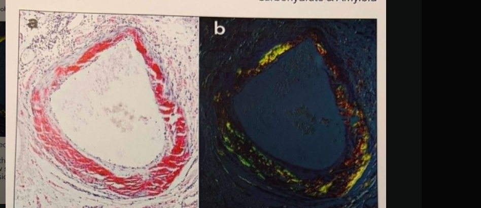
What stain is pictured here?
Staining technique: Congo red
Principle of the technique: Green bifringence following congo red staining most specific technique for demonstration of amyloid.
Purpose of stain: Demonstration of amyloid in tissue
Results:
Amyloid – Deep pink to red under light apple green birefringence under polarized light
Elastic tissue – Pale pink
Nuclei – Blue
Sources of error: Amyloid is a isomer of cellulose and resembles cellulose in chemical reactions. Prolonged storage in 10% formalin will cause gradual decrease in staining intensity
Preferred fixative: Alcohol or Carnoy solution
Preferred thickness: 8-10um
Microscope used: light, polarized light microscope
Control tissue: sections containing amyloid newly formed deposits stain better
Major reagents Purpose
Major reagents | Purpose |
Congo Red Staining Solution | Primary Dye: Green birefringence Is intrinsic property of the amyloid fibril congo red complex and is the function of the parallel alignment of dye molecules and amyloid fibrils |

Staining technique showed
Congo Red
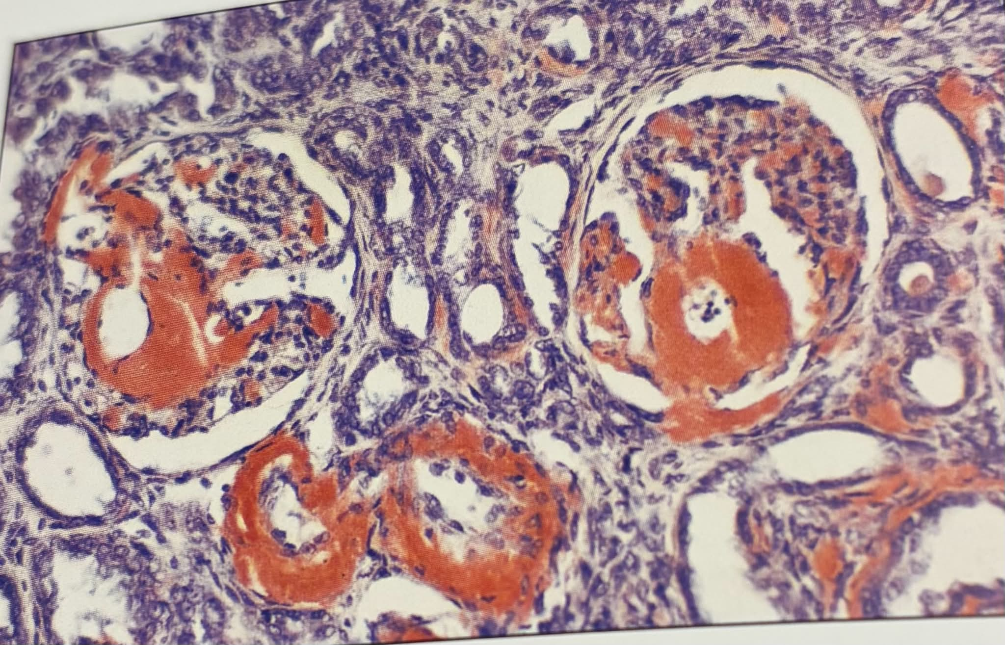
Substance stained red in this image
Amyloid
Other techniques that may demonstrate amyloid ?
Thioflavin T, crystal violet