(L21) IMED2004 - Stem Cells II
 Call Kai
 Learn
Learn Practice Test
 Spaced Repetition
 Match
Match Flashcards
 Knowt Play
1/62

There's no tags or description
Looks like no tags are added yet.
Name | Mastery | Learn | Test | Matching | Spaced | Call with Kai | Chat |
|---|
No analytics yet
Send a link to your students to track their progress
63 Terms
What are the eight learning outcomes for Stem Cells II?
1. Define the features of adult stem cells.
2. Understand "plasticity" in reference to stem cells.
3. Outline the factors governing adult stem-cell plasticity.
4. Discuss the role of the stem-cell niche in regulating activation and quiescence.
5. Name and list the functions of common signalling pathways in stem-cell function.
6. Describe stem-cell activation from quiescent cells → transit-amplifying cells → terminally differentiated cells.
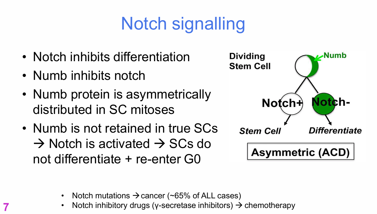
7. Understand how asymmetric cell division maintains the stem-cell pool through uneven distribution of Notch.
8. Describe tissue-specific examples of stem-cell homeostasis in skeletal muscle, gut, blood and liver.
What is the main focus of Stem Cells II?
Adult stem-cell systems and how they maintain mature tissues.
Lecturer explanation:
The lecture focuses on adult stem cells, their niches, signalling, activation and tissue-specific examples.
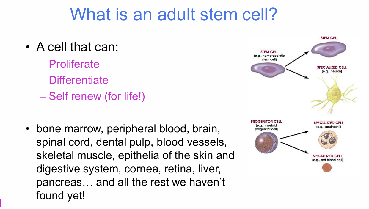
What three properties define an adult stem cell?
An adult stem cell can:
- Proliferate
- Differentiate
- Self-renew for life
Lecturer emphasis:
These same core properties apply to embryonic stem cells, induced pluripotent stem cells and naturally occurring adult stem cells.


Where can adult stem cells be found?
Examples listed:
- Bone marrow
- Peripheral blood
- Brain
- Spinal cord
- Dental pulp
- Blood vessels
- Skeletal muscle
- Epithelia of the skin
- Digestive system
- Cornea
- Retina
- Liver
- Pancreas
- Other tissue populations not yet identified
Lecturer explanation:
Adult stem cells are located in specific tissues because those tissues need mechanisms for long-term maintenance.

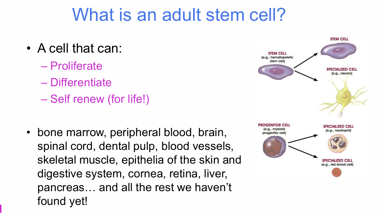
What should you identify in the adult stem-cell examples diagram?
The diagram contrasts:
- Stem cell → specialised cell
- Progenitor cell → specialised cell types
It illustrates that adult stem cells and progenitors generate tissue-specific mature cells.

What does an adult stem cell require to survive long-term?
A protected home:
- The stem-cell "niche"
- Protection from damage
.
The slide states that adult stem cells require "not much else" beyond this protected environment.

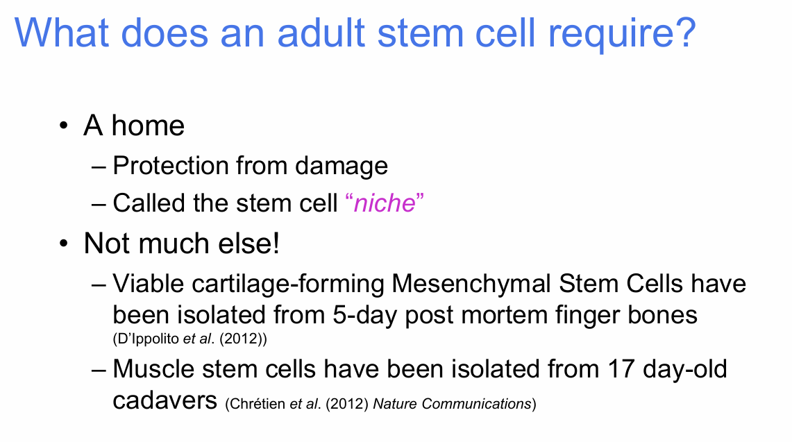
What post-mortem examples illustrate the robustness of adult stem cells?
- Viable cartilage-forming mesenchymal stem cells were isolated from 5-day post-mortem finger bones.
- Muscle stem cells were isolated from 17-day-old cadavers.
.
Lecturer explanation:
The lecturer linked this robustness to the fact that many adult stem cells spend most of their time in a quiescent, low-metabolic state.


What are examples of tissue-specific adult stem cells?
Examples shown:
- Muscle progenitor / satellite-cell lineage in skeletal muscle
- Neuroepithelial / neural stem-cell lineages in the nervous system
.
Key idea:
Adult stem cells reside in specific tissues and generate cell types relevant to those tissues.

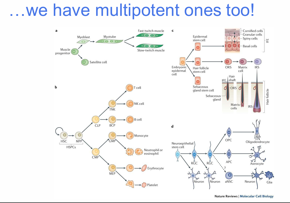
What does it mean that some adult stem cells are multipotent?
Some adult stem cells can generate more than one mature cell type.
Examples shown include:
- Haematopoietic stem cells generating many blood-cell lineages
- Embryonic epidermal-derived stem-cell lineages generating several skin-associated cell types
- Neural stem-cell lineages generating multiple neural cell types

What is stem-cell plasticity?
Plasticity refers to the ability of a stem cell to generate cell types beyond a single restricted fate.
Adult stem cells are located in specific tissues (unlike embyronic stem cells)
,
Examples:
- A muscle stem cell can obviously become muscle.
- If a stem cell can become other cell types, it is described as more plastic.
Plasticity is closely related to potency.
How do bipotential and multipotential stem cells differ in plasticity?
- Bipotential = low plasticity
- Multipotential = high plasticity
.
Lecturer explanation:
Plasticity and potency were described as closely linked and often used in a similar way to describe how many different fates a stem cell can adopt.
What three factors govern adult stem-cell plasticity?
1. Environment
- The stem-cell niche regulates stem-cell functions.
.
2. Signals
- Cells are exposed to different diffusible signals.
.
3. Transducers
- Transcription factors alter which genes are switched on or off.
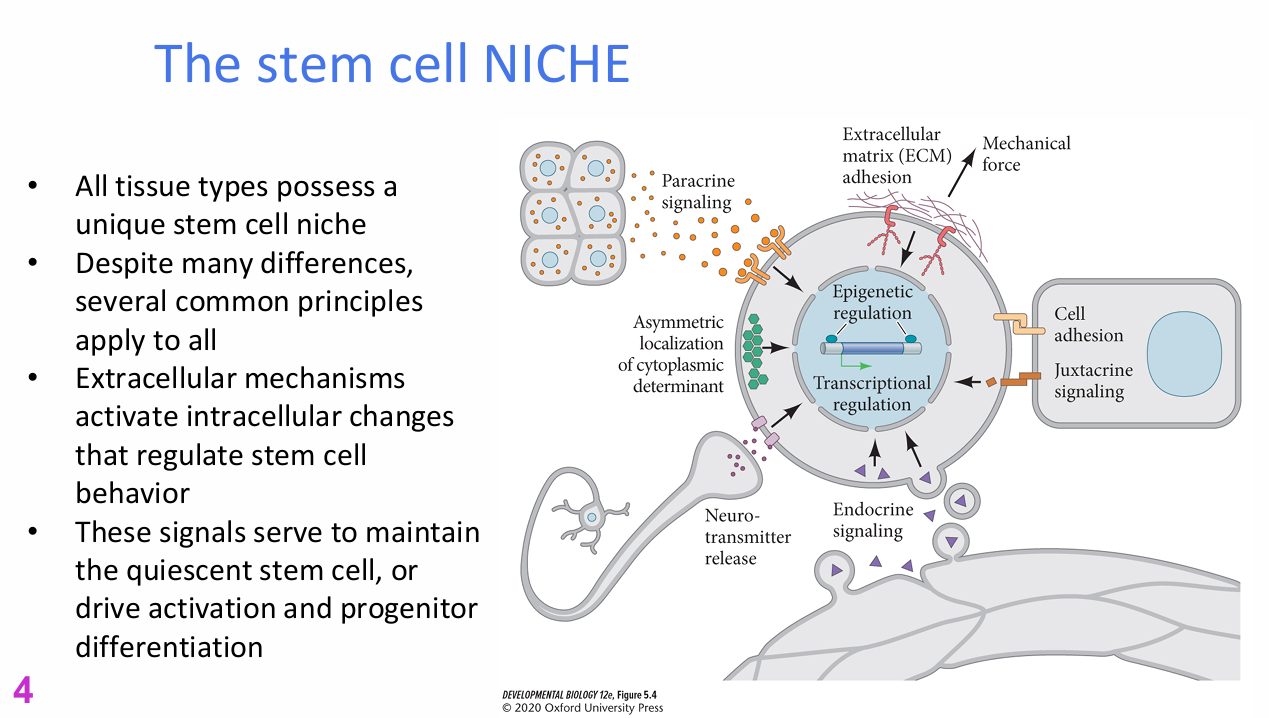
What is the stem-cell niche?
The stem-cell niche is the specialised local environment in which a tissue's stem cells reside.
.
Key principles:
- Every tissue type has a unique stem-cell niche.
- Despite differences, common regulatory principles apply across tissues.
- Extracellular mechanisms trigger intracellular changes that regulate stem-cell behaviour.
- Niche signals can maintain quiescence or drive activation and progenitor differentiation.


What types of niche inputs can regulate a stem cell?
Inputs shown include:
- Paracrine signalling
- Asymmetric localisation of cytoplasmic determinants
- Extracellular matrix (ECM) adhesion
- Mechanical force
- Cell adhesion
- Juxtacrine signalling
- Endocrine signalling
- Neurotransmitter release
.
Intracellular responses shown:
- Epigenetic regulation
- Transcriptional regulation

How can niche signals either maintain quiescence or activate a stem cell?
The niche provides a balance of local and systemic signals.
.
Signals may:
- Tell a stem cell that tissue homeostasis is stable and quiescence should be maintained
OR
- Indicate damage or altered tissue conditions, triggering activation, proliferation and differentiation
.
Lecturer explanation:
A stem cell does not need every possible input at once; different tissues rely on different combinations of niche signals.


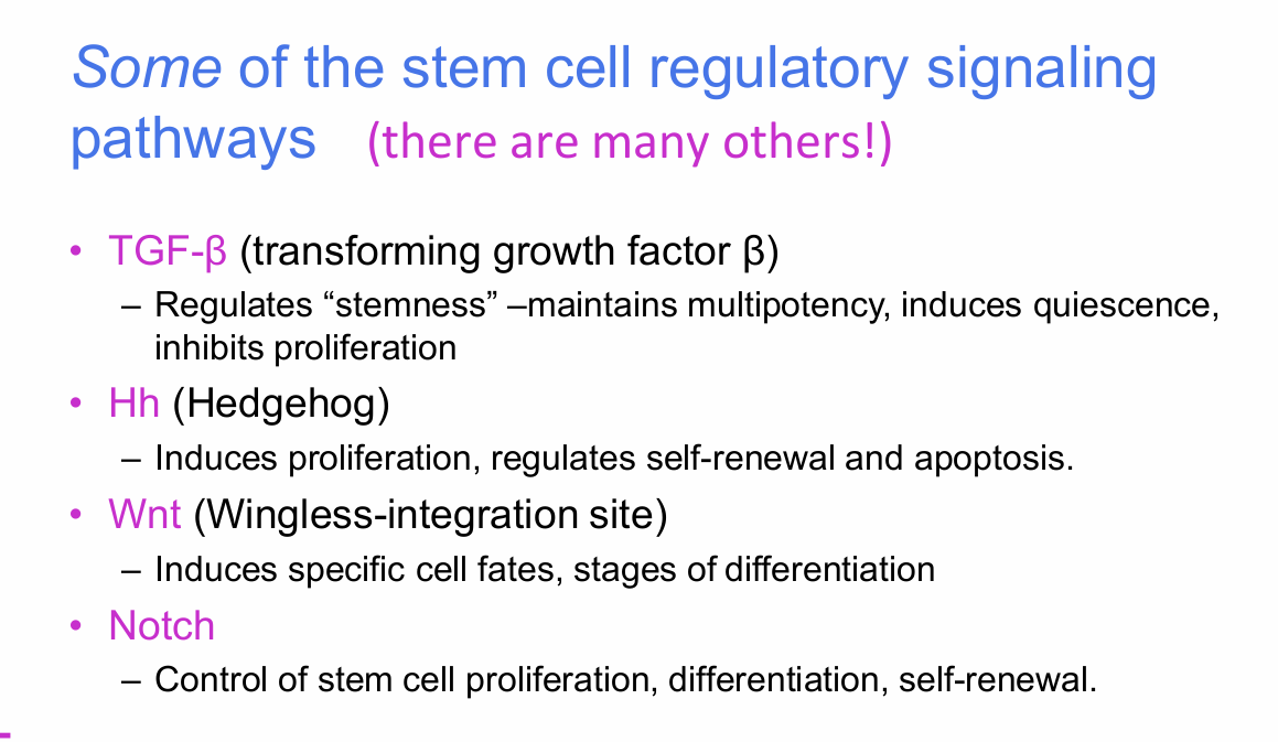
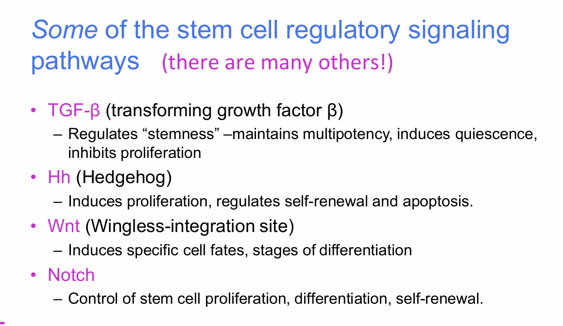
What four signalling pathways were highlighted in adult stem-cell regulation?
1. TGF-β (transforming growth factor β)
2. Hh (Hedgehog)
3. Wnt (Wingless-integration site)
4. Notch

What is the role of TGF-β in stem-cell regulation?
TGF-β:
- Regulates "stemness"
- Maintains multipotency
- Induces quiescence
- Inhibits proliferation
.
keeps them quiet

What is the role of Hedgehog (Hh) in stem-cell regulation?
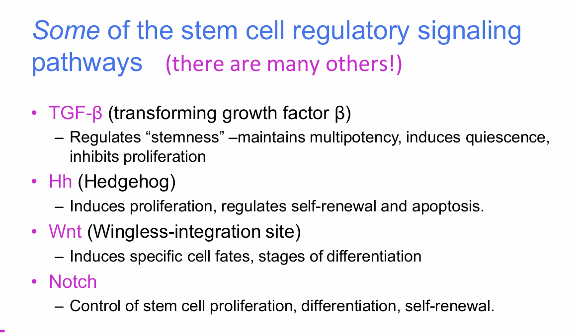
Hedgehog:
- Induces proliferation
- Regulates self-renewal
- Regulates apoptosis
What is the role of Wnt in stem-cell regulation?
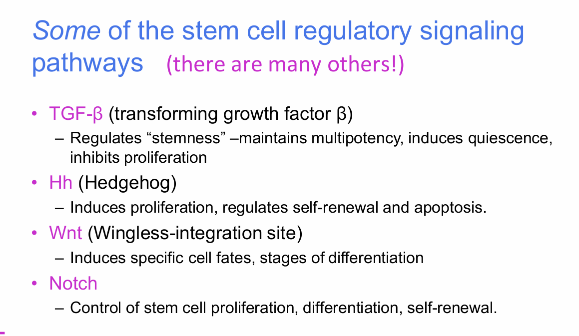
Wnt:
- Induces specific cell fates
- Regulates stages of differentiation


What is the role of Notch in stem-cell regulation?
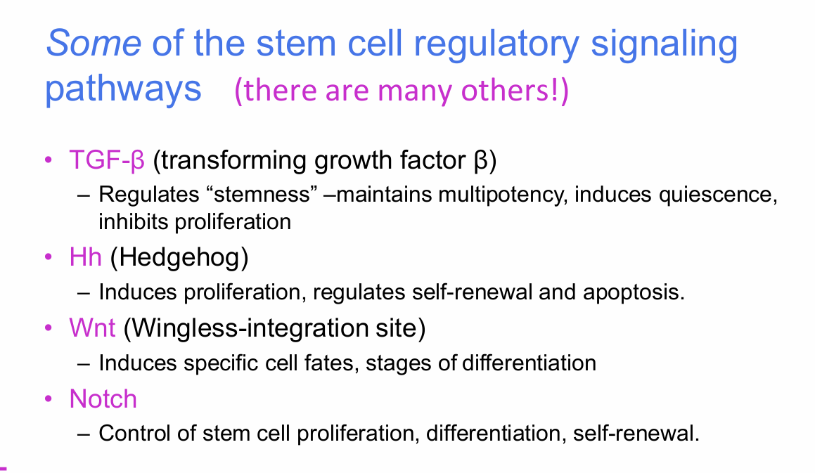
Notch controls:
- Stem-cell proliferation
- Differentiation
- Self-renewal
Lecturer explanation:
Notch was described as particularly important for maintaining a stem-like state and preventing inappropriate differentiation.


What is the key concept shown by the TGF-β, Hedgehog, Wnt and Notch signalling diagram?
Different extracellular signals act through different receptors/pathways and induce different intracellular transducers, including transcription factors.
These different signals regulate transitions from:
- Stem cells
- Progenitors / transit-amplifying cells
- Mature cells
Each arrow in the lineage diagram represents different signalling inputs.

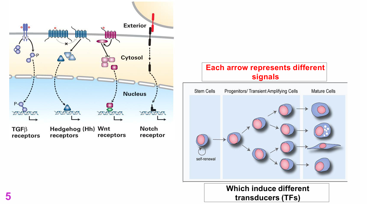
What detailed receptor mechanisms were illustrated for TGF-β, Hedgehog, Wnt and Notch? (NOT ASSESSABLE)
The slide illustrates distinct signalling mechanisms for:
- TGFβ receptors
- Hedgehog (Hh) receptors
- Wnt receptors
- Notch receptor
.
Compartments shown:
- Exterior
- Cytosol
- Nucleus
.
Lecturer explanation:
The lecturer explicitly stated that the detailed molecular mechanisms of these pathways would not be examined in detail and that the main point was that different receptors function in different ways.

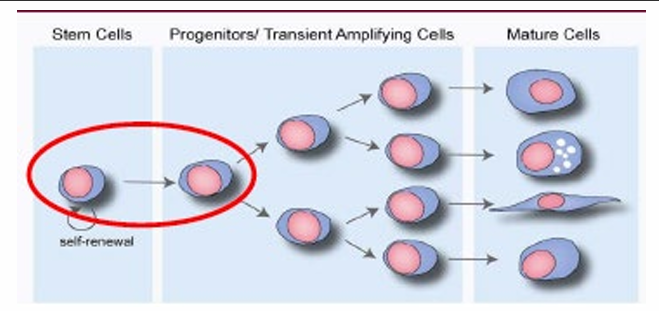
What activates a quiescent adult stem cell?
Stimuli such as:
- Signalling changes
- Disruption of the stem-cell niche
.
These stimuli cause the cell to:
- Exit G0 quiescence
- Re-enter the cell cycle
This activation can take about 24 hours.
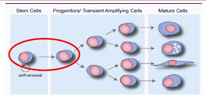
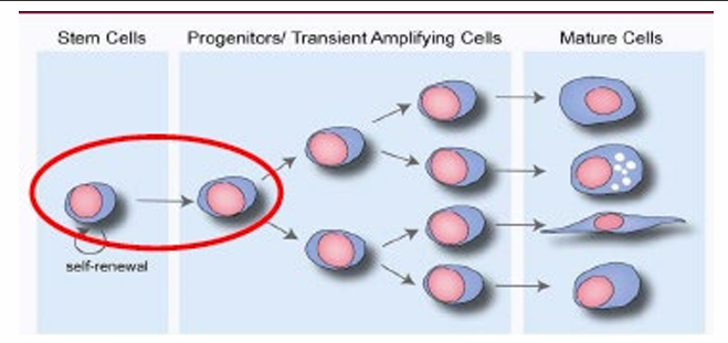
What typically happens in the first divisions after adult stem-cell activation?
The stem cell often undergoes asymmetric division during the first or second round of mitosis.
.
Outcome:
- One daughter retains stem-cell identity
- The other becomes a progenitor / transit-amplifying cell
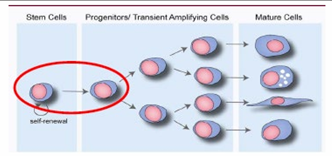

What are transit-amplifying cells and what do they do?
Transit-amplifying (TA) cells:
- Proliferate very rapidly
- Are controlled by signalling
- Are also influenced by contact and local tissue context
.
- Are induced to differentiate
- Undergo fate-specification decisions
- Withdraw from the cell cycle
- Mature into differentiated cells


Why do transit-amplifying cells undergo fate-specification decisions?
TA cells can receive different differentiation signals that direct them toward distinct mature fates.
Lecturer example:
In the haematopoietic system, different signals can direct progenitors toward T cells, B cells, NK cells, erythrocytes and other lineages.


How do true stem cells return to quiescence after activation?
True stem cells regain quiescence by returning to G0.
.
This decision must be local and depends on:
a. Contact signals from the stem-cell niche
b. Short-range signals
c. Asymmetric cell division


What mechanisms can determine which daughter cell remains a stem cell after division?
The slide presents three interacting possibilities:
- External signals
- Intrinsic factors already present within the cell
- Local niche contact
.
Lecturer explanation:
The lecturer suggested that stem-cell fate is probably determined by a combination of these mechanisms rather than any one mechanism alone.


How do Notch and Numb regulate differentiation?
- Notch inhibits differentiation.
- Numb inhibits Notch.
- Numb is distributed asymmetrically during stem-cell mitosis.
- Numb is not retained in the true stem-cell daughter.
- In the daughter retaining Notch activity: differentiation is inhibited and the stem cell re-enters G0.
- In the Numb-rich daughter: Notch is inhibited, permitting differentiation.


How does asymmetric distribution of Numb maintain the stem-cell pool?
1. A dividing stem cell distributes Numb unequally.
2. One daughter has low/no Numb and therefore retains active Notch.
3. Active Notch inhibits differentiation.
4. That daughter retains stem-cell identity and returns to G0.
5. The Numb-rich daughter has Notch inhibited and differentiates.

What clinical associations with Notch were mentioned?
Slide statements:
- Notch mutations → cancer (~65% of ALL cases)
- Notch inhibitory drugs, including γ-secretase inhibitors → chemotherapy
.
Lecturer explanation:
The lecturer presented this as an example of how disruption of a pathway that normally controls stem-cell fate can contribute to cancer.

Which adult stem-cell examples are compared in the second half of the lecture?
- Muscle — simple
- Gut — complex
- Blood — "insane" / highly complex

What are satellite cells in skeletal muscle?
Satellite cells are the tissue-specific stem cells of skeletal muscle.
During growth, stem cells:
- They are active
- They proliferate
- They differentiate
- Their number reduces to approximately 2-5% of the muscle-cell population
.
In adult muscle:
- They are quiescent
- Turnover is very low
.
During regeneration:
- They become strongly activated.

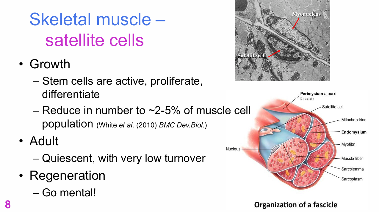
Where are skeletal-muscle satellite cells located?
Satellite cells are located along muscle fibres, associated with the fibre surface beneath/near the basal lamina.
.
Labels shown include:
- Myonucleus
- Satellite cell
- Perimysium around fascicle
- Mitochondria
- Endomysium
- Myofibrils
- Muscle fibre
- Sarcolemma
- Sarcoplasm
- Nucleus

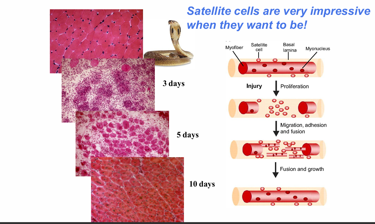
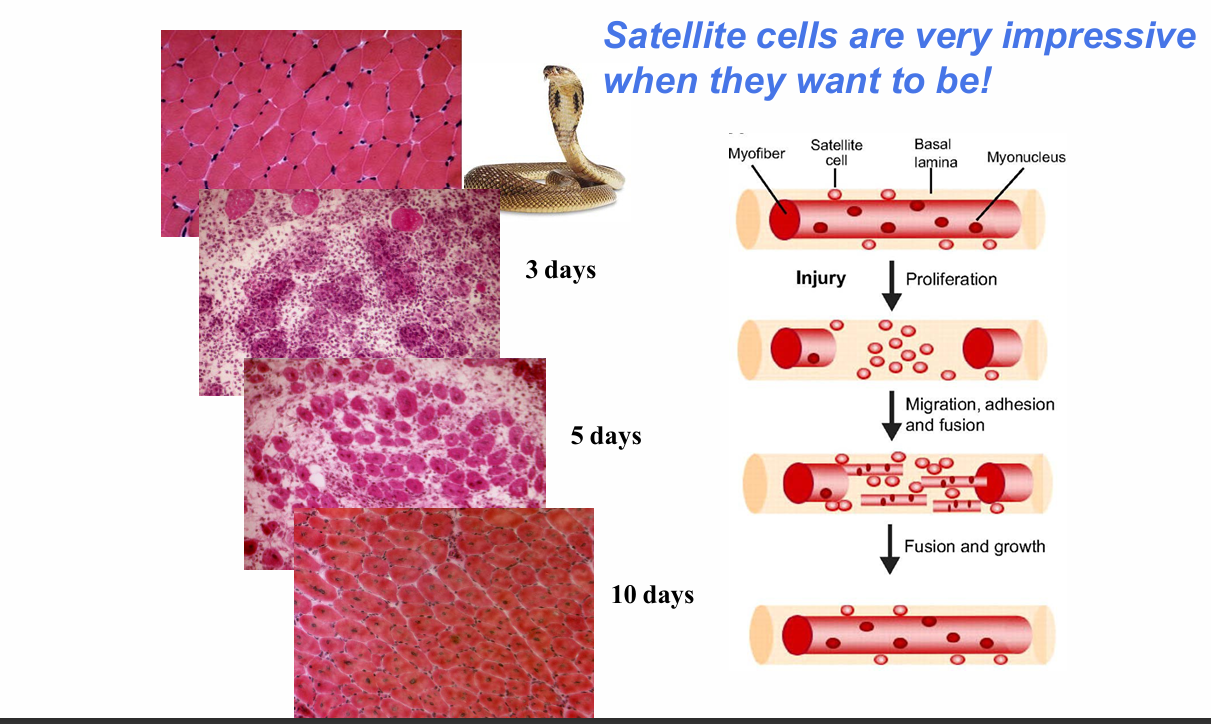
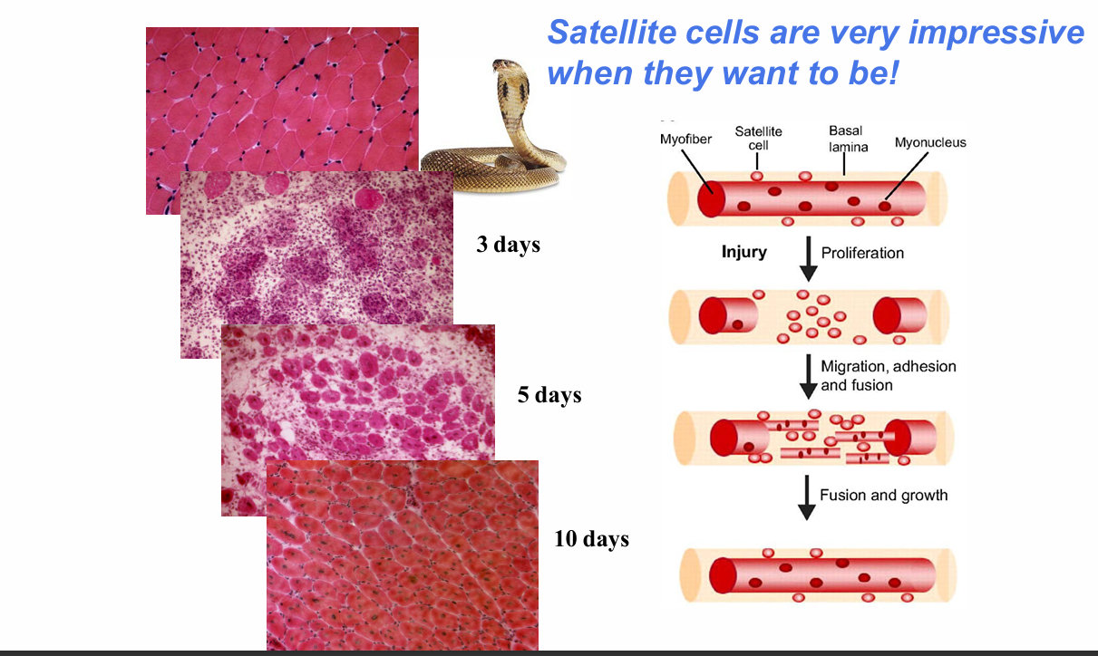
What is the sequence of satellite-cell-mediated muscle regeneration after injury?
1. Injury damages the myofibre.
2. Satellite cells are activated.
3. They proliferate.
4. Their progeny migrate.
5. Cells adhere and fuse.
6. Fusion and growth generate new muscle fibres.
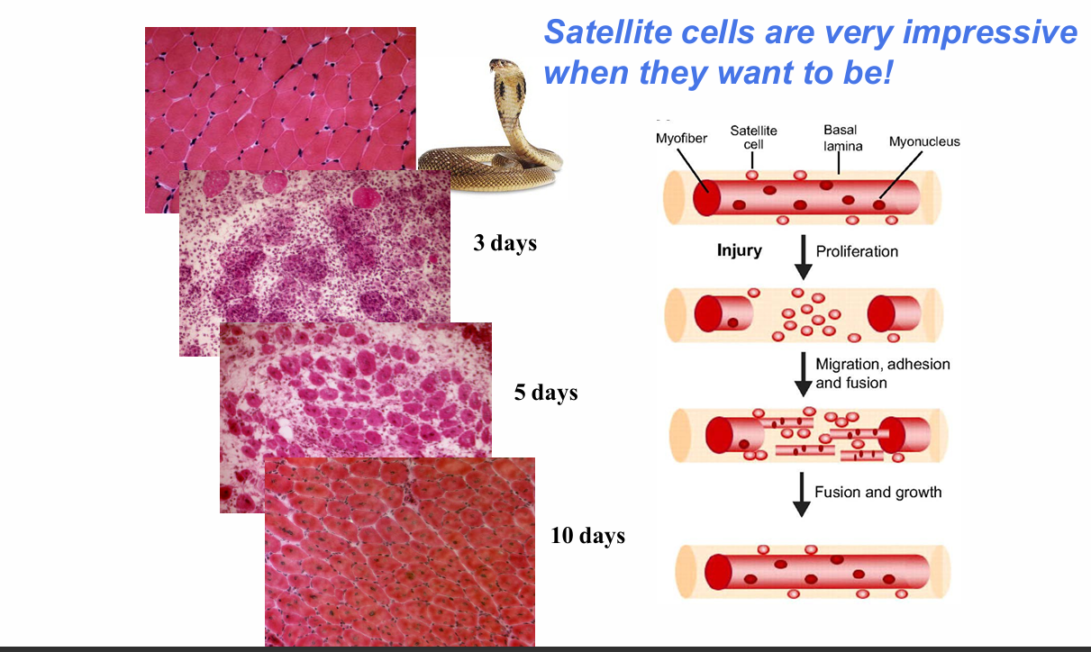
How rapidly can severely damaged skeletal muscle regenerate in the example shown?
After severe injury:
- ~3 days: prominent injury/necrosis and immune-cell response
- ~5 days: new fibres are appearing
- ~10 days: tissue appears close to normal
.
Lecturer explanation:
New fibres can be recognised by centrally located nuclei, unlike mature fibres where nuclei are more peripheral.

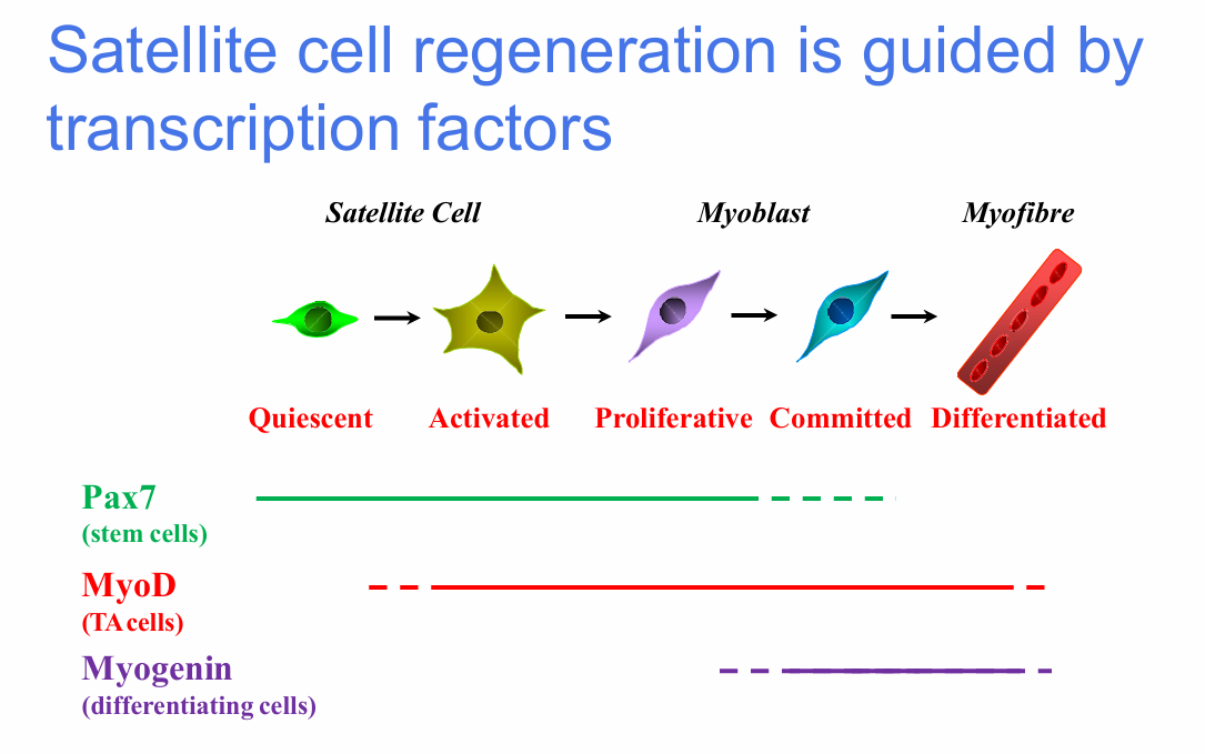
Which transcription factors mark stages of satellite-cell regeneration?
Pax7:
- Stem-cell / quiescent satellite-cell marker
MyoD:
- Transit-amplifying / activated myogenic-cell marker
Myogenin:
- Differentiating-cell marker


What sequence of cell states and transcription factors occurs during muscle regeneration?
Quiescent satellite cell
→ activated / proliferative cell
→ committed myoblast
→ differentiated myofibre
.
Associated factors:
- Pax7: stem-cell stage
- MyoD: activated / TA-cell stage
- Myogenin: differentiating stage

How do Pax7 and MyoD expression change as satellite cells activate and diverge?
Sequence shown:
- Pax7+ cell activates
- Pax7/MyoD+ cells proliferate
- Cells then diverge
.
Self-renewal branch:
- Retains Pax7 expression
.
Differentiation branch:
- Becomes MyoD+
- Then MyoD/Myogenin+ as differentiation progresses


Why does the intestinal epithelium require highly active stem-cell replacement?
Small-intestinal epithelial cells have a turnover time of approximately 2-4 days.
Crypt-base stem cells continuously generate progenitors that:
- Proliferate
- Migrate up the villus
- Differentiate
- Are eventually shed by apoptosis


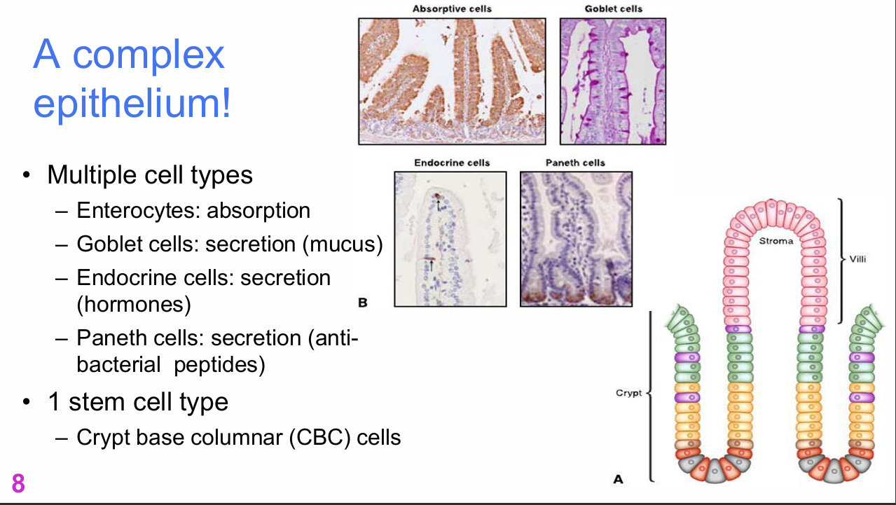
What mature cell types make up the intestinal epithelium and what are their functions?
- Enterocytes: absorption
- Goblet cells: secretion of mucus
- Endocrine cells: secretion of hormones
- Paneth cells: secretion of antibacterial peptides
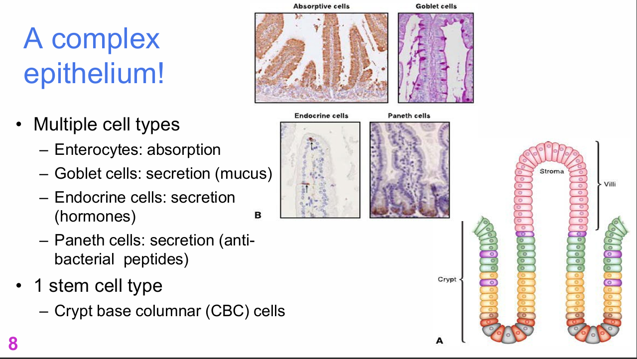

Which stem-cell type produces the multiple cell types of the intestinal epithelium?
Crypt base columnar (CBC) cells.
Despite the gut containing multiple specialised mature cell types, the slide identifies one major stem-cell type: CBC cells.

What are the defining features of crypt base columnar (CBC) stem cells?
CBC stem cells:
- Are multipotent
- Can generate all intestinal cell types
- Are marked by LGR5
- LGR5 is identified on the slide as a Notch pathway member
- Can also produce a reserve +4 stem-cell population for damage
- The reserve +4 cells are also LGR5+


What progenitor branches are shown downstream of LGR5+ CBC stem cells?
The diagram shows:
- Absorptive progenitor → absorptive enterocytes
- DLL1+ secretory progenitor → enteroendocrine, tuft and goblet cells
- LGR5+/LRC Paneth-cell progenitor → Paneth cells
- +4 stem cell as a reserve population


How rapidly do CBC-derived transit-amplifying cells divide?
CBCs produce transit-amplifying cells that:
- Divide every 12-16 hours
- Generate approximately 300 cells per crypt per day
- Sequentially differentiate as they move through the intestinal epithelium


What spatial sequence occurs from intestinal crypt to villus?
At the crypt:
- LGR5+ CBC stem cells and +4 stem cells are located near the base.
- TA cells proliferate.
.
As cells move upward:
- They migrate
- Sequentially differentiate into mature intestinal cell types
.
At the villus tip:
- Cells are lost by apoptosis/shedding

What are the key features of adult haematopoietic stem cells?
Adult haematopoietic stem cells:
- Are located in bone marrow
- Self-renew
- Are multipotent
- Form all blood-cell types

Which blood-cell types are generated from haematopoietic stem cells?
Examples listed:
- Red blood cells
- B cells
- T cells
- Granulocytes
- Monocytes
- Macrophages
- Platelets


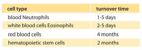
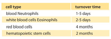
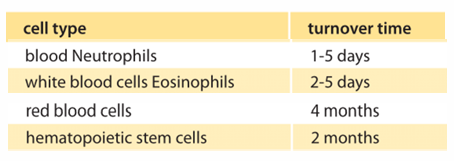
How much new blood does adult bone marrow produce each day according to the lecture?
Approximately:
- 30 mL of new blood per day
- Containing about 100 billion cells per day
.
Lecturer explanation:
Different blood-cell types have different turnover rates, so their production demands differ (most common are neutrophils since they have shortest half life)

What stem-cell populations are shown in the haematopoietic stem-cell niche?
The niche includes:
- Long-term quiescent HSCs
- Short-term active HSCs
- Mobile HSC / progenitor cells
Other labelled niche components include:
- Osteoblasts
- Osteoclasts
- Bone matrix
- Endothelial cells
- Blood vessels
- Macrophages
- Sympathetic nerve
- Endosteal niche
- Perivascular niche


How are long-term and short-term haematopoietic stem cells spatially organised in the niche?
Lecturer explanation:
- Long-term quiescent HSCs are positioned close to the bone/endosteal region and associated with bone-lining cells.
- Short-term active HSCs are positioned closer to blood vessels.
- Mobile HSC/progenitor populations can enter the circulation.

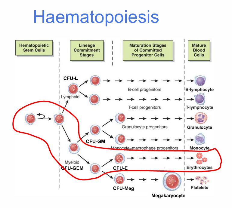
What is the overall sequence of haematopoiesis?
Haematopoietic stem cell
→ lineage commitment
→ maturation of committed progenitor cells
→ mature blood cells
.
Major outcomes shown:
- B lymphocyte
- T lymphocyte
- Granulocyte
- Monocyte
- Erythrocyte
- Megakaryocyte → platelets


Which colony-forming units are labelled in the haematopoiesis diagram?
Labels shown:
- CFU-L
- CFU-GM
- CFU-GEM
- CFU-E
- CFU-Meg
.
Other labels:
- Haematopoietic stem cells
- Lineage commitment stages
- Maturation stages of committed progenitor cells
- Mature blood cells


How do signals determine blood-cell lineage during haematopoiesis?
HSC-derived progenitors can enter different branches depending on the signals they receive and the cytoplasmic factors inherited during division.
The lecture contrasts:
- Lymphoid lineages
- Myeloid lineages
Different signals then direct production of specific mature blood-cell types.


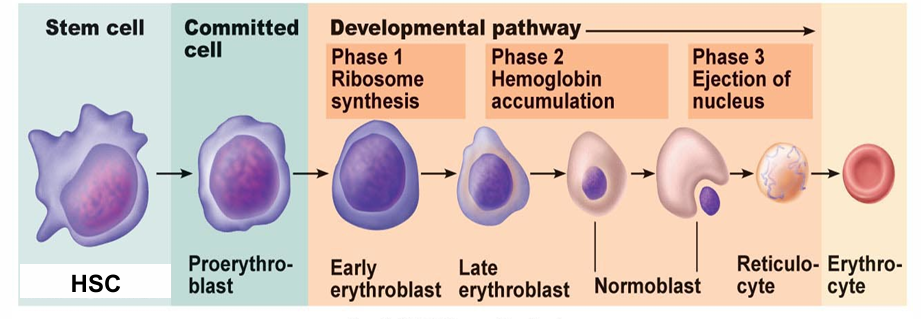
What major cellular changes occur during erythropoiesis?
During erythropoiesis:
- Cell size decreases
- The nucleus and organelles are lost
- Haemoglobin content increases
How long does red-blood-cell maturation take in the lecture example?
- HSC → reticulocyte: approximately 15 days
- Reticulocyte → mature RBC in bloodstream: approximately 2 additional days


What is the erythropoietic sequence shown in the developmental pathway?
HSC
→ proerythroblast
→ early erythroblast
→ late erythroblast
→ normoblast
→ reticulocyte
→ erythrocyte
.
The slide also shows:
- Phase 1: ribosome synthesis
- Phase 2: haemoglobin accumulation
- Phase 3: ejection of nucleus
.
Not assessable:
The lecturer said the individual developmental phase labels were not important to memorise in detail.


What does the reticulocyte index reflect?
The slide links the reticulocyte index to:
- Reticulocyte percentage
- Reticulocyte maturity
- Erythropoiesis
Lecturer explanation:
It is used as an indicator of how actively red-cell production is occurring.

Why must erythropoiesis be tightly regulated?
- Too few RBCs → tissue hypoxia
- Too many RBCs → increased blood viscosity
Therefore, red-cell production must be maintained within an appropriate range.

What is erythropoietin (EPO) and what triggers its release?
EPO = erythropoietin.
- Released by the kidneys
- Released in response to hypoxia
- Stimulates erythropoiesis


How does EPO stimulate erythropoiesis?
EPO:
- Speeds conversion of HSC → proerythroblast
- Speeds maturation of proerythroblasts
- Produces an increased circulating reticulocyte count in approximately 5 days

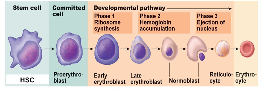
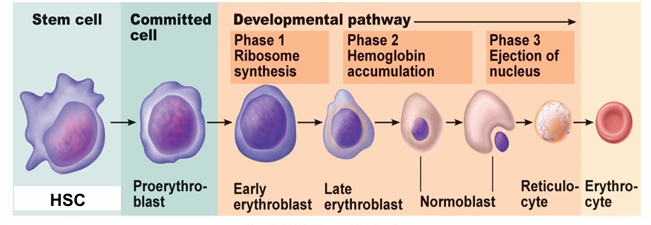
What should you identify in the EPO/erythropoiesis pathway diagram?
Sequence:
- HSC
- Proerythroblast
- Early erythroblast
- Late erythroblast
- Normoblast
- Reticulocyte
- Erythrocyte
.
Key relationship:
Hypoxia → kidney EPO release → accelerated erythropoiesis


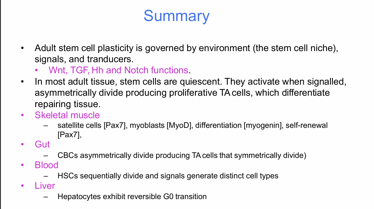
What are the major take-home messages from Stem Cells II?
- Adult stem-cell plasticity is governed by the environment/stem-cell niche, signals and transducers.
- Wnt, TGF, Hh and Notch have distinct regulatory functions.
- In most adult tissues, stem cells are quiescent until activated by signals.
- Activated stem cells asymmetrically divide, producing proliferative transit-amplifying cells that differentiate and repair tissue.
- Skeletal muscle: satellite cells are associated with Pax7; myoblasts with MyoD; differentiation with myogenin; self-renewal retains Pax7.
- Gut: CBC cells asymmetrically divide to produce TA cells, which then symmetrically divide.
- Blood: HSCs sequentially divide and different signals generate distinct blood-cell types.
- Liver: hepatocytes use reversible G0 transition for tissue maintenance and repair.