Reactive Lymph & Infectious Mono
 Call Kai
 Learn
 Practice Test
 Spaced Repetition
 Match
 Flashcards
 Knowt Play
1/11

There's no tags or description
Looks like no tags are added yet.
12 Terms
Lymphocytes Lineage
Stem cells (HSCs)
Common lymphoid progenitors
Lymphoblasts
Prolymphocytes
Mature B Lymphs
Memory B Cells
Plasma cell (effector cells
Mature T Lymphs
Helper T cells (HTLs)
Cytotoxic T cells (CTLs)
NK cells (Precursor is common lymphoid progenitors)
Reactive Lymph Characteristics
Pleomorphic (broad range of sizes & shapes)
Lower N:C ratio, dark blue rims, skirting of RBCs
Reactive lymphocytes seen in the blood smear is activated cytotoxic T (CTL) not infectious B lymphocytes
Historically classifies as Downey cells
Lymphoblast
High N:C ratio
Fine chromatin, prominent nucleoli
Scant basophilic cytoplasm (little cytoplasm, dark blue rims)
Prolymphocyte
More cytoplasm than lymphoblast, chromatin more condensed
Prominent nucleolus

Infectious Mononucleosis
Caused by Epstein Barr’s Virus
Symptoms include fever, sore throat, splenectomy, posterior cervical lymphadenopathy (enlarged lymph at the back of neck)
Clinically associates with reactive lymphocytes, and leukocytosis
Infected B lymphocytes produce heterophile antibodies of type IgM (NOT specific to EBV) that can be tested with Mono-Latex.
Sue-Vue Color Mono Test {Principle, SOP}
Reagent containing animal RBC that have specific antigens induce response to heterophile antibody
NOT specific to EBV, only support but does not confirm diagnosis
SOP
3 circles for positive, negative controls, and patient sample
Add 1 drop of controls or patient sample to the corresponding zones
Add 1 drop of color reagent to all tests, mix and spread (AVOID cross-contamination)
Rock side-to-side for 2 minutes
Positive test shows agglutination and heterophile antibodies are detected. Next step is to consider EBV-specific serology
Plasmodium vs Babesia {Transmission, Life Cycle, Diagnosis}
Plasmodium
Transmitted by Anopheles mosquitoes, common in endemic subtropical regions (Africa, Central & South America, Southeast Asia)
Life cycle involves liver and blood stages
Babesia
Transmitted by Ixodes ticks, common in northeastern U.S
No liver involvement
Malaria {Pathology}
Caused intra/extravascular hemolysis that leads to normochromic-normocytic. Leukopenia & thrombocytopenia
Diagnose with thick & thin smears
4 species (most common → least)
P. vivax: Schüffner’s dots, amoeboid-shape trophozoite, visible schizont
P. falciparum: banana-shaped gametocyte, no schizont or trophozoite, multiple rings per RBC
P. ovale: Schüffner’s dots and enlarged RBC
P. malariae: Schüffner’s dots, band-shape trophozoites
Babesia Life Cycle
Human/Animals (asexual)
Tick introduces Babesia sporozoites into the bloodstream
Sporozoites entered RBC and develop into trophozoites
Asexual multiplication
Ruptured RBCs that caused hemolytic anemia and released merozoites that infect more RBCs
Differentiates into male/female gametes
Ticks (sexual)
Gametes fuse and sexual reproduction
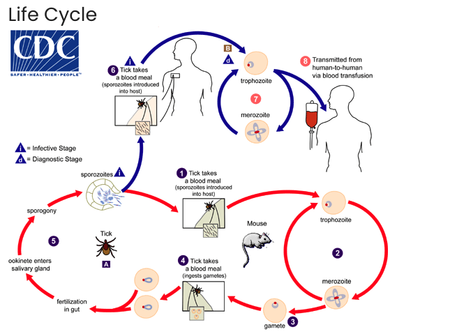
P. Falciparum vs Babesia

Percent Parasitemia {SOP, Formula}
Count between 500(high parasitemia >10%) or 2000 RBC (low parasitemia < 1%)
Use two counters to track infected RBC and total RBC
%parasitemia=total RBCinfected⋅100
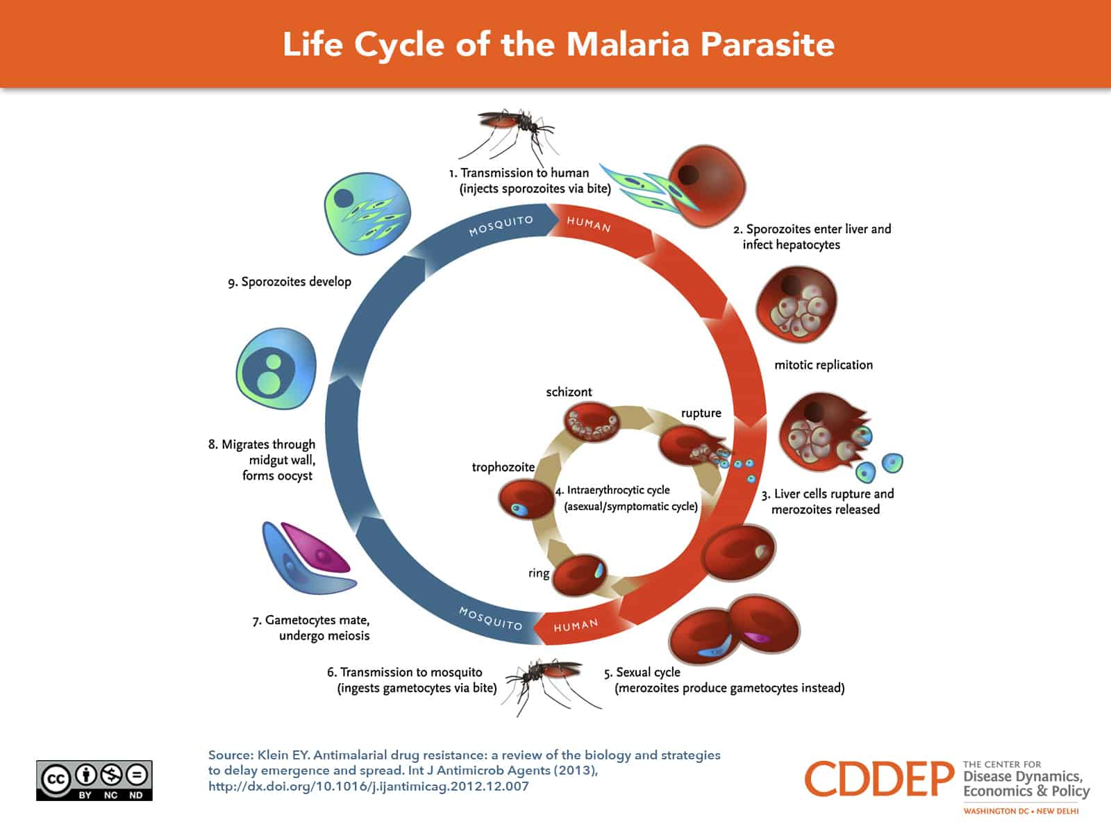
Malaria Life Cycle
Mosquito injects sporozoites into bloodstream
Sporozoites enter liver cells and develop to merozoites (daughter parasite produced by multiplication process)
Liver cells ruptured, merozoites released and infected RBC
Enter RBCs and metabolically develop in the trophozoite stage, undergoes multiplication in the Schizont stage
RBCs ruptured, merozoites released to infect more RBC
Some merozoites produce gametocytes instead