disorders of the vitreous and vitreo-retinal interface
 Call Kai
 Learn
Learn Practice Test
 Spaced Repetition
 Match
Match Flashcards
 Knowt Play
1/41

There's no tags or description
Looks like no tags are added yet.
Name | Mastery | Learn | Test | Matching | Spaced | Call with Kai | Chat |
|---|
No analytics yet
Send a link to your students to track their progress
42 Terms
describe the vitreous (recap) (3)
Type of connective tissue about 99% H20, (also collagen fibrils, hyaluronic acid & other)
Transparent, avascular, ‘Jelly’ like structure
Occupies 67-75% of eye volume (4ml)
functions of the vitreous (5)
•Give shape and form to the eye
•Keep the retina in place
•Provide optical clarity - therefore needs to be optically clear
•Act as a shock absorber
•Inhibits neovascularisation
anatomical points of the vitreous (2)
•Vitreous cortex/posterior hyaloid membrane/surface - condensation of collagen fibrils
•Strong vitreoretinal adhesion at certain points (vitreous base (ora seratta) > optic nerve, scars > blood vessels, macula)
what are the processes involved with the normal ageing of the vitreous (3)
Liquefaction (Synchysis) AND
Fibre density increases (>40 yrs) leading to…
Shrinkage (syneresis) occur
describe liquefaction - synchysis (3)
•Increasing liquefaction from 2nd decade
•Lacunae (fluid filled spaces) develop, increase in size & coalesce
•By 70 years ~50% liquified
Describe the increase of fibre density (2)
•Collagen forms clumps which reduce light transmission
•Vitreous strands and floaters
describe shrinkage (syneresis) (2)
•Vitreous pulls away from the retina - posterior vitreous detachment (PVD)
•NB: Areas of strong vitreoretinal adhesion can lead to abnormalities
describe what vitreous floaters are (2)
•Small, dark or translucent dots and filaments in vitreous
common - Occur in all age groups (usually older due to ageing vitreous)

what are the causes of vitreous floaters (3)
•Most often - normal aging vitreous
•Earlier in myopes, trauma, inflammation, certain diseases
•Always keep in mind serious causes - symptoms in addition to floaters are important !!! - flashing lights as well may indicate something in retina - can be benign but always check
what are the symptoms of vitreous floaters
•Patient reports floating object - dots, lines, spider web, various shapes
•Most obvious against white background

what are the signs of vitreous floaters (1)
•Not always detectable with ophthalmoscope
what is the management of vitreous floaters (3)
Reassurance is main therapy - double check however odd floater here and there is usually okay
if new/large floaters/new shower of floaters - red flag
Can be surgically removed if particularly troublesome - rare
why may congenital vitreous anomalies arise
•Incomplete hyaloid artery regression - leaving anomalies which we can see when examining px eye
name and describe congenital vitreous anomalies we may see ()
Persistent hyaloid artery - Coming from the disc (with or without blood)
Mittendorf dot - Small opacity on posterior lens capsule
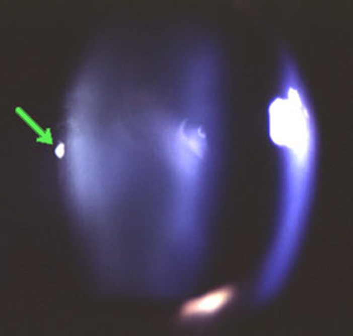
Bergmeister’s papilla - Clump of glial tissue on disc


what is the management of congenital vitreous anomalies
benign and do not require treatment - asymptomatic
state the different vitreous abnormalities to know (5)
Persistent foetal vasculature !
Vitreous Cysts !
Asteroid Hyalosis !!
Synchysis scintillans !!
Vitreous haemorrhage !!!
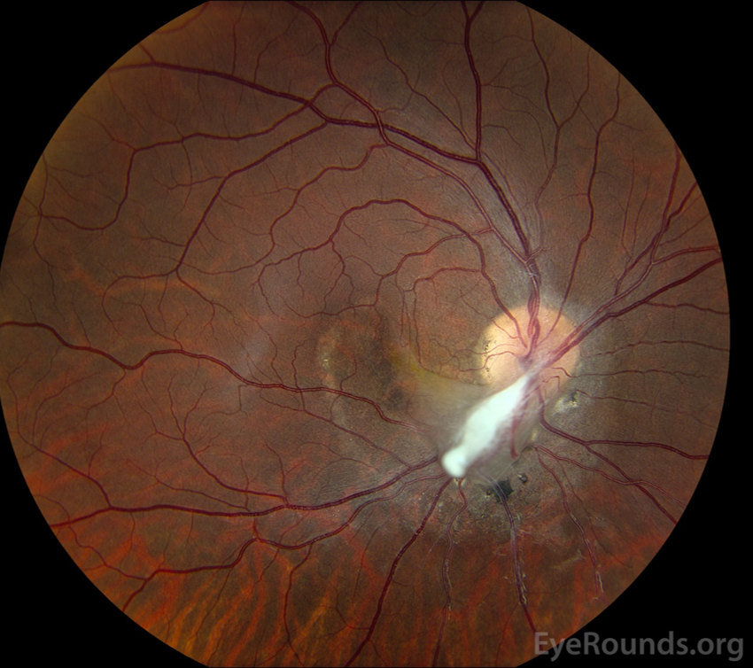
describe what persistent foetal vasculature is (4)
•Also known as - Persistent Hyperplastic Primary Vitreous (PHPV)
•Persistence of the hyaloid system (incomplete regression) resulting in fibrovascular proliferation - rare
•Anterior form: around the lens.
•Posterior form: around the retina and optic disc

what is the management of persistent foetal vasculature
immediate referral and treatment by paediatric ophthalmologist
what are the associations with persistent foetal vasculature
Other congenital anomalies (cataract, glaucoma microphthalmos)
differential diagnosis of persistent foetal vasculature
leukocoria - white/pale pupil appearance
what are vitreous cysts (3)
•Extremely rare
•Normal incidental finding or associated with ocular pathology
congenital/acquired
describe congenital vitreous cysts (3)
•Located at hyaloid canal
•Associated with Mittendorf's dot or Bergmeister's papillae
•Non-pigmented pearly-grey, smooth surface
describe acquired vitreous cysts (4)
•Trauma, inflammation/infection
•Retinal disease (RP, retinoschisis, choroidal atrophy)
•Retinal detachment surgery
•Usually reduced VA
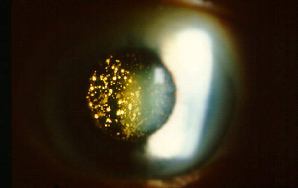
describe asteroid hyalosis (4)
•Sometimes called Benson’s disease
•Fairly common (1 in 100), usually > 55 years old and unilateral
•Small yellow-white spheres
•Composed of calcium-phospholipid complexes suspended throughout the vitreous (stable/static) - as px looks around they stay still

what are the sx and management of asteroid hyaloisis (4)
•Usually asymptomatic
•No effect on vision
•Self-limiting and benign
Patient education - routine review
describe synchisis scintillans (4)
bilateral vitreous disorder in the young - RARE - probably occurs in only blind, severely damaged eyes
Numerous reflective small angular yellow crystals floating throughout vitreous - mobile in vitreous - swirl round as px moves eye
crystals float around in a shower then settle to bottom of vitreous
Composed of cholesterol (not associated with high serum cholesterol)

sx and signs of synchisis scintillans
•Usually asymptomatic - not much management to do
•Thought to follow trauma or inflammation
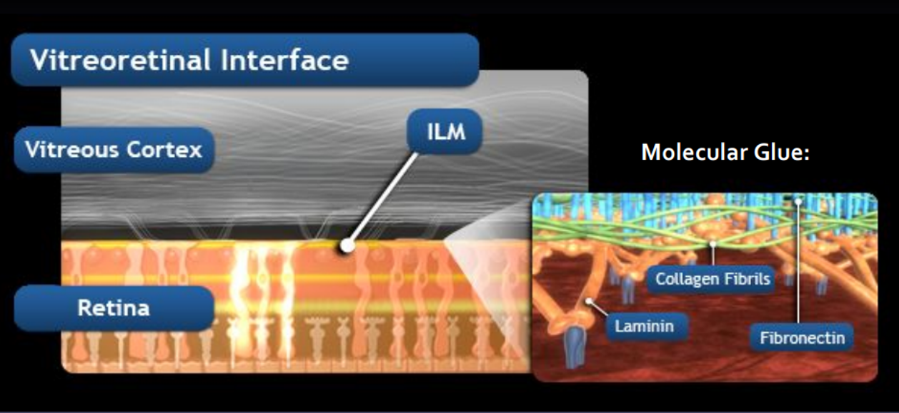
describe the vitreoretinal interface
Vitreous cortex and ILM of retina adhere via a molecular glue
Intervening extracellular matrix (fibronectin, laminin, collagen and other extracellular components)
as we age this adhesion weakens and can lead to abnormalities… like a vitreous haemorrhgae / PVD - in this lecture
describe the aetiology of a vitreous haemorrhage (6)
Blood enters vitreous - Disc vessels, retinal vessels, choroid
Causes:
Diabetic retinopathy (32-54%)
Trauma (12-19%)
Retinal tear or detachment (11-44% of cases)
Posterior vitreous detachment (4-11% of cases)
Others: retinoschisis, vein occlusion, sickle cell retinopathy, AMD etc.

what are the symptoms of a vitreous haemorrhage (4) (variable)
•Painless loss of vision
•Sudden, significant increase in the number and size of floaters (asking about floaters imp!!!)
•Blurry, cloudy or hazy vision (small bleed)
•Large bleed may obscure vision
how can we examine a patient with a vitreous haemorrhage (6)
•Vision/visual acuity
•Slit-lamp examination - looking directly at retina and anterior/posterior vitreous
•Eye pressure - can be reduced/increased
•Dilated eye examination
•Gonioscopy - peripheral retina and anterior angle
•B-scan ultrasound - hospital setting
describe the signs of a vitreous haemorrhage (2:1)
Non-dispersed:
Clear red haem - dense dark red/brown patch
Pre-retinal (boat shaped) - posterior to vitreous

Dispersed:
Obscuration of view - blood disperse through vitreous

describe the prognosis of a vitreous haemorrhage
•Variable depending on cause - sometimes can clear and vision will be restored or…
•Fibrovascular proliferation may result which could lead to scarring and contraction and ultimately retinal detachment
management of a vitreous haemorrhage (1)
•Emergency/same day (within 24 hours) referral
secondary care of a vitreous haemorrhage (4)
•Further examination/Mx
•B-can, Gonioscopy, IOP …. to find cause
•Treat the cause
Monitor or vitrectomy
describe posterior vitreous detachment (5)
Separation of vitreous cortex and neurosensory retina – part of normal ageing process - very common
Incomplete: involves posterior region
Complete: extends to ora serrata (vitreous base)
Most eyes 80+ years - Onset typically 60-70 years
Can be associated with myopia, trauma or inflammation
what are the symptoms of a PVD (3)
Sudden onset of floaters (most common) - ‘Cobwebs’ ‘bugs’ ‘spots’ (not usually a ‘shower’ - which may indicate retinal break) - Change position with eye movement
Photopsia (flashes of light 25-50%) - Dim illumination, temporal VF
Metamorphopsia - distorted vision - (less common) - If vitreo-retinal traction at macula
state and describe the main sign of a vitreous haemorrhage (3)
weiss ring - classic sign - unusually large floater
Grey/black annular ring suspended above disc on posterior hyaloid face - circular, doughnut-shaped floater in vision
Complete or incomplete


state and describe other signs of a PVD (2)
Visible posterior hyaloid face

Peripheral pre-retinal and disc margin haemorrhages
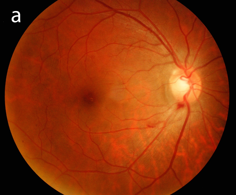
describe complications that can arise with a PVD (5)
Strong association with retinal tears and detachment (10-15%)
Risk of retinal detachment greatest in first 6-8 weeks
Shafer sign (tobacco dust) RPE cells in anterior vitreous (90% have a retinal break)

Vitreous haemorrhage (70% have a retinal break)
Macular hole formation (tractional)
optometric management of a PVD
All acute onset PVDs require:
Detailed H & S - risk factors
Examination of the anterior vitreous - pigment cells
Dilated fundus examination, using an indirect technique - peripheral retinal breaks
Unable to carry out an adequate examination, you must refer to a practitioner who is competent to do so
Patient education - signs/symptoms of retinal detachment - give written and verbal explanation - imp to go to A&E if sx persist or new sx !!
•Re-examination within 6-8 weeks of first report of symptoms may be indicated (not a requirement)
•Retinal break, vitreous haemorrhage or Tobacco dust - same day referral to ophthalmologist
•Follow local protocols and keep full and accurate records of all patient contact
NICE guidelines
