PSIO 201 - Unit III
 Call Kai
 Learn
Learn Practice Test
 Spaced Repetition
 Match
Match Flashcards
 Knowt Play
1/82

There's no tags or description
Looks like no tags are added yet.
Name | Mastery | Learn | Test | Matching | Spaced | Call with Kai | Chat |
|---|
No analytics yet
Send a link to your students to track their progress
83 Terms
what do all 3 muscle tissues have in common?
they all exhibit a quality called excitability as their plasma membranes can change their electrical states (from polarized to depolarized) and send an electrical wave called an action potential along the entire length of the membrane

skeletal muscle completely depends on what?
signaling from the nervous system to work properly
contractility
allows muscle tissue to pull on its attachment points and shorten with force.
actin
a protein pulled by myosin (contracting)
what is the basic function of all three types of muscle tissue?
generate tension
skeletal muscle functions include what?
locomotion (movement), facial expressions (chewing and showing emotion), posture and body position (balance and stabilization), body temp regulation (for homeostasis)
when we say skeletal muscle is voluntary what do we mean by that?
requires nervous system input, but we can “override” it. (e.g. blinking)
muscle contraction
protein filaments interact which shortens the length of the muscle (overlap of fibrils changes)

muscle action
skeletal systems transform muscle contraction into movement, how the skeleton moves as the muscles do.

where do muscles pull?
on bone
origin
where muscle “starts” on a bone (stationary)
insertion
where muscle “ends” on a bone (moves toward origin)

muscle action is based on the movement of what?
the movement of insertion towards origin

correctly naming an action or movement requires what two pieces of information?
the action + the name of the region that moves

movement to reduce the angle of the joint
flexion

movement to increase the angle of the joint
extension

movement away from the midline
abduction

movement toward the midline
adduction

medial rotation
rotation of limb toward the midline (inwards)
lateral rotation
rotation of limb away from midline (outwards)
to move palm up
supination (spine of hand on the bottom)
to move palm down
pronation (p, palm down)
reverse muscle action
when the origin moves towards the insertion

muscle primarily responsible for a movement “prime mover”
agonist

muscle which opposes the action of the agonist
antagonist

assists agonist; makes action more efficient or powerful
synergist

special synergists which help prevent movement at muscle origin
fixator
how do you determine the prime mover?
by identifying the muscle that produces the most dominant force during a specific joint action or movement

how do lever systems work?
muscles act to facilitate movement at a fulcrum by exerting force on a lever.
lever
rigid bar e.g. bone
fulcrum
fixed point around which the rigid bar (bone) moves (joint)
first class levers
effort, fulcrum, load - not very common in the human body

second class levers
force, lever, effort - not many examples, sacrifices speed and range of motion for force

third class levers
force, effort, load - most common, favors speed and range of motion over force

deep fascia
covers groups of muscles

what covers/ surrounds each muscle?
epimysium


what surrounds fascicles? (label 2)
perimysium

what surrounds individual muscle fibers (cells)? (label 6)
endomysium

what holds muscle fibers and is surrounded by endomysium? (label 5)
fascicles

what is label 4?
muscle fibers or the cells!
structural organization of skeletal muscle
organ: muscle
tissue: fascicles
cell: muscle fibers
sub cellular: myofibrils
what does it mean when we say that muscle fibers multinucleate?
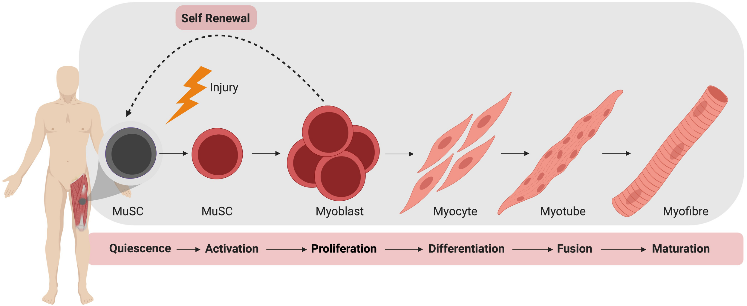
arises from fusion of myoblasts
muscle fibers cannot undergo what after birth?
mitosis
myoblasts turn into what after birth?
become myocytes (mature muscle cells!)
hyperplasia
increased number of cells

hypertrophy
increase cell size (big boi)

after birth, growth of muscle fibers happens by hyper_____ rather than hyper____.
happens by hypertrophy rather than hyperplasia.
satellite cells DO undergo mitosis after birth and aid in what?
muscle regeneration
functional organization of skeletal muscle
conduction of electrical signals
control of muscle contraction
contraction

conduction
how information travels via electrical signals
sarcolemma (plasma membrane) surrounds sarcoplasm (cytoplasm)
transverse tubules (t-tubules) arise from sarcolemma
control
regulation of activities of the SR
sarcoplasmic reticulum (SR) surrounds myofibrils
stores Calcium which helps generate muscle contractions
contraction
shortening of myofibrils inside the cytoplasm of skeletal muscle fibers
longitudinal bundles of protein filaments inside muscle fiber (actin and myosin)
highly organized into repeating units (sarcomeres)

sarcomeres
repeating units that are highly organized. the sarcomeres are what make the striations visible when looking under a microscope.

I band
thin only (actin)

A band
thick/overlap or not

h zone
thick only (myosin)

m line
middle line

z disc
edges of sarcomeres with thin filaments
what happens to the sarcomere during contraction?
A-band stays the same
i-band shortens
z lines come closer together
greater area of overlap
crossbridge
when myosin heads bind to actin
what causes thin filaments to slide along thick filaments?
a conformational change charged by ATP hydrolysis
name contractile proteins
actin and myosin
name regulatory proteins
troponin and tropomyosin
name structural proteins
titin, dystrophin, alpha-actinin, myomesin, nebulin

actin
thin filaments, has bonding site for myosin

myosin
motor protein in thick filaments, myosin head binds to actin forming cross bridge during contraction

tropomyosin
in thin filaments, covers myosin binding sites when muscle is relaxed.

troponin
in thin filaments, holds tropomyosin in place when muscles are relaxed.
during contraction, ca binds to troponin and causes a conformational change that shifts tropomyosin away from myosin binding sites on actin → exposing binding sites to allow for crossbridge formation.
titin
stabilizes position of thick filament, gives muscle elasticity and extensibility.
gives sarcomere bounce back needed once stretched out.
dystrophin
links thin filaments to sarcolemma, transmits tension from sarcomeres to tendons. aids in allowing for the movement of bone alongside muscle.
nebulin
anchors thin filaments to z disc
alpha-actinin
found in z disc, binds to actin to titin
myomesin
found in m line, binds to titin and thick filaments that connect them at the M line.
where does crossbridge cycling occur?
only in the area of overlap! where actin and myosin are binded!
how does the contraction cycle begin?
when calcium is released from the sarcoplasma reticulum - which causes the tropomyosin to move leaving myosin binding sites on actin to be exposed
before binding to actin, what happens with myosin head?
atp binds to the head, causes a conformational change “cocking” the myosin head, the atp is now into ADP-Pi, making it ready to bind.

what is the power stroke?
when the myosin heads pivot toward the center of the sarcomere. “pulling action”
how does the myosin head detach from the actin?
atp binds to the head which causes the crossbridge to break
how does the cycle continue?
myosin heads hydrolyze ATP used to break the crossbridge, and this energy is used to become reoriented, recocked, and then a new crossbridge begins.

rigor complex
attached head group after the power stroke
rigor mortis
rigor of death because of the lack of ATP (energy) to help detach the crossbridge.
what is needed for the binding of actin and myosin TO BEGIN?
calcium attachment to the troponin
what is needed for the ACTUAL binding of actin and myosin?
ATP on the myosin head that is hydrolyzed into ADP-Pi.