Lecture 13 - Globins: Myoglobin and Hemoglobin
 Call Kai
 Learn
 Practice Test
 Spaced Repetition
 Match
 Flashcards
 Knowt Play
1/101

There's no tags or description
Looks like no tags are added yet.
102 Terms
Why globins are a model system
Well-studied proteins that show how 3D structure determines function: O₂ transport/storage, cooperativity, allosteric regulation, conformational change, evolution (gene duplication, fetal vs adult Hb), disease (sickle cell, thalassemia), and techniques (X-ray crystallography, spectroscopy, O₂-binding curves)
Hemoglobin/myoglobin "firsts"
First protein crystallized (1849)
first with mass accurately measured
first studied by ultracentrifugation
first linked to a physiological condition
first shown to have a disease-causing point mutation
first X-ray structures solved
Myoglobin (Mb) function
In rapidly respiring muscle, increases O₂ solubility and facilitates O₂ diffusion from capillaries (diffusion alone is slow because O₂ solubility is low)
also stores O₂
Evidence that Mb stores O₂
Mb concentration is about 10x higher in diving mammals (whales, seals) than in land mammals
Hemoglobin (Hb) function
Transports O₂ from lungs to tissues
needed because O₂ diffusion alone is too poor for transport in large animals

Hb subunit composition
α₂β₂ tetramer (a dimer of αβ dimers)
each subunit is structurally similar to myoglobin
O₂ concentration in plasma vs bound to Hb
Plasma: about 10⁻⁴ M (low solubility)
Bound to Hb in blood: about 0.01 M, roughly 100x higher
Hemocyanin
Alternative O₂ transporter protein that contains copper (Cu)
Hemerythrin
Alternative O₂ transporter protein with non-heme iron

Heme group
Prosthetic group in each Hb/Mb subunit that binds one O₂
a heterocyclic porphyrin derivative, specifically protoporphyrin IX with a central Fe

Required oxidation state of heme iron
Fe(II), the ferrous or reduced form
only Fe(II) binds O₂
LEO the lion says GER
Loss of Electrons = Oxidation
Gain of Electrons = Reduction

Fe ligands 5 and 6 in heme
Four porphyrin nitrogens in the plane
5th ligand = proximal His F8 (below the plane)
6th ligand = O₂ on the distal side (above the plane)

Origin of Hb's color
Electrons move between Fe(II) d orbitals (eg and t₂g)
Fe(II) is d⁶ low spin
O₂ binding rearranges the electrons and changes the d-orbital energy gap, so the absorption spectrum shifts
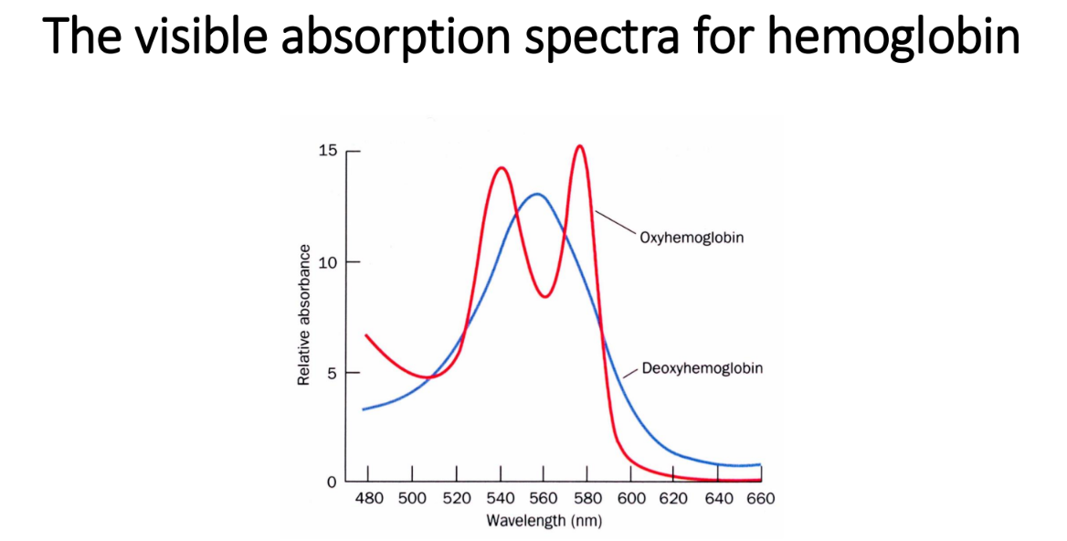
Absorption spectra: oxy vs deoxy Hb
Oxy-Hb: two peaks, about 540 and 578 nm (red)
Deoxy-Hb: one broad peak, about 555–560 nm (bluish)
Use of absorbance at 578 nm
Easy way to measure the percent of O₂ bound to Hb (fractional saturation)
Methemoglobin
Hb with oxidized Fe(III).
Brown, has water as the 6th ligand, and cannot bind O₂.
Gives dried blood and old meat their brown color
Methemoglobin reductase
Enzyme that reduces methemoglobin, Fe(III), back to normal Hb, Fe(II)
Ascorbic acid and meat
Butchers use it to reduce methemoglobin so meat looks fresh and red
Why free heme can't bind O₂ reversibly
O₂ bridges two hemes (Fe–O–O–Fe heme dimer), which oxidizes Fe(II) to Fe(III)
Picket-fence (sterically hindered) heme
Bulky groups on one face of the heme block the heme–O₂–heme dimer, so reversible O₂ binding can occur
This is the model for what the globin does
Two functions of the globin protein
(a) Modulates O₂-binding affinity
(b) Makes reversible O₂ binding possible by preventing Fe oxidation
Globin–heme "hamburger" analogy
Globin surrounds the heme like a bun
only the propionic acid side chains face the solvent
Mutations in the heme pocket can cause autooxidation to methemoglobin
Mb + O₂ equilibrium and Kd
Mb + O₂ ⇌ MbO₂
Kd = [Mb][O₂] / [MbO₂]
Fractional saturation (YO2)
Bound / total = [MbO₂] / ([Mb]+[MbO₂]) = [O₂] / (Kd + [O₂])

pO₂
Partial pressure of O₂ (O₂ tension), used as the measure of O₂ concentration

YO2 for Mb in terms of P₅₀
Y = pO₂ / (P₅₀ + pO₂)

P₅₀
The pO₂ at which YO2 = 0.50 (half-saturation)

Meaning of P₅₀ for affinity
Lower P₅₀ = higher O₂ affinity
Higher P₅₀ = lower affinity
Shape of Mb's O₂-binding curve
Hyperbolic (non-cooperative)
Shape of Hb's O₂-binding curve
Sigmoidal (cooperative)
P₅₀ of myoglobin
2.8 torr
Pressure unit conversions
1 torr = 1 mm Hg = 0.133 kPa
760 torr = 1 atm
Arterial vs venous pO₂
Arterial ≈ 100 torr
venous ≈ 30 torr
Mb saturation from arterial to venous pO₂
Y drops only from 0.97 to 0.91, so Mb releases little O₂ over the physiological range
(poor transporter, good storage)
Hb saturation from arterial to venous pO₂
0.95 at 100 torr to 0.55 at 30 torr, so it releases about 40% of its O₂
P₅₀ of Hb
About 26 torr in whole blood (normal BPG); about 12 torr for stripped Hb

Dashed curve on the O₂ saturation plot (slide 20)
A hypothetical non-cooperative Hb with about the same P₅₀ as real Hb
Between arterial (100 torr) and venous (30 torr) pO₂ it would unload only about 0.27 of its O₂, versus about 0.40 for real cooperative Hb
This shows why cooperativity matters for O₂ delivery

Hill equation setup
E + nS ⇌ ESn
(E = protein, S = ligand such as O₂, n = small number)
K = [E][S]ⁿ/[ESn]
Measure of COOPERATIVITY

Hill equation (general)
Ys = [S]ⁿ / (K + [S]ⁿ)
Same as the Mb equation except for the exponent n

Hill equation for O₂
YO2 = (pO₂)ⁿ / ((P₅₀)ⁿ + (pO₂)ⁿ), where K = (P₅₀)ⁿ
Hill constant (n)
Non-integral parameter giving the degree of cooperativity among interacting ligand-binding sites or subunits
Interpreting n
n > 1: positive cooperativity (larger n = more cooperative)
n = 1: non-cooperative
n < 1: negative cooperativity

Hill plot equation
log [ Y / (1−Y) ] = n · log [ S ] − log K
(form y = mx + b)
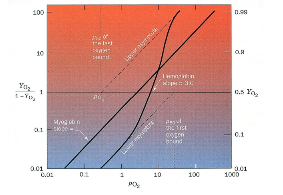
What the Hill plot gives you
Slope = n
X-intercept (−b/m) = log P₅₀, the point where Y = 0.5

Hill plot slopes for Mb and Hb
Mb slope = 1 (non-cooperative)
Hb slope ≈ 3.0 at its steepest point near Y = 0.5

Hill plot asymptotes for Hb
Both asymptotes have slope 1
The lower asymptote gives P₅₀ of the first O₂ bound
the upper asymptote gives P₅₀ of the last O₂ bound

First vs last O₂ binding to Hb
First O₂: subunits bind independently with low affinity
Last O₂ (3 sites full): binds independently with high affinity
The 4th O₂ binds about 100x more tightly than the first, a ΔG difference of 11.4 kJ/mol
"All or none" behavior of Hb
When one O₂ binds the rest tend to bind
when one is released the rest tend to be released
Positive cooperativity (definition)
The ligand-binding state of one heme raises the ligand-binding affinity of another heme
Mb vs Hb at venous pO₂
Hb gives up O₂ much more easily than Mb, and Hb's binding is cooperative
Who solved the globin structures
John Kendrew (myoglobin) and Max Perutz (hemoglobin), 1959–1968

Myoglobin dimensions and size
44 × 44 × 25 Å
single subunit
153 residues, 121 of them in α-helices

Globin helix naming
Helices labeled A–H
Loops between them are named for the two helices (e.g., CD, EF, FG)
Residues are named by helix plus position (His F8 = 8th residue of helix F) or by absolute number
Part of the EF segment forms the F′ helix

Heme in the myoglobin structure


Residues invariant in all vertebrate Hb and Mb
Thr C4, Phe CD1, Leu F4, His F8, Tyr HC2

Sequence alignment table color key
Blue = identical in both Hb chains
purple = identical in both Hb chains and Mb
dark purple = invariant in all vertebrate Hb and Mb

Hemoglobin dimensions and symmetry
Roughly spherical, 64 × 55 × 50 Å, with a twofold rotation axis
The similar α and β subunits sit at the corners of a tetrahedron

Hb α chain structural difference
The α chain has no D helix

Hb subunit interfaces
α1β1 (and α2β2): 35 residues in contact
α1β2 (and α2β1): 19 residues in contact
Extensive interactions between unlike subunits

T state
Tense, deoxy, low-affinity conformation of Hb
individual subunits are called "t"
R state
Relaxed, oxy, high-affinity conformation of Hb
individual subunits are called "r"
Quaternary change on oxygenation
The α1β1 dimer rotates about 15° relative to the α2β2 dimer

Fe position in deoxy vs oxy heme
Deoxy: Fe sits about 0.6 Å out of the heme plane, toward His F8
O₂ binding pulls Fe into the plane (Oxy)
Fe moves into the plane and drags the attached His F8, which moves the whole F helix like a lever on a fulcrum
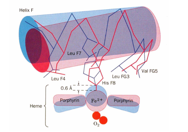
Color code in the F-helix figure (slide 46)
Blue = deoxy (T) position: Fe 0.6 Å out of plane, porphyrin domed
Red = oxy (R) position: Fe in plane, F helix shifted toward the heme

Close contact in deoxy Hb (slide 44)
In the T state, His F8 is in close contact with the porphyrin ring
If Fe moved into the heme plane without His F8 reorienting, the His would bump into the porphyrin

Residues packed around His F8
Leu F4, Leu F7, Leu FG3, Val FG5, and Leu H19 pack tightly around the proximal His
Their packing is why His F8 can't reorient unless the whole F helix shifts
How binding at one heme raises affinity at others
Shifting the α1β2 contacts moves the distal His E7 and Val E11 out of O₂'s path to the Fe in other subunits, increasing affinity of the heme toward oxygen.

α1β2 contact as a binary switch
The contact has two stable positions held by different but equivalent sets of H-bonds, one for T and one for R
No stable in-between state exists
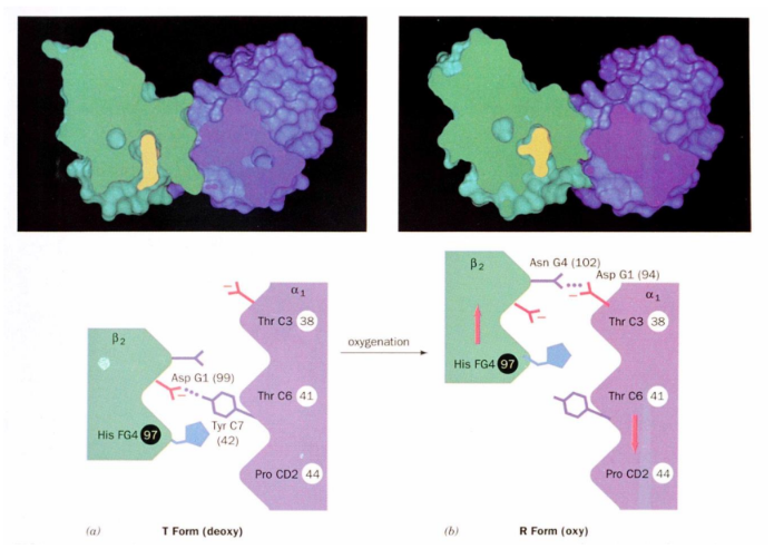
What drives the T→R transition
Energy released by forming the Fe–O₂ bond
Chain of events coupling Fe to the quaternary shift
Fe can't move into the plane unless His F8 reorients.
His F8 is packed too tightly to reorient unless the F helix shifts across the heme
The F helix can only shift along with the quaternary change that steps the α1C–β2FG contact one turn along the α1C helix
Why all subunits switch together
The α1β1 and α2β2 interfaces are rigid, so the shift must happen at α1β2 and α2β1 at the same time
No single subunit or dimer can change alone

Structural basis of cooperativity
Once the tetramer switches to R, the remaining unliganded subunits become R even without O₂ bound, and R subunits have much higher O₂ affinity
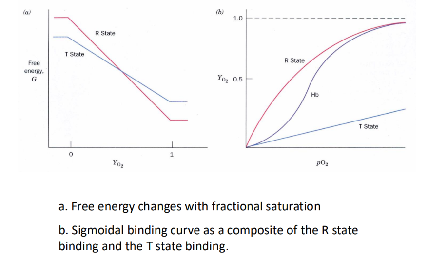
Free energy vs YO2 plot
At low saturation the T state has lower free energy (more stable)
at high saturation the R state does
The lines cross partway through binding

Sigmoidal curve as a composite
Hb's curve shifts from the low-affinity T-state curve (nearly flat) at low pO₂ to the high-affinity, hyperbolic R-state curve at high pO₂

Bohr effect
Higher pH (lower [H⁺]) makes Hb bind O₂ more tightly
lower pH (higher [H⁺]) makes Hb release O₂ more easily

pH and P₅₀
pH up → P₅₀ down (higher affinity)
pH down → P₅₀ up (lower affinity; curve shifts right)

Bohr effect magnitude example
At 20 torr, about 10% more O₂ is released when pH drops from 7.4 to 7.2

CO₂ hydration reaction
CO₂ + H₂O ⇌ H⁺ + HCO₃⁻, catalyzed by carbonic anhydrase in red blood cells

Carbonic anhydrase role
Rapidly converts CO₂ from respiring tissue (about 0.8 mol CO₂ per O₂ consumed) to bicarbonate
without it, CO₂ bubbles would form in blood

Link between CO₂ transport and O₂ release
Hb takes up H⁺ from CO₂ hydration, which causes it to release more O₂.
That proton uptake also pulls the reaction toward bicarbonate, aiding CO₂ transport

Carbamate formation
R-NH₂ + CO₂ ⇌ R-NH-COO⁻ + H⁺
CO₂ reacts with N-terminal amino groups of Hb (carbamates)
CO₂ bound directly to Hb
About 5% of blood CO₂ is bound to Hb, but this accounts for about 50% of the CO₂ exchanged
In the lungs, O₂ binding makes Hb release CO₂
Molecular origin of the Bohr effect
The T→R transition changes the pKa of several groups
N-terminal amino groups: 20–30% of the effect
His146β: about 40%, via a T-state salt bridge with Asp94 that is lost in the R state

Salt bridges and Cl⁻ at the subunit interface (slide 51)
In the T state, intersubunit salt bridges hold the deoxy structure together. These include the C-terminal His146β pairing with Asp94β and Cl⁻ bridging positively charged groups. They break in the R state, which is why Cl⁻ and H⁺ stabilize T and lower O₂ affinity

BPG
D-2,3-bisphosphoglycerate
Lowers Hb's O₂ affinity by binding and stabilizing the T (deoxy) state

BPG binding stoichiometry and affinity
Binds 1 BPG per Hb tetramer with K = 1×10⁻⁵ M to deoxy-Hb
binds only weakly to oxy-Hb

BPG binding site
Central cavity of deoxy-Hb, lined with positively charged groups (His, Lys, N-termini of β chains) that pair with BPG's negative charges
In the R state the cavity is too narrow for BPG to fit

Stripped Hb vs whole blood
Stripped Hb (BPG removed) has a much higher O₂ affinity
its curve sits left of whole blood's

Effect of 4.7 mM BPG on stripped Hb
Raises P₅₀ from 12 to 22 torr

Order of O₂ curves, left to right (high to low affinity)
Stripped Hb → Hb + CO₂ → Hb + BPG → Hb + BPG + CO₂ → whole blood
Total O₂ release explained
Hb releases about 40% of its O₂ between arteries (95% saturated) and veins (55%)
Without BPG little O₂ is released
BPG, CO₂, H⁺, and Cl⁻ together account for Hb's O₂-binding behavior in blood
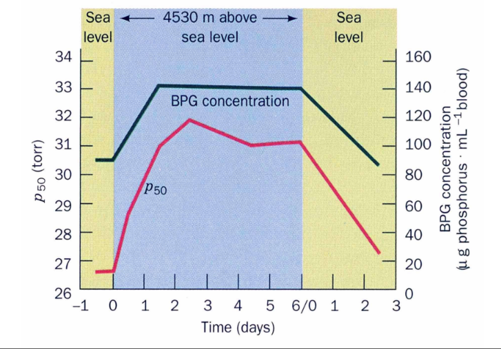
High-altitude adaptation (BPG)
At 4530 m, BPG rises over about 2 days and P₅₀ rises from about 27 to about 31 torr
Both return toward normal after going back to sea level
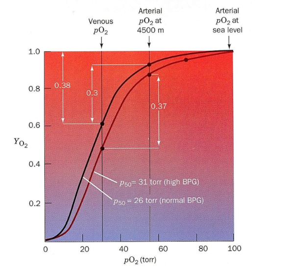
Why BPG helps at altitude
Arterial pO₂ at 4500 m is only about 55 torr
With normal BPG (P₅₀ 26) Hb unloads only 0.30
With high BPG (P₅₀ 31) it unloads 0.37, close to the 0.38 delivered at sea level

BPG binds specifically to the __________ and stabilizes it in the ______. In the ______ the central cavity is too narrow for BPG to fit or bind.
deoxy state; T state; R state
Fetal hemoglobin (HbF)
α₂γ₂: the γ subunit replaces β.
Binds BPG poorly, so it has higher O₂ affinity than maternal HbA and O₂ moves from mother to fetus
Number and nature of Hb variants
About 500 variants, 95% of them single amino acid substitutions.
About 5% of the world's population carries a variant
Four classes of Hb mutations
Surface charge changes
changes in internal residues
changes that stabilize methemoglobin
changes at the α1β2 contact
Surface mutations
Rarely change Hb function
Sickle cell is the major exception
Internal-residue mutations
Distort Hb's shape and binding properties
Cause Heinz bodies, which lead to hemolytic anemia (red cell lysis)