688 L 8/17 Principles of Muscle Energy
 Call Kai
 Learn
 Practice Test
 Spaced Repetition
 Match
 Flashcards
 Knowt Play
1/50

There's no tags or description
Looks like no tags are added yet.
51 Terms
Somatic Dysfunction
the impaired or altered function of related components of the body framework system
TART
tissue texture changes
asymmetry
restricted motion
tederness
5 Other Features to Look for in Physical Exam
temperature
texture
moisture
edema
erythema
Acute Somatic Dysfunction Skin Temperature
hot
Chronic Somatic Dysfunction Skin Temperature
cold
Acute Somatic Dysfunction Skin Texture
boggy/rigid
Chronic Somatic Dysfunction Skin Texture
ropy/stringy
Acute Somatic Dysfunction Skin Moisture
increased/moist
Chronic Somatic Dysfunction Skin Moisture
decreased/dry
Acute Somatic Dysfunction Skin Edema
present
Chronic Somatic Dysfunction Skin Edema
not present
Acute Somatic Dysfunction Skin Erythmea
increased prolonged redness
Skin Erythmea Exception
the skin can be red after a technique, but it is only a problem if it is in one spot or hurts
Chronic Somatic Dysfunction Skin Erythmea
prolonged blanching
Active Motion
movement produced voluntarily by the patient (the patient is active)
Passive Motion
motion induced by the osteopathic practitioner while the patient remains relaxed (patient is passive)
Active OMT Methods
any technique in which the patient voluntarily performs an osteopathic practitioner-directed motion (patient does work)
Passive OMT Methods
any technique in which the patient is instructed to restrain from voluntary muscle contraction (physician does the work)
Barriers
every motion has a barrier (the limit of motion), but not everyone has a somatic dysfunction
abnormal restriction of motion is what we are checking for
5 Barriers
anatomic
physiologic
elastic
pathologic
restrictive
Anatomic Barrier
the limit of motion imposed by anatomic structure, the limit of passive motion, going beyond this results in damage to the tissues
Physologic Barrier
the normal limit of active motion
Elastic Barrier
the range between the physiologic and anatomic barriers in which passive ligamentous stretching occurs before tissue disruption
Pathologic Barrier
restriction of joint motion associated with pathologic change of tissues (like osteophytes)
Restrictive Barrier
a functional limit that abnormally diminishes the normal pathologic range, this is the one you are looking for when screening for somatic dysfunction
Osteophytes
boney lumps that grow on the bones (on the zygapophyseal joints of the spine or other joints)
Normal Joint Rotation Diagram

Barrier with Somatic Dysfunction Diagram
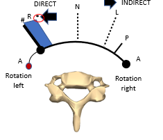
Restrictive Barrier Zone
position through which direct techniques move where the somatic dysfunction is, starts with the feather’s edge of the barrier and ends with the hard edge
Feather’s Edge of the Barrier
the spot where you can just barely feel resistance (the very edge)
Hard Edge
the end of the restrictive barrier zone where the joint locks out and will not move any further
Position of Laxity
position at which the tissues are as soft/loose as they can be, moving away from this position will increase the tension in the tissues
Direct Technique
the restrictive barrier is engaged and a final activating force is applied to correct somatic dysfunction
Indirect Technique
the restrictive barrier is disengaged and the dysfunctional body part is moved away from the restrictive barrier
Muscle Energy Technique
a form of omt in which the patient’s muscles are actively used on request, from a precisely controlled positon, in a specific direction, and against a distinctly executed physician counterforce
an active, direct method
Dr. Fred Mitchell DO
created muscle energy, named because of its reliance on active patient effort through muscle contraction
Dr. Ed Stiles
a guy that is still alive that took one of Dr. Fred Mitchell’s class, said that mitchell wasn’t going to teach the class again if the people didn’t get it
6 Proposed Mechanisms of Muscle Energy
*post-isometric relaxation
*joint mobilization using muscle force
respiratory assistance
oculocephalogyric reflex
reciprocal inhibition
crossed entensor reflex
Post-Isometric Relaxation: Goal
muscle relaxation
Post-Isometric Relaxation: Physiological Basis
after performing this contraction, the neuromuscular apparatus is in a refractory state in which passive stretching may be performed without encountering a myotatic reflex
physician resists contraction then “takes up the slack”
Isometric Contraction
length of muscle does not change during the contraction
Isotonic Contraction
the length of the muscle changes during contraction but the force/weight does not
Post-Isometric Relaxation: Force of Contraction
gentle pressure, 10-20 lbs of pressure at most
Myotatic Reflex
stretch reflex, an involuntary automatic response to a muscle being stretched, makes the muscle think it might be torn
we want to avoid this reflex for muscle energy
Triggers of Myotatic Reflex Must Overcome:
the amount of stretch
rate of stretch
Principles of Diagnosis Using Muscel Energy Technique
get an accurate diagnosis (without an accurate diagnosis, you cannot perform an appropriate treatment)
check the motion one plane at a time, then learn to “stack” the barriers to treat multiple planes of motion at the same time
Treatment Using MET
After obtaining diagnosis, physician positions the body part to be treated to its initial resistance (feather’s edge of the restrictive barrier)
Patient is instructed to provide an appropriate muscle effort in a specific direction (away from the barrier) as the physician provides counterforce for 3-5 seconds – isometric contraction
Instruct the patient to completely relax for 3-5 seconds
After the patient has completely relaxed, the physician repositions the patient to the new feather’s edge of the restrictive barrier
Repeat steps 2-4 until maximum level of improvement is achieved, typically 3-5 times*
final stretch to make sure it all of the somatic dysfunction was treated
Return the patient to neutral and then recheck the somatic dysfunction
Indications for MET
presence of somatic dysfunction in absence of contradictions
Relative Contraindictions for MET
patient is uncooperative or unresponsive
language barrier
infection, hematoma, or tear of involved muscle
fracture or dislocation of involved joint
rheumatologic conditions causing instability of the cervical spine
undiagnosed joint swelling of involved joint
positioning that comprises vasculature
severe osteoporosis
primary or secondary neoplastic process in the area to be treated
patient is too young to cooperate*
hypermobility – can make it hard to feel the barrier (we’ll screen for this in lab)
2 Absolute Contraindictions for MET
no somatic dysfunction
patient refusal
Safety and Efficacy of MET
safe if applied correctly, avoid vigorous contractions because that can make met less effective and result in spasms of soreness
localized appropriately → go to the feather’s edge of a barrier