Lec. 7&8: Bacterial Cell Envelopes, Cytology and Division
 Call Kai
 Learn
 Practice Test
 Spaced Repetition
 Match
 Flashcards
 Knowt Play
1/59

There's no tags or description
Looks like no tags are added yet.
60 Terms
Where are the peptidoglycan’s polymers synthesized and why?
Cytoplasmic Synthesis: peptidoglycan polymers are too large to be assembled inside the cell; NAG and NAM are synthesized in the cytoplasm
What facilitates the transport of NAG-NAM-pentapeptide building blocks across the hydrophobic plasma membrane into the periplasmic space
Lipid Carrier
How are cross-links distributed during transpeptidation to mitigate structural fragility?
Scattered Cross-Linking (Meshwork Formation): Transpeptidase forms peptide cross-links at scattered positions across adjacent glycan chains
Where does transpeptidation occur?
Periplasmic space; once building blocks pass through the membrane, the transpeptidase enzyme complex may begin the process
How does peptidoglycan synthesis proceed directionally?
From plasma membrane outward; as new layers are added underneath, older outer layers are pushed upward, gradually erode, and are shedded
What is teichoic acid’s role in gram-positive cell wall?
Solidifying and reinforcing the thick peptidoglycan cell wall
Teichoic acid composition:
Polymers of glycerol or ribitol
What is Lipoteichoic Acid’s role in gram-positive?
Anchor peptidoglycan cell wall to plasma membrane (its hydrophobic lipid tail embeds directly into the plasma membrane)
How is the gram-negative cell wall anchored?
Braun's Lipoproteins: Lipoprotein’s protein component is attached to the peptidoglycan while lipid tail is embedded in the outer membrane
How do porins in the outer membrane differ from plasma membrane transporters?
Porins are non-specific, allowing any molecule below a certain size threshold to pass between external environment and periplasm. In contrast, plasma membrane transporters are molecule-specific carrier proteins
Outer-membrane:
Asymmetric bilayer consisting of;
Inner leaflet; mainly phospholipids
Outer leaflet; mainly lipopolysaccharide (LPS)
Why is LPS medically termed endotoxin?
During systemic bloodstream infection, gram-negative bacterial lysis releases LPS whose Lipid A component causes severe immune system response, leading to septic shock
Anatomy of lipopolysaccharides (LPS):
Lipid A (hydrophobic)
Core Polysaccharide (hydrophilic)
O-side chain (O-antigen)
What is Lipid A composed of?
Two phosphorylated glucosamine rings + long-chain fatty acids
Structural and functional roles of Lipid A:
Its fatty acids anchor it to the outer membrane and it is the endotoxin’s toxic component
How does core polysaccharide expression compare to O-antigen expression across bacterial species and strains?
Core polysaccharide displays moderate structural conservation; O-Side Chain (O-Antigen) is extensively variable among bacterial species and even strains
Why does O-Side Chain (O-Antigen) extend outward into the extracellular environment and why is this polysaccharide variable?
Immune Evasion: gram-negative bacteria alter O-antigen sugar composition to evade host antibody recognition.
How do Lipid A and O-side chain differ in terms of hydrophobicity?
Lipid A is hydrophobic; O-side chain is hydrophilic
Why is lipopolysaccharide exclusively characteristic of gram-negative and not gram-positive?
Because LPS is a structural component of the outer membrane, and only Gram-negative bacteria have an outer membrane.
Capsule chemical composition:
Polysaccharides
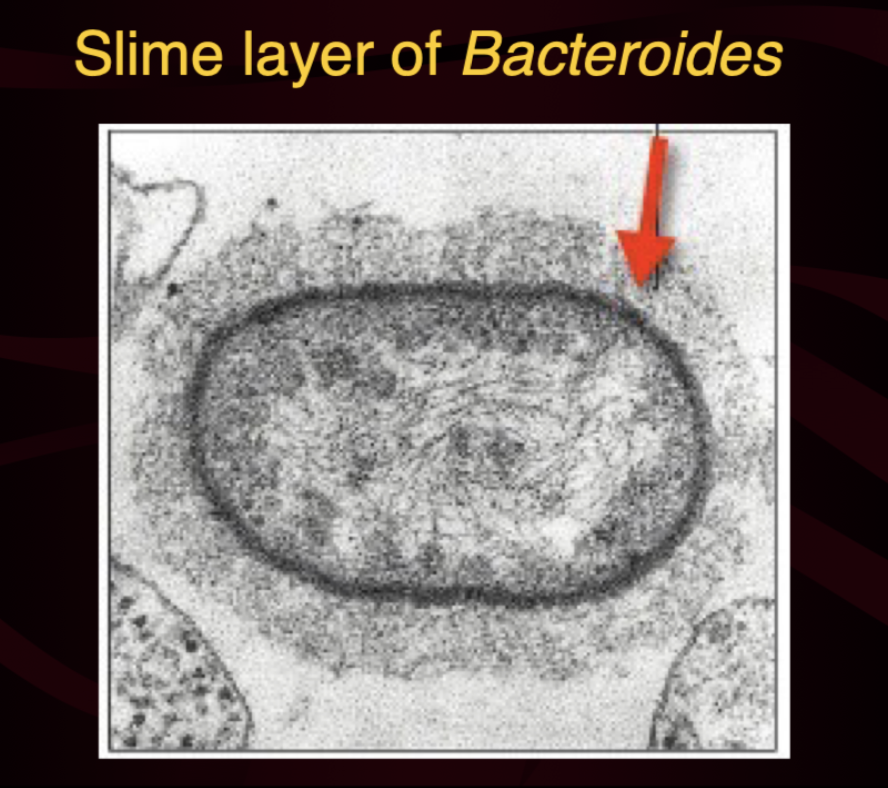
Slime layer chemical composition:
Polysaccharides + glycoproteins or glycolipids
Surface layer (S-layer) chemical composition:
Proteins or glycoproteins
Capsule structural organization:
Thick, dense, well-organized

Slime layer structural organization:
Thin, loose, unorganized
Surface layer (S-layer) structural organization:
Highly organized

What similarities do capsules and S-layers share?
attachment strength: not easily removed
microscopic visibility: clearly visible by microscopy
Slime layer attachment strength:
Easily removed
Slime layer microscopic visibility:
Difficult to visualize via microscopy
Capsule primary biological role:
Virulence factor (shields antigens)
Slime layer primary biological role:
Surface attachment & protection
Surface layer primary biological role:
Extreme structural robustness (pH/UV/pressure)
How do external layers (capsule, slime layer, s-layer) participate in Bacteriophage defense?
Bacteriophages must bind to surface receptors to infect bacterial cells; external layers shield these receptors, preventing bacteriophages from attaching and injecting viral DNA
How does capsule act as a virulence factor?
Masks cell surface antigens/epitopes from host immune cells
Does flagellar motility require an energy source?
Yes; flagellar motion requires proton motive force (ATP/proton gradient)
Monotrichous:
A single flagellum located at one cell pole
Amphitrichous:
A single flagellum at each of the two opposite cell poles
Lophotrichous:
A cluster of two or more flagella located at one or both poles
Peritrichous:
Flagella distributed all over the entire cell surface
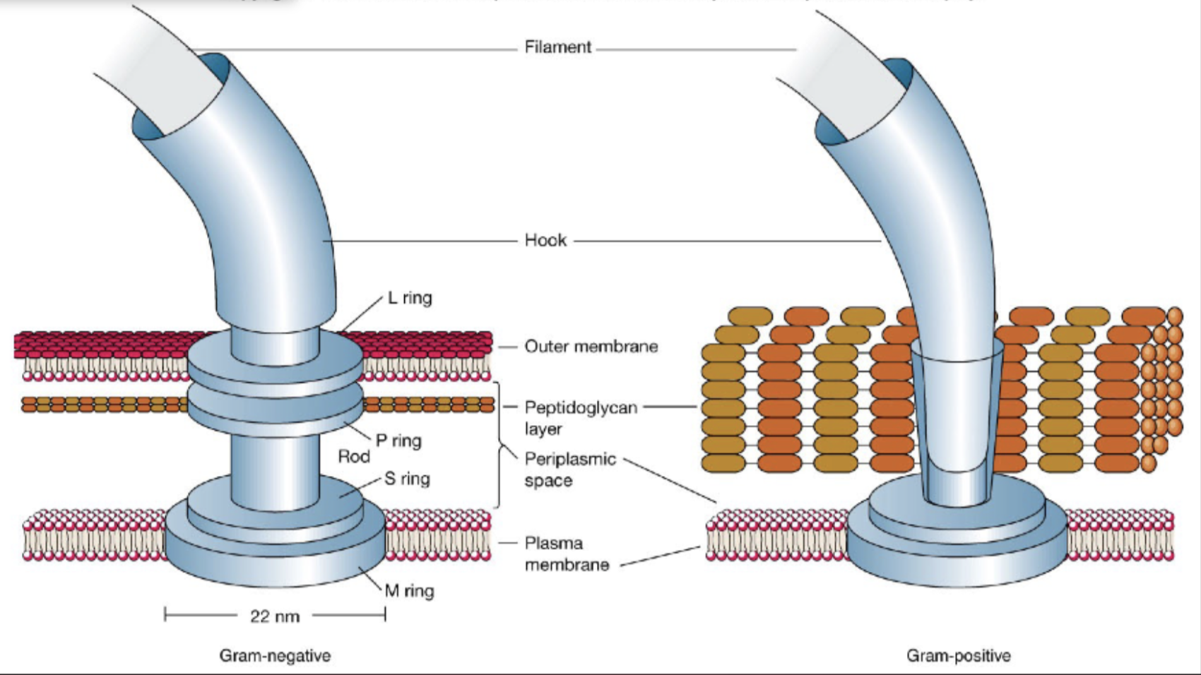
Why do gram-negative flagellar motors have 4 rings while gram-positive flagellar motor contains 2?
Gram-positives have more simple flagellar motor architecture due to lack of outer membrane
How do bacterial flagella facilitate runs and tumbles?
Counterclockwise (CCW) Rotation → run
Clockwise (CW) Rotation → tumble

Chemotaxis:
Directional movement towards chemical attractants (eg. amino acids) or away from repellants (eg. toxins)
Magnetosome:
Iron-containing inclusion bodies that enable cells to perform magnetotaxis
The primary site of metabolism:
Cytoplasm
Cytoskeleton:
Protein filaments (less complex than eukaryotic cytoskeletons) that help maintain cell shape and direct internal molecular traffic
What feature makes pathogens such as C. difficile (hospital-acquired diarrhea) and Bacillus anthracis (anthrax) exceptionally difficult to eradicate with standard hospital disinfectants?
Endospore formation
Three endospore adaptations for resistance:
Extreme dehydration
Calcium-Dipicolinic Acid (Ca-DPA) Complex
Small Acid Soluble Proteins (SASPs)
How does dehydration increase endospore resistance?
Removing free water (content drops from 90% to ~ 15%) prevents hydrolysis and heat damage (thermal denaturation of DNA and proteins).
How does Ca-DPA increase endospore resistance
Stabilizes the spore’s DNA, preventing genome degradation
How do SASPs increase endospore resistance?
Specialized proteins bind to spore DNA, protecting it from UV radiation damage and chemical modifiications
Sporulation (vegetative cell → endospore)
An irreversible 8-stage genetic program initiated by nutrient starvation or toxic environmental stress that takes 7-10 hours to complete
Which event is associated with mature endospore release?
Mother cell (sporangium) lyses
How many spores are produced by one vegetative cell?
One Spore Per Cell: sporulation is a survival mechanism, not a reproductive mechanism (no net increase in cell number occurs).
Multi-Layered Spore Envelope:
Core: dehydrated cytoplasm containing DNA, ribosomes, Ca-DPA, and SASPs
Cortex: peptidoglycan
Spore Coat: keratin-like proteins
Exosporium: lipid-protein-polysaccharide membrane
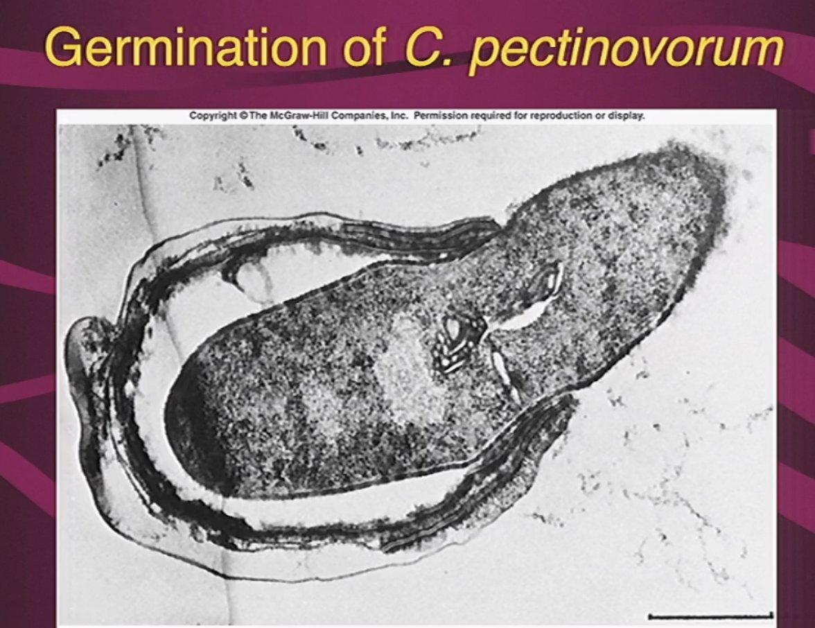
Germination (Endospore → vegetative cell)
Rapid process taking only ~15 minutes ("just add water") when favorable nutrient conditions return
Three stages of endospore germination:
Sensing: pre-existing receptors on spore coat detect environmental nutrients without requiring metabolism or ATP synthesis
Rehydration: permeability of spore changes, leading to massive water influx
Outgrowth: core swells, breaking the spore coat. A germ tube is what emerges and develops into vegetative cell
Binary Fission (Primary Mode):
A single cell elongates, replicates its chromosome and divides near the midpoint into two equal, identical daughter cells

Budding:
Outgrowth forms on the parent cell that enlarge, and detache.
Modes of Cellular Division:
Binary Fission (Primary Mode)
Budding
Fragmentation (Filamentous Growth)
Exospore Formation: Spores formed externally (distinct from internal endospores)
Steps in Bacterial Binary Fission
chromosome anchoring: circular chromosome is attached to specific site on cytoplasmic membrane
Replication & Elongation: as cell grows, chromosome replicates
Chromosome Segregation: duplicated daughter chromosomes separate toward opposite poles as the cell elongates.
Septum Formation: new membrane and peptidoglycan wall material invaginate at cell midsection, fusing to divide cytoplasm equally

What determines the characteristic spatial arrangements of spherical bacteria (cocci)?
The orientation of division planes