Week 7 - DNA Replication
 Call Kai
 Learn
 Practice Test
 Spaced Repetition
 Match
 Flashcards
 Knowt Play
1/49

There's no tags or description
Looks like no tags are added yet.
50 Terms
What evidence connects DNA to heredity and cell division?
DNA is found in the cell nucleus and chromosomes.
The amount of DNA doubles during S phase of the cell cycle.
Diploid cells generally contain twice as much DNA as corresponding haploid cells before replication.
Following DNA replication, each replicated chromosome consists of two sister chromatids, each containing one DNA double helix.
During mitosis, sister chromatids separate, producing two genetically identical daughter cells under normal circumstances.

Why must DNA replication be highly accurate and rapid?
Accuracy: Genetic information must be copied with very few errors to preserve DNA sequences across cell divisions.
Even an error rate of 1 in 1 million nucleotides would produce approximately 6,400 errors when copying a human-sized genome of 6.4 billion nucleotides.
Speed: DNA must be copied quickly enough for cells to divide.
In E. coli:
The genome contains approximately 4.6 million base pairs.
DNA replication proceeds at approximately 1,000 nucleotides per second.
The error rate is less than 1 in 1 billion nucleotides.
Under optimal conditions, E. coli can divide approximately every 20 minutes.
What are the three proposed models of DNA replication, and how do they differ?
1. Semiconservative replication:
Each parental DNA strand serves as a template for a new complementary strand.
Each daughter DNA molecule contains one parental strand and one newly synthesized strand.
2. Conservative replication:
The two parental strands remain together in one daughter DNA molecule.
A second DNA molecule consists of two newly synthesized strands.
3. Dispersive replication:
Parental DNA is distributed throughout both daughter DNA molecules.
Each daughter strand contains intermixed segments of old and new DNA.
- The experimentally supported mechanism is semiconservative DNA replication

What was the purpose of the Meselson–Stahl experiment, and how was it performed?
Meselson and Stahl investigated which model of DNA replication was correct: conservative, semiconservative, or dispersive.
They initially grew E. coli bacteria in a medium containing heavy nitrogen (¹⁵N), producing heavy DNA.
The bacteria were then transferred to a medium containing light nitrogen (¹⁴N).
Researchers collected DNA after successive rounds of replication.
Density-gradient centrifugation was used to distinguish DNA molecules based on their densities.
The resulting DNA banding patterns were compared with predictions from the three replication models.
What were the results of the Meselson–Stahl experiment, and what did they demonstrate?
Before transfer (generation 0): All DNA was heavy (¹⁵N/¹⁵N).
After one round of replication: All DNA had an intermediate density (¹⁵N/¹⁴N).
After two rounds of replication: Both intermediate-density DNA (¹⁵N/¹⁴N) and light DNA (¹⁴N/¹⁴N) were detected.
These results supported the semiconservative model of DNA replication.
Conclusion: Each daughter DNA molecule contains one parental strand and one newly synthesized strand.

How did the Meselson–Stahl experiment distinguish between the three DNA replication models?
Conservative replication: Predicts separate heavy and light DNA bands after the first round.
Semiconservative replication: Predicts one intermediate band after the first round, followed by intermediate and light bands after the second.
Dispersive replication: Predicts one intermediate band after the first round and a single, progressively lighter band after subsequent rounds.
The observed results matched the semiconservative model, demonstrating that each daughter DNA molecule retains one parental strand.

How does semiconservative DNA replication produce two identical daughter DNA molecules?
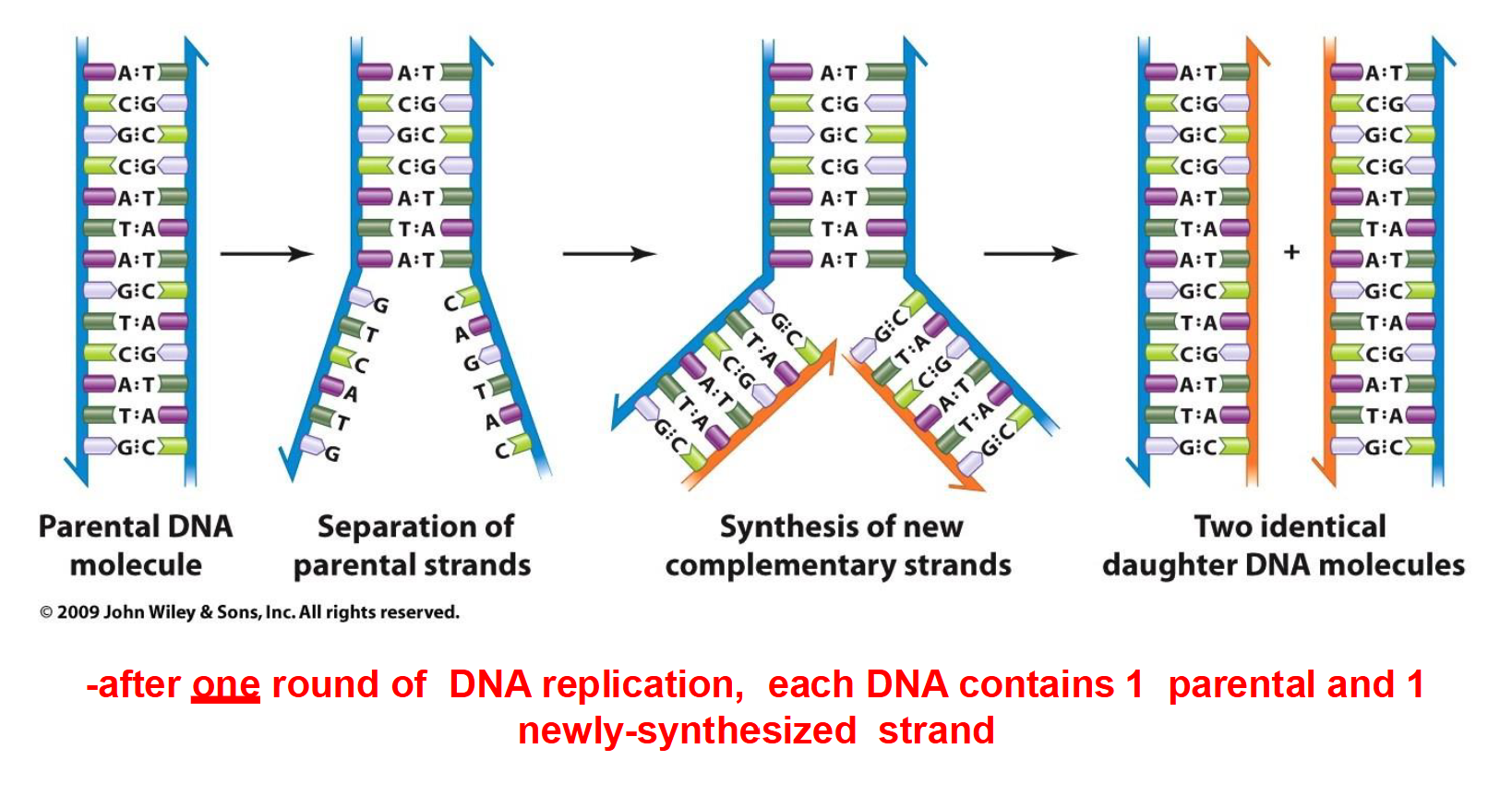
The two strands of the parental DNA double helix separate.
Each parental strand acts as a template for synthesizing a new complementary strand.
New nucleotides are added according to complementary base pairing (A–T and G–C).
Two daughter DNA molecules are produced.
Each daughter molecule contains one original (parental) strand and one newly synthesized strand.
DNA replication involves the coordinated activity of multiple proteins.
What is theta replication, and how does it occur?
Theta replication (θ) is a mode of DNA replication that occurs in most circular DNA molecules, including bacterial chromosomes.
Replication typically begins at one origin of replication.
DNA unwinds, forming a replication bubble with two replication forks.
Replication is bidirectional, meaning the forks move in opposite directions around the circular DNA.
The process produces two circular daughter DNA molecules.
Example: The approximately 4.6-million-base-pair chromosome of E. coli.

What is rolling-circle replication, and how does it occur?
Rolling-circle replication is a specialized mode of DNA replication that occurs in the F factor (F plasmid) and certain viruses.
Replication begins at a single origin of replication, where one DNA strand is nicked.
The free 3′ end is extended by DNA polymerase, using the intact strand as a template.
As DNA synthesis proceeds, the original nicked strand is displaced.
The displaced DNA can serve as a template for synthesis of a complementary strand.
Replication is unidirectional.
Rolling-circle replication can produce multiple copies of circular DNA.

How does rolling-circle replication facilitate the transfer of the F factor during bacterial conjugation?
An F⁺ donor cell attaches to an F⁻ recipient cell through an F pilus, forming a conjugation bridge.
One strand of the F plasmid is nicked and transferred into the recipient cell.
Rolling-circle replication replaces the transferred strand in the donor cell.
The recipient synthesizes a complementary DNA strand to form a double-stranded F plasmid.
Once transfer and replication are complete, both cells are F⁺.

How does linear chromosome replication occur in eukaryotic cells?
Linear chromosome replication occurs in the linear chromosomes of eukaryotic cells.
Replication begins at multiple origins of replication distributed along each chromosome.
Each origin produces a replication bubble, with two replication forks moving in opposite directions.
Therefore, replication is bidirectional.
As replication continues, neighbouring replication bubbles expand and eventually merge.
The process produces two linear daughter DNA molecules.
Example: According to your professor's slide, mice have approximately 25,000 origins of replication, with each replicon containing about 150,000 nucleotides.

Comparison Table

What are the common features of the three modes of DNA replication?
All three modes of DNA replication begin at an origin of replication.
DNA unwinds to expose the parental strands, which serve as templates for synthesizing new DNA.
Newly synthesized DNA is produced in the 5′ → 3′ direction.
DNA replication involves two types of newly synthesized strands:
Leading strand: Synthesized continuously.
Lagging strand: Synthesized discontinuously in short fragments.
What are the four essential requirements for DNA replication?
1. DNA-dependent DNA polymerase: Enzyme that synthesizes new DNA using a DNA template.
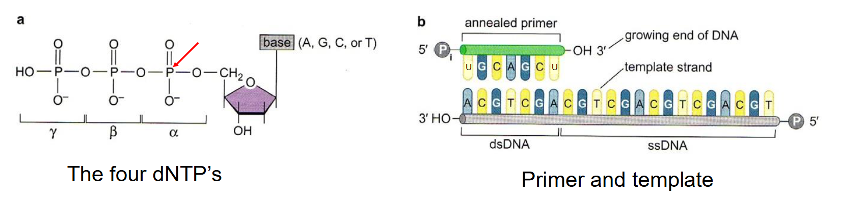
2. Four deoxyribonucleoside triphosphates (dNTPs): dATP, dTTP, dCTP, and dGTP, which serve as the building blocks for new DNA.
3. Single-stranded DNA template: Provides the sequence used to synthesize a complementary DNA strand.
4. RNA primer: Provides a free 3′-OH group that DNA polymerase requires to begin DNA synthesis.
In which direction is DNA synthesized, and how is the new strand related to its template?
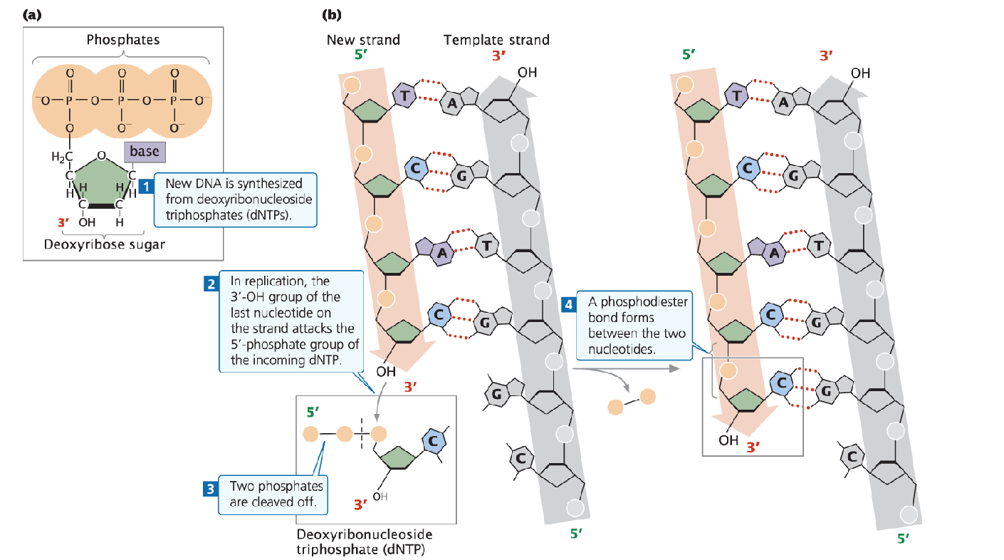
DNA is always synthesized in the 5′ → 3′ direction.
DNA polymerase adds new nucleotides to the free 3′-OH group of the growing strand.
The newly synthesized DNA strand is complementary to the parental template strand (A–T and G–C).
The new strand is also antiparallel to its template.
Complementary DNA strands are held together by hydrogen bonds between their nitrogenous bases.
What are the three main stages of DNA replication?
1. Initiation:
The DNA double helix is unwound and separated into single strands.
RNA primers are synthesized to provide starting points for DNA synthesis.
2. Elongation:
DNA polymerase synthesizes new complementary DNA strands using each parental strand as a template.
3. Termination:
DNA synthesis ends when replication is completed.

What is the function of DNA helicase during replication?
DNA helicase is an enzyme that unwinds the DNA double helix during initiation.
It separates the two parental DNA strands by disrupting the hydrogen bonds between complementary bases.
This exposes the bases on each strand, allowing them to serve as templates for DNA synthesis.
The region where the DNA strands separate is called the replication fork.

What is the function of topoisomerase during DNA replication?
Topoisomerase prevents excessive twisting and supercoiling of DNA ahead of the replication fork.
As helicase unwinds DNA, torsional stress builds up in the remaining double helix.
Topoisomerase relieves this stress by temporarily breaking and rejoining DNA strands.
This allows DNA unwinding and replication to continue.

What is the function of single-stranded binding proteins (SSBs) during DNA replication?
Single-stranded binding proteins (SSBs) bind to the separated DNA strands.
They stabilize the single-stranded DNA and prevent the strands from reannealing (base-pairing again).
This keeps the DNA templates accessible for replication.

What is primase, and why are RNA primers required for DNA replication?
Primase is an enzyme that synthesizes short RNA primers complementary to the DNA template.
DNA polymerase cannot initiate DNA synthesis independently.
The RNA primer provides a free 3′-OH group, allowing DNA polymerase to begin adding DNA nucleotides.
RNA primers are required to initiate synthesis on both the leading and lagging strands.

What is the difference between the leading and lagging strands during DNA replication?
Leading strand:
Synthesized continuously in the same direction as replication fork movement.
Requires one RNA primer to initiate synthesis.
Lagging strand:
Synthesized discontinuously in the opposite direction of replication fork movement.
Requires multiple RNA primers and produces short DNA segments called Okazaki fragments.
Both strands are synthesized in the 5′ → 3′ direction because DNA polymerase can only add nucleotides to the 3′ end.

Summary Image of Initation

How does DNA polymerase III function during the elongation stage of DNA replication?
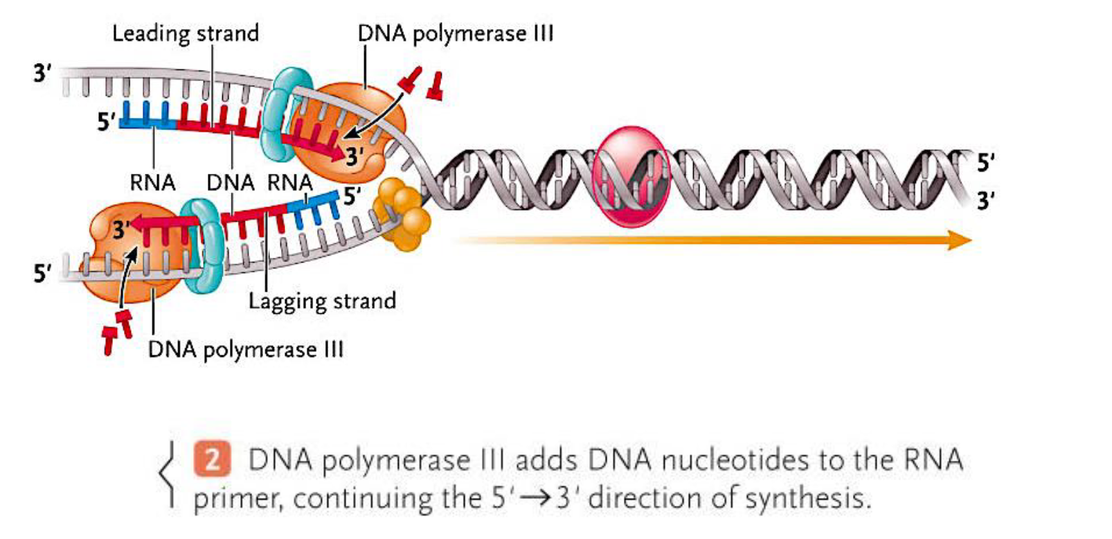
DNA polymerase III extends RNA primers by adding complementary DNA nucleotides.
It synthesizes new DNA exclusively in the 5′ → 3′ direction.
On the leading strand, DNA polymerase III synthesizes DNA continuously toward the replication fork.
On the lagging strand, it synthesizes DNA discontinuously, forming Okazaki fragments.
Each Okazaki fragment begins with a new RNA primer synthesized by primase.
What are Okazaki fragments, and how are they formed?
Okazaki fragments are short segments of DNA synthesized discontinuously on the lagging strand.
They form because DNA polymerase can only synthesize DNA in the 5′ → 3′ direction, while the lagging strand must be copied away from the replication fork.
Primase repeatedly synthesizes RNA primers.
DNA polymerase III extends each primer, producing an Okazaki fragment.
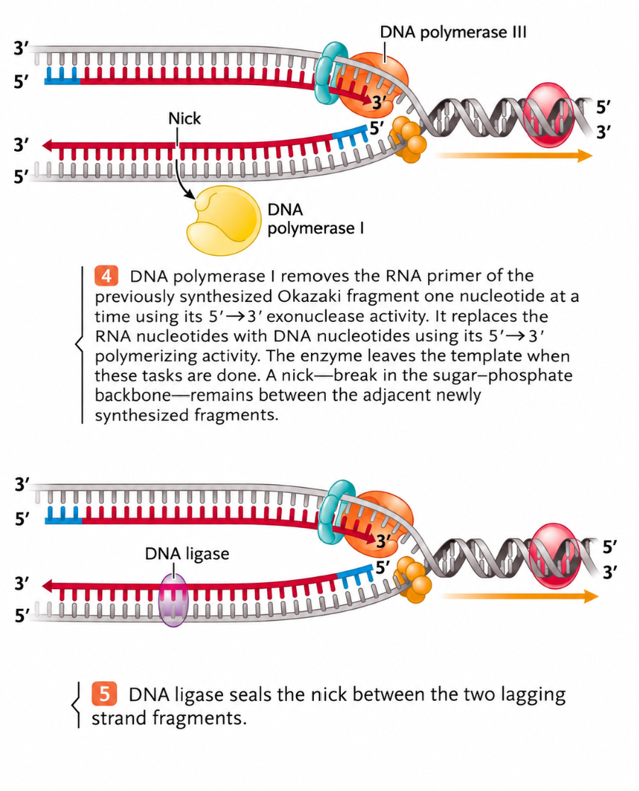
DNA polymerase I subsequently removes the RNA primers and replaces them with DNA.

What are the functions of DNA polymerase I and DNA ligase during DNA replication?
DNA polymerase I:
Removes the RNA primers used to initiate DNA synthesis.
Replaces the RNA nucleotides with DNA nucleotides in the 5′ → 3′ direction.
DNA ligase:
Seals the remaining nicks between adjacent DNA fragments.
Forms phosphodiester bonds in the sugar-phosphate backbone.
Joins Okazaki fragments into a continuous DNA strand.
How do the leading and lagging strands continue to be synthesized as the replication fork moves?
Helicase continues unwinding the parental DNA.
Leading strand: DNA polymerase III continuously adds nucleotides in the same direction as fork movement.
Lagging strand:
Primase repeatedly synthesizes new RNA primers.
DNA polymerase III extends each primer to form Okazaki fragments.
DNA polymerase I replaces RNA primers with DNA.
DNA ligase seals the remaining nicks.
Both strands are synthesized 5′ → 3′, producing complementary DNA strands.
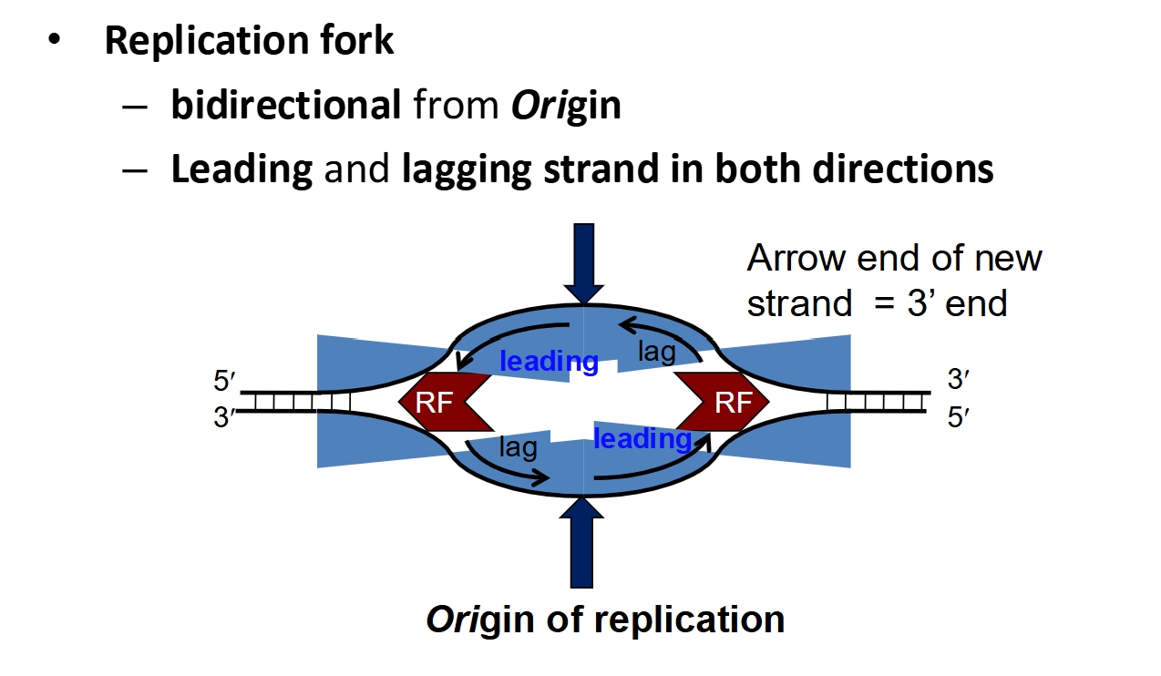
What is a replication fork, and how are leading and lagging strands arranged during bidirectional replication?
A replication fork is the Y-shaped region where DNA unwinds, exposing the parental strands for replication.
During bidirectional replication, two replication forks move in opposite directions away from the origin.
Each replication fork contains:
A leading strand, synthesized continuously in the same direction as fork movement.
A lagging strand, synthesized discontinuously in the opposite direction of fork movement.
Both strands are synthesized exclusively in the 5′ → 3′ direction.
Why are RNA primers required during DNA replication, and what does it mean for DNA polymerase to be processive?
RNA primers are short RNA sequences synthesized by primase, complementary to the DNA template.
They provide a free 3′-OH group, allowing DNA polymerase to begin DNA synthesis.
One primer is required to initiate the leading strand, while multiple primers are required for the lagging strand (one per Okazaki fragment).
DNA polymerase is processive, meaning it can add many nucleotides to a growing DNA strand each time it binds to DNA.
This contributes to rapid and efficient DNA replication.
What is the end-replication problem, and why does it occur in eukaryotic chromosomes?
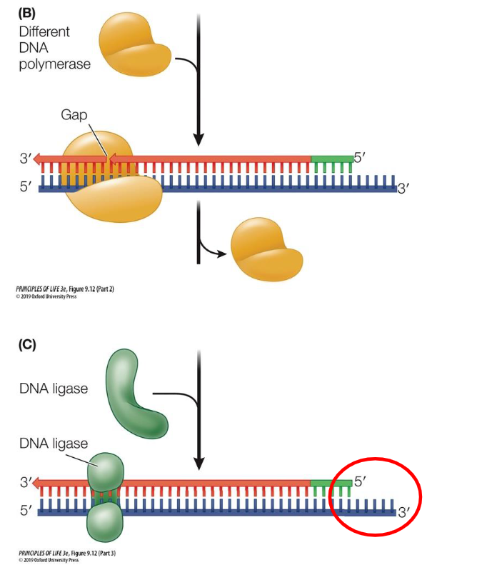
The end-replication problem occurs because DNA polymerase cannot completely replicate the ends of linear chromosomes.
During lagging-strand synthesis, DNA polymerase requires an RNA primer with a free 3′-OH group.
When the final RNA primer is removed, there is no available 3′-OH end to extend and replace it with DNA.
This leaves an unreplicated region at the chromosome end, resulting in a single-stranded DNA overhang.
Consequently, linear chromosomes can become progressively shorter after repeated cell divisions.
What are telomeres, and what are their functions?
Telomeres are repetitive, noncoding DNA sequences located at the ends of linear eukaryotic chromosomes.
In humans, they contain repeated TTAGGG sequences.
Telomeres function as protective caps that:
Protect protein-coding DNA from being lost as chromosomes shorten.
Provide extra DNA at chromosome ends, allowing some shortening without immediately affecting genes.
Prevent DNA repair mechanisms from mistakenly joining chromosome ends together.
Telomeres help maintain chromosome stability during cell division.

What is telomerase, and how does it maintain telomere length?
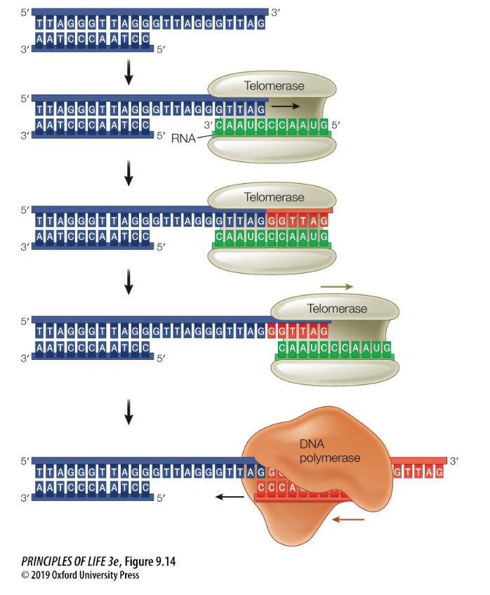
Telomerase is an enzyme that extends telomeres at the ends of eukaryotic chromosomes.
It contains its own RNA template, which base-pairs with the single-stranded telomeric DNA overhang.
Telomerase acts as an RNA-dependent DNA polymerase, synthesizing new DNA using its RNA template.
It adds repetitive telomeric DNA sequences to the 3′ end of the chromosome.
The extended DNA provides a template for additional lagging-strand synthesis, helping compensate for telomere shortening.
This helps maintain chromosome length and protects important genetic information.
Which cells express telomerase, and why is its activity important?
Telomerase is active in certain cells that require continued cell division, including:
Stem cells, such as bone marrow stem cells.
Many cancer cells.
Telomerase maintains telomere length, allowing these cells to undergo additional divisions without excessive telomere shortening.
According to your professor's slide, approximately 85% of cancer cells express telomerase.
Telomerase activity is tightly regulated because its activation can support the continued division of cancer cells.
How are telomeres and telomerase related to cellular aging and cancer?
Telomeres gradually shorten during repeated rounds of DNA replication.
When telomeres become critically short, cells may undergo senescence (stop dividing) or apoptosis (programmed cell death).
Telomere shortening is associated with cellular aging.
Telomerase maintains telomere length in certain continuously dividing cells, including stem cells.
Approximately 85% of cancer cells express telomerase, helping them maintain their telomeres and continue dividing.
Drugs that affect telomerase activity are being investigated for potential applications in cancer treatment and aging-related research.

How are telomere length, cellular aging, and cancer related?
Short telomeres are associated with cellular senescence (permanent cell-cycle arrest) and cell death.
Certain premature-aging disorders, including progeria and Werner syndrome, are associated with telomere dysfunction or shortening.
Some cancer cells have high telomerase activity, allowing them to maintain telomere length and supporting continued cell division.

What is oriC, and how does DnaA initiate DNA replication in E. coli?
oriC is the origin of replication in E. coli, approximately 245 base pairs long.
It contains:
An AT-rich region, which is relatively easy to unwind.
Four DnaA binding sites.
DnaA proteins are initiator proteins that bind to oriC.
DnaA binding causes a short region of DNA to unwind, initiating DNA replication.

How is DNA helicase recruited during the initiation of prokaryotic DNA replication?
DnaA initiator proteins bind to oriC and cause a short region of DNA to unwind.
DNA helicase is then loaded onto the exposed single-stranded DNA.
Helicase disrupts hydrogen bonds between complementary bases, allowing the DNA double helix to unwind further.
This creates replication forks where DNA synthesis can begin.

What are the functions of DNA helicase, single-strand binding proteins, and DNA gyrase during prokaryotic DNA replication?
DNA helicase:
Travels along the lagging-strand template in the 5′ → 3′ direction.
Unwinds DNA by disrupting hydrogen bonds between complementary base pairs.
Single-strand binding proteins (SSBs):
Bind to exposed single-stranded DNA.
Prevent reannealing and the formation of secondary structures, such as hairpins.
DNA gyrase:
A bacterial topoisomerase that relieves torsional stress ahead of the replication fork.
Temporarily breaks both strands of DNA and rejoins them, preventing excessive twisting caused by helicase activity.
Useful Image

What is the function of primase during prokaryotic DNA replication?
Primase is an RNA polymerase that synthesizes short RNA primers (10–12 nucleotides) complementary to the DNA template.
These primers provide a free 3′-OH group, allowing DNA polymerase to begin synthesizing DNA.
Primase associates with DNA helicase to initiate primer synthesis.
The leading strand requires one initial primer, while the lagging strand requires a new primer for each Okazaki fragment.
Unlike DNA polymerase, RNA polymerase does not require an existing primer to initiate synthesis.

What are the three main enzymatic activities associated with DNA polymerases?
5′ → 3′ polymerase activity: Adds DNA nucleotides to the 3′ end of a growing strand, synthesizing new DNA.
3′ → 5′ exonuclease activity: Removes incorrectly incorporated nucleotides from the 3′ end, allowing proofreading.
5′ → 3′ exonuclease activity: Removes nucleotides from the 5′ end, allowing RNA primer removal.
What are the functions and enzymatic activities of DNA polymerase I and DNA polymerase III in E. coli? (“CHECK TO SEE IF FULL TABLE IS NEEDED)
DNA polymerase III:
The main DNA polymerase responsible for synthesizing new DNA.
Has 5′ → 3′ polymerase activity to extend DNA strands.
Has 3′ → 5′ exonuclease activity for proofreading.
DNA polymerase I:
Removes RNA primers and replaces them with DNA.
Has 5′ → 3′ polymerase activity for DNA synthesis.
Has 3′ → 5′ exonuclease activity for proofreading.
Also has 5′ → 3′ exonuclease activity to remove RNA primers.
How are RNA primers removed and Okazaki fragments joined during prokaryotic DNA replication?
When DNA polymerase III reaches the RNA primer of a previously synthesized Okazaki fragment, it is replaced by DNA polymerase I.
DNA polymerase I uses its 5′ → 3′ exonuclease activity to remove the RNA primer.
It then uses its 5′ → 3′ polymerase activity to replace the removed RNA with DNA.
A nick remains in the sugar-phosphate backbone between adjacent fragments.
DNA ligase seals the nick by forming a phosphodiester bond between the 3′-OH and 5′-phosphate groups.
This joins the Okazaki fragments into one continuous DNA strand.

Summary Table
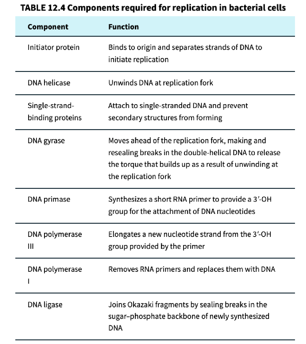
How is DNA replication initiated and regulated in eukaryotic cells?
Replication begins at autonomously replicating sequences (ARS), which are generally AT-rich regions.
Eukaryotic chromosomes contain multiple origins of replication, with replication proceeding bidirectionally from each origin.
Initiation involves the MCM helicase complex, which is loaded onto double-stranded DNA.
DNA replication occurs during the S phase of the cell cycle.
During G1 phase, replication licensing factors prepare the origins for replication through origin licensing.
Origin licensing helps ensure that the entire genome is replicated once and only once per cell cycle.
What are the functions of DNA polymerases α, ε, and δ during eukaryotic DNA replication?
Eukaryotic cells contain approximately 15 different DNA polymerases, many of which function in DNA repair.
DNA polymerase α (alpha):
Associated with primase activity, initiating DNA synthesis by generating RNA primers.
DNA polymerase ε (epsilon):
Primarily responsible for leading-strand DNA replication.
DNA polymerase δ (delta):
Primarily responsible for lagging-strand DNA replication.
What are the key differences between prokaryotic and eukaryotic DNA replication?
Prokaryotes:
Typically replicate from a single origin.
Helicase is loaded onto single-stranded DNA.
DNA polymerase III performs most DNA synthesis.
Have relatively longer RNA primers and Okazaki fragments.
Eukaryotes:
Replicate from multiple origins along each chromosome.
The MCM helicase complex is loaded onto double-stranded DNA.
DNA replication is restricted to S phase.
Use Pol α, Pol ε, and Pol δ for initiation and DNA synthesis.
Have shorter RNA primers and Okazaki fragments.
Must also coordinate DNA replication with nucleosome disassembly and reassembly.
Telomeres shorten during repeated rounds of eukaryotic DNA replication.
What is the nucleosome problem during eukaryotic DNA replication, and how is it resolved? (revist)
Eukaryotic DNA is packaged into nucleosomes, consisting of DNA wrapped around histone proteins.
During DNA replication, nucleosomes must be disassembled or displaced to allow the replication machinery to access DNA.
After replication, nucleosomes must be reassembled on the newly synthesized DNA.
Histone proteins and their modifications must also be maintained or restored because they influence DNA packaging, stability, and accessibility.
Chaperone proteins help transfer and organize histones and their modifications during replication.
Chromatin-assembly factors facilitate nucleosome reassembly.
Newly assembled nucleosomes contain a mixture of old and new histones.
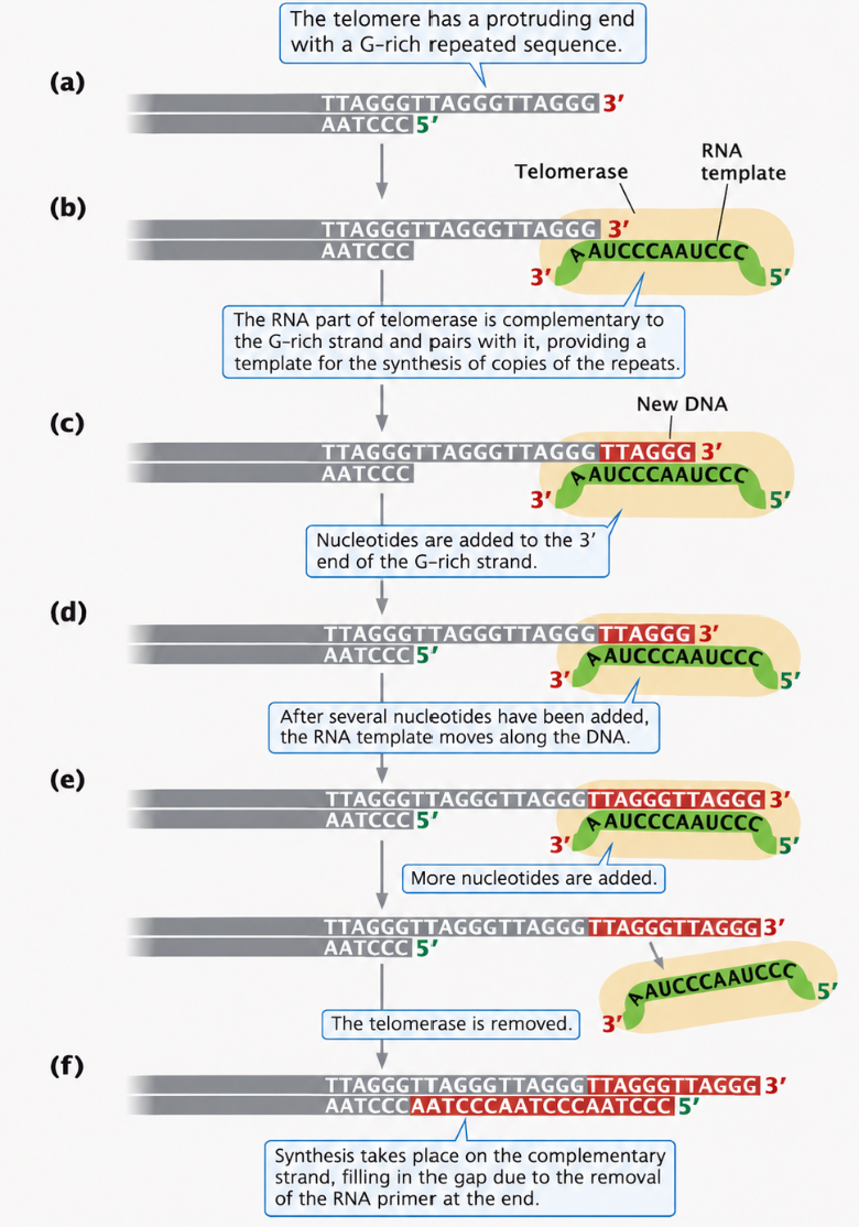
How does telomerase extend the ends of eukaryotic chromosomes?
Telomerase binds to the single-stranded 3′ overhang at the end of a chromosome.
It contains its own RNA template, which base-pairs with the telomeric DNA.
Telomerase acts as an RNA-dependent DNA polymerase, adding repetitive DNA sequences to the chromosome's 3′ end.
It can reposition and add additional telomeric repeats.
The extended DNA serves as a template for complementary lagging-strand synthesis.
Although a short gap or overhang remains, telomerase extension helps prevent overall chromosome shortening.
How does telomerase activity differ between various types of eukaryotic cells?
Highly dividing cells, including certain stem cells and germ cells, can express telomerase to maintain their telomeres.
In many somatic cells, telomerase activity is low or absent, allowing telomeres to shorten over repeated cell divisions.
Telomere shortening is associated with cellular aging and senescence.
Many cancer cells express telomerase, helping maintain telomere length and supporting continued cell division.
Summary Table
