Immunology Module 2
 Call Kai
 Learn
 Practice Test
 Spaced Repetition
 Match
 Flashcards
 Knowt Play
1/102

There's no tags or description
Looks like no tags are added yet.
103 Terms
Functions of B Cells
Antigen Presentation (phagocytosis)
Autoantibody production
Cytokine Production
Stages of B cell Development
HSC
Pro-B
Pre-B
Immature B
Mature B
Activated B
Blast B
Plasma B
What are B cell developements outlined by?
cell surface markers (CD)
Transcription factor expression
immunoglobulin DNA arrangements
Role of Stromal Cells
cells in the bone marrow that provide support, cytokines, growth factors to instruct HSC to differentiate into B cells
ex. CXC112 and IL7
Antibody/Immunoglobulin structure
2 heavy chains and 2 light chains
held together by intra and inter-sulfide binds
Variable and constant regions
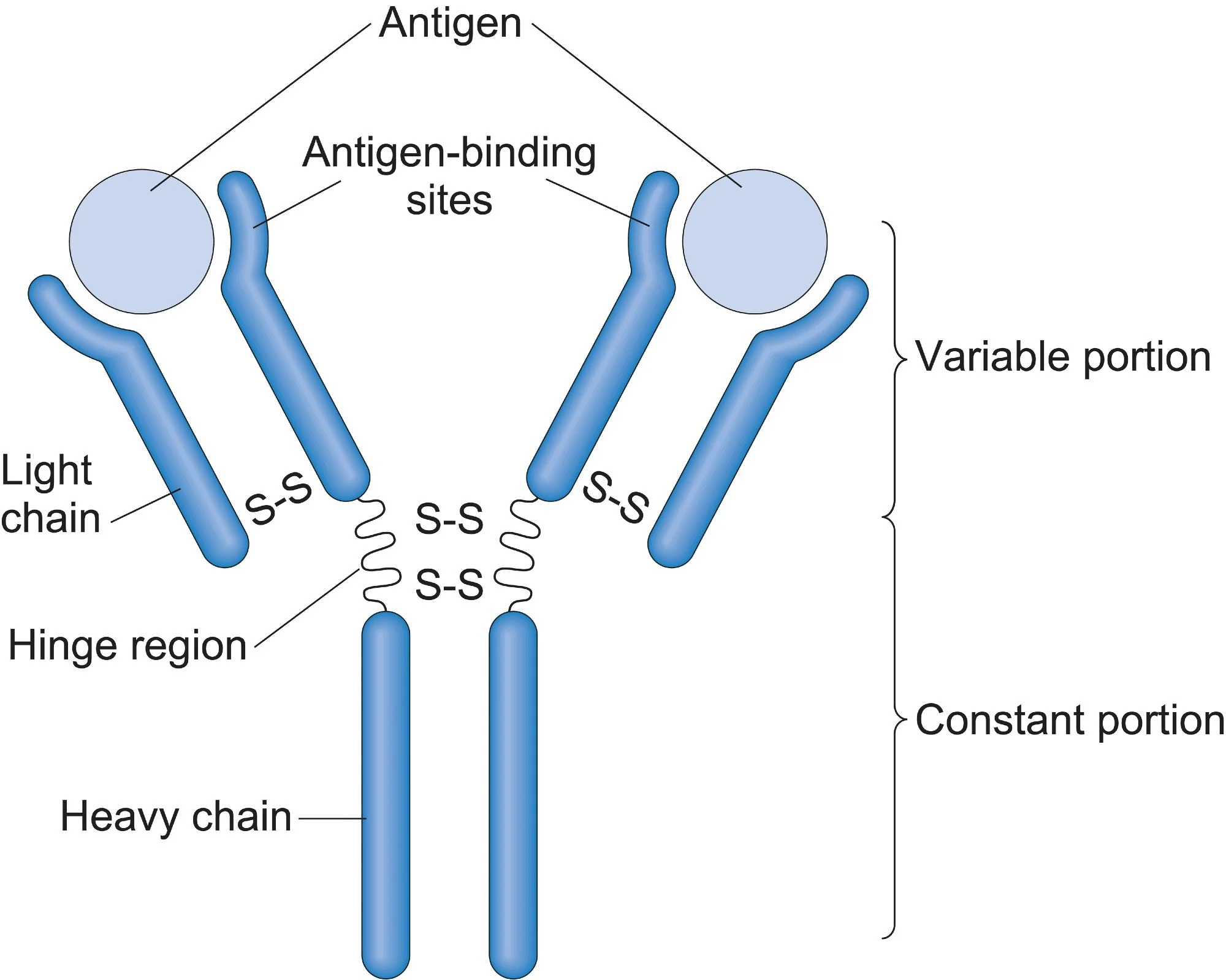
Variable Region
Region near antigen binding site; the first 110 amino acids are highly variable
Constant Region
Everywhere except the first 110 amino acids; highly conserved
Germ line theory
States that one gene codes for one anitbody
later disproven, too many antibodies (10^11) compared to genes
Somatic Recombination Theory
B cells have limited germline antibody genes, but mutational process in B cells alters genes
Tonegawa Experiment
collected DNA from B cells and Liver cells
Cut DNA with BamHI
labeled light chain with radioactive nucleic acid proteases to track DNA (this tags what codes for the BCR)
measure migration/size of DNA
Found that B cell dna cut and recombines, the pieces are smaller than liver cells
Early Pro B cells
DJ recombination of the heavy chain occurs
starts VDJ of heavy chain recombination
self-renewal property is eliminated
K first rule
kappa chain is the first to be used in a BCR, if it fails, the lamda light chain is used
Two light chains increases diversity
ProB/PreB
VDJ heavy chain recombination is completed
B cells undergo clonal expansion and proliferation
VJ recombination of the light chain
Immature B cell
expresses IgM
Negative selection for cells that bind to self
Cells undergo deletion, receptor editing, anergy
undergoes tolerance
CXC112
Pre-pro B cells binds to CXCL12
Homing factor, keeps Pre/pro B cells in the bone marrow
IL7
B cell stromal factor
Pro survival signal
activates VDJ recombination
enables pro-B cell proliferation (allows clonal expansion)
What happens during somatic recombination mechanism?
B cell shuffles gene segments and permantly removes DNA
C region gets smaller
Epitope:
region of antiGEN that antibody binds to
E -pitope binds to antig-E-n
Paratope
antigen binding site, where the antigen binds to on a antibody
VDJ recombination of Heavy chain
Recombination signal sequences (RSS): DNA sequences that flank each Antibody gene segment
Gene segments are joined by Rag 1/2 recombinases (recombination activation gene)
Hydroxyl (OH-) at the nicked end and phosphate group forms a hairpin
Signal end joining ligates end
Ku70/80 recruited as scaffold for the enzymes
Artemis opens the hairpin
happens asystemically, increases variance
P nucleotides added complementary overhands
exonucleus trims overhand
terminal deoxynucleotide transferase (TDT) adds nucleotides
ligase attaches DNA segments
VJ Recombination (Light chain specific)
Recombination signal sequences (RSS): DNA sequences that flank each Antibody gene segment
RSS contains a heptomer, spacer (either 12 or 23 bp) and a nonamer
Gene segments are joined by Rag 1/2 recombinases (recombination activation gene)
Hydroxyl (OH-) at the nicked end and phosphate group forms a hairpin
Signal end joining ligates end
Ku70/80 recruited as scaffold for the enzymes
Artemis opens the hairpin
happens asystemically, increases variance
P nucleotides added complementary overhands
ligase attaches DNA segments together
Junctional Diversity
variation of the number of nucleotides and how they come together
Artemis variable hairpin opening
exonuclease trimming
Tdt adds p-nucleotides randomly
Combinatorial Diversity
diversity generated via ability to combine various genes
multiple gene segment for antibodies
different light and heavy chain combinations
Parts of RSS
heptomer, spacer (either 12 or 23 bp) and a nonamer
Central tolerance
happens during immature B stage in the bone marrow
self antigen in presented to immature B cells by other cells in bone marrow (especially stromal cells)
If no binding occurs, immature B proceeded to secondary lymphoid organs (most go to spleen)
If binding occurs, the cell undergoes clonal deletion, anergy, or receptor editing
Clonal deletion
cell death via apoptosis that occurs if BCR binds to self antigen during central tolerance
Anergy
permanent quiescence/unresponsiveness, cell cannot activate
Receptor editing
last ditch effort to save BCR if B cell binds to self antigen in central tolerance, genes are rearranged in the light chain using RAG 1 & 2 or switch to lambda chain if RAG 1 & 2 don’t work
Nemenzee Experiment
looks to see if receptor editing occurs
used transgenic mice with BCR against Kk antigen:
Group 1 had stromal cells that expressed Kd antigen, BCR didn’t bind and B cells in the periphery had a BCR specific to Kk
Group 2 had stromal cells that expressed Kd and Kk antigen, BCR did bind to stromal cells. Still, a small number of B cells found in periphery with a modified light chain that didn’t recognize kk
Transition Phase
B cells enter the white pulp of the spleen
75% of B cells that passed central tolerance become transitional B cells; other 25% go to different secondary lymphoid organs or stay in the bone marrow
Order of transition phase
Immature B to T1 to T2
Differences between T1 and T2
T2 had higher IgD, less IgM, and increased BCR surface expression than T1
Peripheral tolerance categories
split into T1 and T2 tolerance
T1 Selection
removes 55-75% of cells
Negative selection: B cells that bind to self antigen are removed
occurs in the periateriolar sheath of the spleen
T2 Selection
Positive selection: low level signaling indicates survival
Indicative via tonic signaling and the upregulation of BAFF receptor
Tonic Signaling
low levels of BCR signaling in absence of an antibody binding; BCR stimulates cell via intracellular signaling
Causes a higher concentration of DAG (diacylglycerol) in the inner leaflet of the plasma membrane of T2 cells
BAFF
BAFF receptor on B cells binds to BAFF; a pro survival signal
BAFF binding to BAFFR increases from T1 to T2
BAFF binding to BAFFR causes a decrease in DAG in inner leaflet of T1 cells
Effect of DAG on survival
low DAG leads to death; high DAG leads to survival
DAG concnetrations in T1 and T2 lead to phenotypic changes
Plasma Cells
immunoglobulin producing machines
have many ribosomes on rough ER
includes long-lived plasma cells/memory plasma cells that live in the bone marrow
B2 Cells/Mature follical cells
95% of mature B cells
highly migratory across all secondary lymphoid organs
high V region diversity due to lots of somatic recombination
high memory
T cell activation required
B1 Cells
5% of mature B cells
found mainly in the peritoneal and perivascular cavities
low V region diversity
moderate memory
independent of T cells
produces natural IgM, cytokines, and clears apoptic cells with IgM
Marginal Zone Cells
<1% of mature B cells
found in teh marginal zone of the spleen
low V region diversity
memory is unknown (newer cell discovered)
can be both T cell independent or dependent
responds to blood borne antigens and produce IgM
T cell dependent activation in B2 Cells steps
antigen binds to BCR. (3 steps)
T cells form an immunological synapse, activates B cell
Steps of BCR on B cell binding to antigen
follicular dendritic cells can present antien and secrete BAFF or B cells detect free floating antigen
antigen binding causes membrane changes, and the antigen in phagocytosed
process pieces of the antigen are displayed on MHC class II
Steps of immunological synapse with T cell
TCR binds to MHC class II with the amtigen togehter
CD4+ binds to only MHC class II (no antigen)
CD40 on a B cell binds to CD40L on a T cell
T cell releases cytokines to activate B cell
T Cell independent activation in B1 and Marginal Zone cells
Includes TI-1 and TI-2 (thymus independent antigens); TI1= extra activation signaling; TI-2 = extensive activation signaling
TI-1 Antigen
celled extra activation signaling
antigen binds to B cell receptor and PRR (commonly TLR4 or LPS)
TI-2 Antigen
called extensive activation signaling
antigen cross liks and binds with a large number of B cell receptors
What does activation of a B cell cause
clonal expansion, somatic hypermutation, afinity maturation, and isotype switching
Clonal Expansion
massive proliferation of B cell, occurs in dark zone of terminal center
Somatic hypermutation
AID (activation induced cytidine deaminase) causes point mutations in V regions of the BCR to form QUasi Clonal B cells
Affinity Maturation
Quasi clones enter the blood stream, process to the light zone of the germinal celnter
the BCR that binds best has increased survival and becomes a plasma B cell
BCR with weak or no binding die via apoptosis
Isotype switching
in plasma cells, the Fc portion changes to accomodate antibody function
Which Ig is secreted first from Plasma B cell
IgM is secreted first
Long Lived Plasma Cells
secrete antibodies for long period of time (up to a life time)
reside in the bone marrow
Memory B Cells
respond to previously encountered antigen
upon reexposure, the rapidly reactivate (reach peak antibody production in 5 days and produce 100 times more antibodies than the first response)
survive a lifetime due to anti-apoptosis gene
found in germinal centers, lymphoid tissue, blood, lymph, and MALTs
How can antibodies lead to pathogen clearance?
agglutination, neutralization, opsonization, complement activation (CDC), antibody dependent cell mediated cytotoxicity (ADCC)
Agglutination
clumps/holds the antigens together, prevents it from traveling and spreading
Neutralization
blocks attachment of pathogen to the cell (binds to the pathogen, which blocks it from binding to cells = reduced virulence)
Opsonization
labels antigens to be phagocytosed
Complement dependent cytotoxicity (CDC)
leads to MAC complex and cell lysis
Antibody dependent cell mediated cytotoxicity (ADCC)
causes antigen to be destroyed by macrophages, NK cells, and eosinophils
IgM
pentamer
secreted first, present at high levels at initial immune response
high avidity and low affinity
is both membrane bound and secreted
cannot cross placenta
sometimes coexpressed with IgD

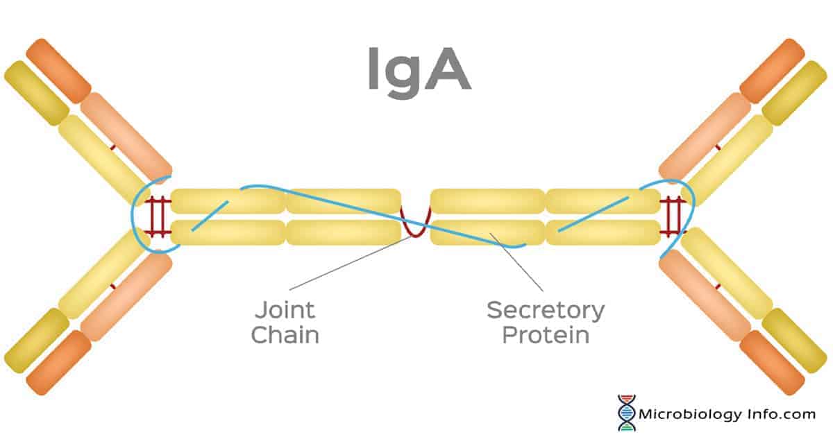
IgA
dimer, Fc regions are unexposed
found in mucosa and fluids
many people lack IgA
protease resistant
long half life
IgG
monamer
most common Ig (70-80% of Ig)
has several subclasses (G1 and G2)
can cross blood vessels and placenta
leads to opsonization, ADCC, CDC
CD16 on NK cells binds to he Fc Portion on antibodies

IgE
monamer
associated with mainly mast cells but also eosinophils and basophils
involved in allergy and helminth production and worm infections
Causes ADCC
leads to the release of chemokines, cytokines, reactive oxygen species, antimicrobial peptides, histamine
leads to smooth muscle contraction during anaphalaxis
IgD
monamer
coexpressed with IgM when B cell matures and leaves the bone marrow
Important during B cell activation in T2 Stage
Why are blood cancers mostly B cell
60-70% of blood cancers are of the B cell
this is because of VDJ recombination and diversity —> DNA breaks and mutations cause some to be cencerous
Multiple myeloma
cancer of a plasma cell
leads to uncontrolled rgowth of cells and secretion of antibodies
B cell lymphomas
cencer in the lymphnodes or spleen
includes hodgkins (more specific and less common; predictable spread behavior) and non-hodgkins (more common and less specific; unpredictable)
Isotype Switching
naive mature B cells start off with IgM and IgD on their surface
if B cell becomes acctivated by T cell and cytokines anre present, B cell will isotype switch and secretes IgD, IgE, IgA, or IgG
cytokines and environment determines which isotype the plasma cell will secrete
Autoimmune Encephalitis Pathology method
B cell response against the N-Methyl D aspartate receptor (NMDAR)
Encephylitis
inflammation in the brain
NMDAR
major receptor in the brain that is important in synaptic plasticity, learning, and memory
NMDA
binds to NMDAR, mimics glutamate (neuroexcitory)
In vitro
in cells
in vivo
in animal
ex vivo
extracted from animal
Anti-NMDAR Encephalitis
antibody against GluN1 subunit of NMDAR that leads to psychiatric and neurological symptoms
antibodies are found in cerebral spinal fluid
Epidemiology of Anti NMDAR encephalitis
4 times more likely in females, and more likely in women with teratomas
clinical triggers: ovarian teratomas and herpes simpler encephalitis
Vaccine used in the Primary Literature Paper parts
GluN1(359-378) peptide induces autoantibody production
complete Freund’s Adjuvant (CFA): an emulsion containing M. Tuberculosis, which enhances immune response and slowly releases antigens over time
Pertussis Toxin: disrupts the blood brain barrier to allow immune cells and anti GluN1 antibodies to impact central nervous system
Flow Cytometry
used to determine if a specific cell type expresses a marker of interest on its surface or determine the quantity of cells in a sample that express a specific marker
Dot Blot
Used to determine if a protein is present in a sample and its relative quantity, but cannot determine the size of the protein
Histological Immunofluorescence
used to determine if a target protein/marker is present in a sample, determine the location of the target (also target movement in live samples), and relative quantity
Split into direct and indirect
direct: primary antibody with tag binds to antigen
indirect: secondary antibody binds to the Fc region of an antibody binded to an antigen
Monoclonal antibody depletion
used to selectively eliminate specific cell types to find out the role of the eliminated cells
Antibodies do this by causing cell death through the CDC or ADCC pathway
Neuron Ca2+ assay
used to quantify ion channel and neuron signaling activity
does this by measuring intracellular Ca2+ using Fura-2-AM and video microscopy
Elevated O maze
Behavioral test to determine/identify anxiety
staying in closed regions means greater anxiety, while exploring open regions indicates less anxiety
Forced Swim Test
greater immobility duration indicates depressive-like despair
Y Maze
Exploring new areas indicates better spatial memory, while exploring the original arm indicates lower spatial memory
Actimitry:
measures horizontal and vertical motor activity
Immunization using GluN1(359-378) peptide key findings
vaccine blocks NMDA-dependent Ca2+
depletion of B cells using monoclonal antibody prevents memory loss and behavior despair, which is presented as a poteintial therapy
pathology is caused by B cell trafficking through meninges and ventricles through cerebral spinal fluid and more plasma CD138+ cells without T cell recruitment
Bepi prep linear epitope prediction
predicts surface accessibility, hydrophilicity, and amino acid propensity to guess which part of a protein an antibody/BCR will bind to on an antigen
Flair MRI
darkens cerebral spinal fluid, used to see lesions in the brain
Evans Blue
measure sthe blood brain barrier permeability, the bluer the brain the more permeable