AE 1.7 - Tricuspid valve/TS/TR, Pulmonic valve/PS/PI, Endocarditis

0.0(0)
Studied by 11 people
call kaiCall Kai
learnLearn
examPractice Test
spaced repetitionSpaced Repetition
heart puzzleMatch
flashcardsFlashcards
GameKnowt Play
Card Sorting

1/34

encourage image

There's no tags or description

Looks like no tags are added yet.

35 Terms

1
New cards

Normal tricuspid valve anatomy

  • 3 leaflets

    • 1. Anterior

      • longest, largest and greatest motion

    • 2. Posterior

      • usually smallest, multiple scallops

    • 3. Septal

      • least mobile

  • open during diastole

  • biggest valve (6-7cm2)

  • separates RA from RV


<ul><li><p>3 leaflets</p><ul><li><p><span style="background-color: transparent;">1. Anterior</span></p><ul><li><p><span style="background-color: transparent;">longest, largest and greatest motion</span></p></li></ul></li><li><p><span style="background-color: transparent;">2. Posterior</span></p><ul><li><p><span style="background-color: transparent;">usually smallest, multiple scallops</span></p></li></ul></li><li><p><span style="background-color: transparent;">3. Septal</span></p><ul><li><p><span style="background-color: transparent;">least mobile</span></p></li></ul></li></ul></li><li><p>open during diastole</p></li><li><p>biggest valve (6-7cm<sup>2</sup>)</p></li><li><p>separates RA from RV</p></li></ul><p></p>
2
New cards

Tricuspid annulus characteristics

  • little fibrous support

    • very different from mitral annulus

    • very pre/afterload dependent

  • saddle-shaped, but flattens as it dilates

  • dilates in the unsupported lateral and posterior directions (very flexible) 

  • large valve orifice

  • sits lower in the heart than MV (closer to apex)


<ul><li><p><span style="background-color: transparent;">little fibrous support</span></p><ul><li><p><span style="background-color: transparent;">very different from mitral annulus</span></p></li><li><p><span style="background-color: transparent;">very pre/afterload dependent</span></p></li></ul></li><li><p><span style="background-color: transparent;">saddle-shaped, but flattens as it dilates</span></p></li><li><p><span style="background-color: transparent;">dilates in the unsupported lateral and posterior directions (very flexible)&nbsp;</span></p></li><li><p><span style="background-color: transparent;">large valve orifice</span></p></li><li><p><span style="background-color: transparent;"><u>sits lower in the heart than MV</u> (closer to apex)</span></p></li></ul><p></p>
3
New cards

Standard echocardiographic views used to evaluate the tricuspid valve

  • PLAX RVIT

  • PSAX

  • A4C RV focus

  • Apical reverse 4C

  • SUBX 4C


<ul><li><p>PLAX RVIT</p></li><li><p>PSAX</p></li><li><p>A4C RV focus</p></li><li><p>Apical reverse 4C </p></li><li><p>SUBX 4C</p></li></ul><p></p>
4
New cards

Primary Tricuspid Regurgitation

Primary

  • Rheumatic

  • Carcinoid

  • Traumatic (acute) TV rupture - due to its anterior location

  • TV prolapse – isolated is rare, usually found with MVP

    • TVP best diagnosed from the PLAX view at the level of the aorta

  • Iatrogenic (caused by accident)

  • device leads

  • endocarditis


Ebstein’s Anomaly

  • Rare congenital malformation

  • Failure of the developing RV leaflets to split from the inlet portion of the ventricle

  • Exaggerated apical displacement of septal leaflet

  • Small functional RV

  • May be associated with ASD/PFO

  • displacement of leaflet is > 8mm


5
New cards

Functional Tricuspid Regurgitation

Functional

  • secondary (annular dilation)

  • Left heart disease

    • LV dysfunction

  • RV dysfunction

    • RV ischemia/volume overload/cardiomyopathy

    • Dilation

  • Pulmonary hypertension

    • Chronic lung disease

    • Pulmonary thromboembolism, L to R shunt

  • Right atrial abnormalities

    • Afib


6
New cards

Common causes of Tricuspid Regurgitation

most common = annular dilation (secondary)

  • structurally normal TV leaflets

  • results in incomplete coaptation of the TV


<p>most common = annular dilation (secondary) </p><ul><li><p>structurally normal TV leaflets</p></li><li><p>results in incomplete coaptation of the TV</p></li></ul><p></p>
7
New cards

Causes of Tricuspid Stenosis

Congenital: 

  • Rare, usually associated with other congenital cardiac defects

  • Ex: maldeveloped leaflets, shortened or malformed chords, annular hypoplasia, papillary muscle abnormalities


Acquired:

  • Rheumatic

    • most common cause of TS

    • TS almost never isolated, occurs with MS/AS

    • Features resemble MS (commissures fused)

  • Carcinoid - valve looks stuck open

    • Caused by rare, malignant neuroendocrine tumor

    • Secretes excessive amounts of serotonin which damages TV and PV

    • Plaque-like deposits on leaflets cause the valve to become thickened, retracted and rigid

    • Creates stenosis and regurgitation

      • most common valvular issue is TR

  • Other:

    • Cor triatriatum dexter

    • Large masses (TV vegetations, right heart thrombus or tumors)


8
New cards

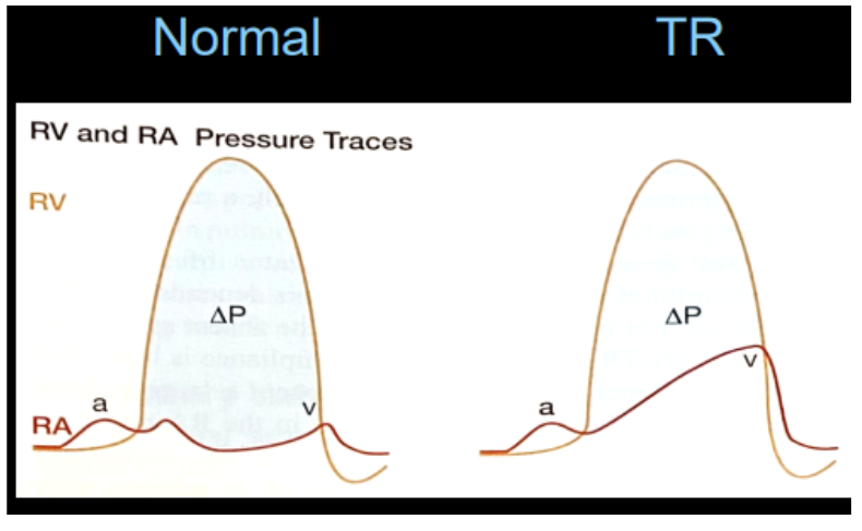
Hemodynamic consequences of Tricuspid Regurgitation

  • backward flow of blood from RV → RA

  • volume backup into the venous system

    • flow reversal in hepatic veins & IVC

  • volume overload into RV (regular + regurgitant blood volume)

  • RV pressure increases


<ul><li><p>backward flow of blood from <span style="background-color: transparent;">RV → RA</span></p></li><li><p>volume backup into the venous system </p><ul><li><p>flow reversal in hepatic veins &amp; IVC</p></li></ul></li><li><p>volume overload into RV (<em>regular + regurgitant blood volume</em>) </p></li><li><p>RV pressure increases </p></li></ul><p></p>
9
New cards

Hemodynamic consequences of Tricuspid Stenosis

  • obstructed blood flow from RA → RV

  • increased pressure in RA

  • systemic venous congestion

  • jugular vein distension

  • ascites (fluid buildup)

  • peripheral edema


<ul><li><p>obstructed blood flow from <span style="background-color: transparent;">RA → RV</span></p></li><li><p><span style="background-color: transparent;">increased pressure in RA </span></p></li><li><p><span style="background-color: transparent;">systemic venous congestion </span></p></li><li><p><span style="background-color: transparent;">jugular vein distension </span></p></li><li><p><span style="background-color: transparent;">ascites (fluid buildup) </span></p></li><li><p><span style="background-color: transparent;">peripheral edema </span></p></li></ul><p></p>
10
New cards

Expected chamber remodeling with pressure overload vs. volume overload

RV volume overload

  • flattening of septum during diastole


RV pressure overload

  • flattening of septum during systole


<p>RV <strong>volume </strong>overload </p><ul><li><p>flattening of septum during <strong>diastole </strong></p></li></ul><p></p><p>RV <strong>pressure </strong>overload </p><ul><li><p>flattening of septum during <strong>systole </strong></p></li></ul><p></p>
11
New cards

Clinical findings associated with right-sided valve disease

  • systemic venous congestion

  • ascites

  • peripheral edema

  • enlarged, tender liver (congestion backup) 

  • jugular vein distension

  • RA dilation

  • RV dilation with LV septal shift

  • Dilation of inferior vena cava

  • heart murmur that increases with inspiration


12
New cards

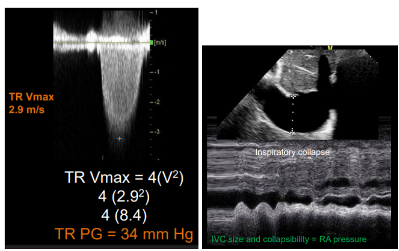
How Tricuspid Regurgitation is used to estimate right ventricular systolic pressure (RVSP)

RVSP = Tricuspid Regurgitation Pressure Gradient + RA pressure
<p>RVSP = Tricuspid Regurgitation Pressure Gradient + RA pressure</p>
13
New cards

Components needed to calculate RVSP

RVSP = Tricuspid Regurgitation Pressure Gradient + RA pressure


RAP = IVC size & collapsibility

<p><span style="background-color: transparent;"><strong>RVSP = Tricuspid Regurgitation Pressure Gradient + RA pressure</strong></span></p><p></p><p>RAP = IVC size &amp; collapsibility </p>
14
New cards

Basic interpretation of calculated pulmonary pressures

normal is less than 35

severe is greater than 60

<p>normal is less than 35 </p><p>severe is greater than 60</p>
15
New cards

Clinical situations that may affect Doppler interrogation & alternate imaging windows (Pulmonary Hypertension & RVSP)

  • The peak TR velocity does not reflect the severity of TR

    • It reflects the pressure difference between the RV and RA during systole

    • Ex. there can be mild TR and very high TR velocities or severe TR with very low TR velocities

  • In Severe TR cases, this cannot accurately be estimated by using the TR velocity technique


  • PLAX RVOT

  • PSAX

  • high PSAX

  • SUBX short axis

  • anterior tilt A5C

  • PSAX PV


16
New cards

Normal pulmonic valve anatomy

  • 3 components

    • annulus, cusps, commissures

  • 3 cusps

    • anterior, left, right

    • not supported by IVS

  • separates RVOT from PA

  • most anterior & superior valve

  • separated from the TV by a muscular fold


<ul><li><p>3 components </p><ul><li><p>annulus, cusps, commissures </p></li></ul></li><li><p>3 cusps </p><ul><li><p>anterior, left, right </p></li><li><p>not supported by IVS</p></li></ul></li><li><p>separates RVOT from PA </p></li><li><p>most anterior &amp; superior valve </p></li><li><p>separated from the TV by a muscular fold</p></li></ul><p></p>
17
New cards

Congenital causes of Pulmonic Stenosis

  • most common cause

  • valvular

    • 1) Typical dome-shaped PV

      • Most common type of congenital PS

      • Preserved valve mobility, narrowed central opening, raphes

    • 2) Dysplastic (abnormally developed) PV

      • Trileaflet with deformed and thickened leaflets, poor mobility

      • Frequently associated with Noonan syndrome

    • 3) Unicuspid/Bicuspid PV

      • Fusion between commissures of leaflets

      • Bicuspid are frequently found with tetralogy of Fallot

  • subvalvular (infundibular)

    • Often associated with Tetralogy of Fallot

    • May be associated with other complex congenital heart lesions

  • supravalvular

    • Can be isolated or in association with tetralogy of Fallot or other syndromes such as Noonan’s

    • May result secondary to residual scarring of the MPA following removal of a PA band

  • branch PA Stenosis

    • May be diffuse or single lesion

    • Often associated with other syndromes


<ul><li><p><strong>most common cause </strong></p></li><li><p>valvular </p><ul><li><p><span style="background-color: transparent;">1) Typical dome-shaped PV</span></p><ul><li><p><span style="background-color: transparent;"><strong>Most common type of congenital PS</strong></span></p></li><li><p><span style="background-color: transparent;">Preserved valve mobility, narrowed central opening, raphes</span></p></li></ul></li><li><p><span style="background-color: transparent;">2) Dysplastic (abnormally developed) PV</span></p><ul><li><p><span style="background-color: transparent;">Trileaflet with deformed and thickened leaflets, poor mobility</span></p></li><li><p><span style="background-color: transparent;">Frequently associated with Noonan syndrome</span></p></li></ul></li><li><p><span style="background-color: transparent;">3) Unicuspid/Bicuspid PV</span></p><ul><li><p><span style="background-color: transparent;">Fusion between commissures of leaflets</span></p></li><li><p><span style="background-color: transparent;">Bicuspid are frequently found with tetralogy of Fallot</span></p></li></ul></li></ul></li><li><p>subvalvular (<span style="background-color: transparent;">infundibular<strong>)</strong></span></p><ul><li><p><span style="background-color: transparent;">Often associated with Tetralogy of Fallot</span></p></li><li><p><span style="background-color: transparent;">May be associated with other complex congenital heart lesions</span></p></li></ul></li><li><p>supravalvular </p><ul><li><p><span style="background-color: transparent;">Can be isolated or in association with tetralogy of Fallot or other syndromes such as Noonan’s</span></p></li><li><p><span style="background-color: transparent;">May result secondary to residual scarring of the MPA following removal of a PA band</span></p></li></ul></li><li><p><span style="background-color: transparent;">branch PA Stenosis</span></p><ul><li><p><span style="background-color: transparent;">May be diffuse or single lesion</span></p></li><li><p><span style="background-color: transparent;">Often associated with other syndromes</span></p></li></ul></li></ul><p></p>
18
New cards

Acquired causes of Pulmonic Stenosis

  • rare – usually carcinoid or rheumatic cases

  • other causes:

    • HOCM

    • Masses

    • Tumors/thrombus


19
New cards

Common causes of Pulmonic Insufficiency (regurg)

  • Primary

    • Carcinoid

    • Congenital

    • Endocarditis

    • Rheumatic (rare)

    • Trauma (rare)


  • Secondary (functional) – most common cause

    • PA dilatation

    • RV cardiomyopathy

    • RV infarct

    • PHTN


20
New cards

Hemodynamic effects of Pulmonic Stenosis

  • RV pressure increase

  • RVH & dilation

  • RVEDP increases

  • increased afterload

  • RV failure

  • pulmonary artery systolic pressure increase


<ul><li><p>RV pressure increase </p></li><li><p>RVH &amp; dilation </p></li><li><p>RVEDP increases </p></li><li><p>increased afterload </p></li><li><p>RV failure </p></li><li><p><span style="background-color: transparent;">pulmonary artery systolic pressure increase</span></p></li></ul><p></p>
21
New cards

Hemodynamic effects of Pulmonic Insufficiency

  • backflow from PA → RV

  • RV volume overload

  • TV annulus dilated → TR

  • RVEDP increases

  • right heart failure

    • peripheral edema

  • diastolic flow reversal in pulmonary branches


<ul><li><p>backflow from PA <span style="background-color: transparent;">→ </span>RV</p></li><li><p>RV volume overload </p></li><li><p>TV annulus dilated <span style="background-color: transparent;">→ TR </span></p></li><li><p><span style="background-color: transparent;">RVEDP increases </span></p></li><li><p><span style="background-color: transparent;">right heart failure </span></p><ul><li><p>peripheral edema </p></li></ul></li><li><p>diastolic flow reversal in pulmonary branches </p></li></ul><p></p>
22
New cards

Expected right ventricular remodeling associated with chronic disease

PI

  • RV volume overload

  • RV/RA dilation

  • right heart failure

  • PA dilation


PS

  • RVH

  • RV dilates

  • decrease in contractility

  • RV failure


23
New cards

Pulmonary Artery Systolic Pressure

  • In the absence of RVOT/PV obstruction: 

    • PASP = RVSP

  • In the presence of RVOT obstruction (PS) use:

    • PASP = RVSP – peak PS gradient


<ul><li><p><span style="background-color: transparent;">In the absence of RVOT/PV obstruction:&nbsp;</span></p><ul><li><p><span style="background-color: transparent;">PASP = RVSP</span></p></li></ul></li><li><p><span style="background-color: transparent;">In the presence of RVOT obstruction (PS) use:</span></p><ul><li><p><span style="background-color: transparent;">PASP = RVSP – peak PS gradient</span></p></li></ul></li></ul><p></p>
24
New cards

Risk factors for infective endocarditis

  • Pre-existing heart conditions

    • Acquired heart disease (ex. Rheumatic)

    • Degenerative heart disease (ex. Prolapse)

    • Congenital Heart disease (ex. Bicuspid)

    • Prosthetic Valves


  • Intracardiac Devices

    • Pacemaker/ICD wires

    • LVAD

    • Catheters


  • Patient Factors

    • Age >60 yrs

    • Male

    • IV drug use

    • Dental procedures


25
New cards

Common causative organisms

staph

strep

enterococci

<p>staph </p><p>strep </p><p>enterococci </p>
26
New cards

Role of transthoracic echocardiography (TTE) in diagnosis

  • first step

  • aids in diagnosis of IE

  • assess the functional abnormalities of the valve(s) affected

  • assess the secondary consequences of the valvular disease

  • identify complications of IE

  • surveillance during and after therapy


  • If pathology found:

    • assessment of the degree

    • severity of valvular obstruction and/or regurgitation

    • evaluation of chamber dimensions

    • ventricular systolic function

    • estimation of the pulmonary pressures


27
New cards

Positive Echo for IE

  • Vegetations

  • Abscess formation

  • New partial dehiscence of prosthetic valve

  • Pseudoaneurysm

  • Leaflet perforation

  • Valve aneurysm/fistula


28
New cards

Modified Duke Criteria

Major Findings

  • + blood cultures

  • Endocardial involvement

  • Positive echo findings

  • New regurgitation


Minor Findings

  • Predisposition (IV drug use, valve disease, etc)

  • Vascular phenomena (hemorrhages)

  • Fever ≥ 38° C

  • Immunological phenome (Osler’s nodes)


Definite

  • 2 major

  • 1 major + 3 minor

  • 5 minor


<p><span style="background-color: transparent;"><strong>Major Findings</strong></span></p><ul><li><p><span style="background-color: transparent;">+ blood cultures</span></p></li><li><p><span style="background-color: transparent;">Endocardial involvement</span></p></li><li><p><span style="background-color: transparent;">Positive echo findings</span></p></li><li><p><span style="background-color: transparent;">New regurgitation</span></p></li></ul><p></p><p><span style="background-color: transparent;"><strong>Minor Findings</strong></span></p><ul><li><p><span style="background-color: transparent;">Predisposition (IV drug use, valve disease, etc)</span></p></li><li><p><span style="background-color: transparent;">Vascular phenomena (hemorrhages)</span></p></li><li><p><span style="background-color: transparent;">Fever ≥ 38° C</span></p></li><li><p><span style="background-color: transparent;">Immunological phenome (Osler’s nodes)</span></p></li></ul><p></p><p><strong>Definite </strong></p><ul><li><p>2 major </p></li><li><p>1 major + 3 minor </p></li><li><p>5 minor </p></li></ul><p></p>
29
New cards

Typical echocardiographic appearance of Vegetations

  • minimum size of a vegetation that is identifiable on transthoracic echocardiography is 3mm

  • located on the underside of the valve

    • atrial side of MV/TV

    • ventricular side of AV/PV

    • Where blood flows from 

  • hypermobile, echogenic mass

  • independent high frequency motion (use m-mode to demonstrate)

  • acoustic properties

    • New: similar to myocardium

    • Old/healed: echogenic


<ul><li><p><span style="background-color: transparent;">minimum size of a vegetation that is identifiable on transthoracic echocardiography is <strong>3mm</strong></span></p></li><li><p><span style="background-color: transparent;"><u>located on the underside of the valve</u></span></p><ul><li><p><span style="background-color: transparent;">atrial side of MV/TV</span></p></li><li><p><span style="background-color: transparent;">ventricular side of AV/PV</span></p></li><li><p><span style="background-color: transparent;">Where blood flows from&nbsp;</span></p></li></ul></li><li><p><span style="background-color: transparent;">hypermobile, echogenic mass</span></p></li><li><p><span style="background-color: transparent;">independent high frequency motion (<strong>use m-mode to demonstrate</strong>)</span></p></li><li><p><span style="background-color: transparent;">acoustic properties</span></p><ul><li><p><span style="background-color: transparent;"><strong>New</strong>: similar to myocardium</span></p></li><li><p><span style="background-color: transparent;"><strong>Old/healed</strong>: echogenic</span></p></li></ul></li></ul><p></p>
30
New cards

Abscess

  • Complication of IE

  • Extension and penetration of infection to surrounding tissues

  • Contains pus

  • Non-pulsatile

  • Do not communicate with the circulation

  • Most common with prosthetic valve & AV IE

  • Pus-containing infection of the heart's myocardium or valve tissue


<ul><li><p><span style="background-color: transparent;">Complication of IE</span></p></li><li><p><span style="background-color: transparent;">Extension and penetration of infection to surrounding tissues</span></p></li><li><p><span style="background-color: transparent;">Contains pus</span></p></li><li><p><span style="background-color: transparent;">Non-pulsatile </span></p></li><li><p><span style="background-color: transparent;"><strong>Do not communicate with the circulation</strong></span></p></li><li><p><span style="background-color: transparent;">Most common with prosthetic valve &amp; AV IE</span></p></li><li><p><span style="background-color: transparent;">Pus-containing infection of the heart's myocardium or valve tissue</span></p></li></ul><p></p>
31
New cards

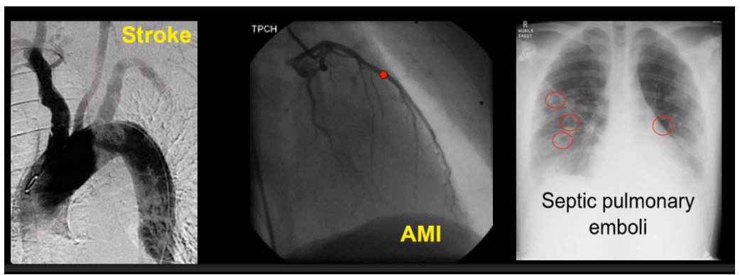
Embolic events

  • obstruction of a blood vessel by an infected thrombus/vegetation (septic emboli)

  • may cause injury by ischemia & infection


  • right side vegetation

    • goes to lungs

  • left side vegetation

    • goes to body & brain


  • life threatening

  • echo can play a key role in predicting embolic events

  • increased risk:

    • large mobile vegetation (> 10 mm)

    • vegetation on MV

    • ↑ size of vegetation while antibiotic therapy is given

    • multivalvular infection

    • previous embolic event

    • infection w/specific organisms (Staph, strep, and Candida species)

<ul><li><p><span style="background-color: transparent;">obstruction of a blood vessel by an infected thrombus/vegetation (septic emboli)</span></p></li><li><p><span style="background-color: transparent;">may cause injury by ischemia &amp; infection</span></p></li></ul><p></p><ul><li><p><span style="background-color: transparent;">right side vegetation </span></p><ul><li><p>goes to lungs </p></li></ul></li><li><p>left side vegetation </p><ul><li><p>goes to body &amp; brain </p></li></ul></li></ul><p></p><ul><li><p><span style="background-color: transparent;">life threatening</span></p></li><li><p><span style="background-color: transparent;">echo can play a key role in predicting embolic events</span></p></li><li><p><span style="background-color: transparent;">increased risk:</span></p><ul><li><p><span style="background-color: transparent;">large mobile vegetation (&gt; 10 mm)</span></p></li><li><p><span style="background-color: transparent;">vegetation on MV</span></p></li><li><p><span style="background-color: transparent;">↑ size of vegetation while antibiotic therapy is given</span></p></li><li><p><span style="background-color: transparent;">multivalvular infection</span></p></li><li><p><span style="background-color: transparent;">previous embolic event</span></p></li><li><p><span style="background-color: transparent;">infection w/specific organisms (Staph, strep, and Candida species)</span></p></li></ul></li></ul><p></p>
32
New cards

Valve perforation

  • interruption of endocardial tissue continuity that can lead to valve perforating

  • hole in the body of the leaflet

  • color jet will arise from site other than valve tip


<ul><li><p><span style="background-color: transparent;">interruption of endocardial tissue continuity that can lead to valve perforating</span></p></li><li><p><span style="background-color: transparent;">hole in the body of the leaflet</span></p></li><li><p><span style="background-color: transparent;">color jet will arise from site other than valve tip</span></p></li></ul><p></p>
33
New cards

Valve aneurysm

  • weakening of valve tissue & loss of elastic components of valve

  • leads to a saccular outpouching of valvular tissue


<ul><li><p><span style="background-color: transparent;">weakening of valve tissue &amp; loss of elastic components of valve</span></p></li><li><p><span style="background-color: transparent;">leads to a saccular outpouching of valvular tissue</span></p></li></ul><p></p>
34
New cards

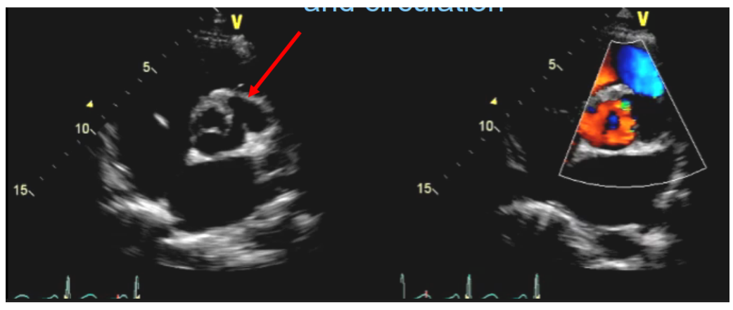
Pseudoaneurysm

  • pulsatile

  • echo free space

  • with flow between the cavity and circulation

<ul><li><p><span style="background-color: transparent;">pulsatile</span></p></li></ul><ul><li><p><span style="background-color: transparent;">echo free space </span></p></li></ul><ul><li><p><span style="background-color: transparent;">with flow between the cavity <strong>and circulation</strong></span></p></li></ul><p></p>
35
New cards

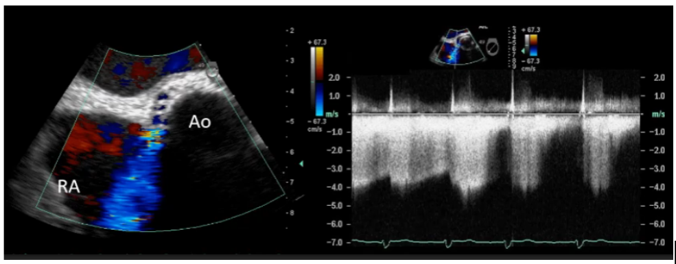
Fistula formation

  • communication between neighboring cavities via a perforation

  • communication confirmed by Doppler (treat as a shunt)

<ul><li><p><span style="background-color: transparent;">communication between neighboring cavities via a perforation</span></p></li><li><p><span style="background-color: transparent;">communication confirmed by Doppler (treat as a shunt)</span></p></li></ul><p></p>