Anatomy Practical Week 4
 Call Kai
 Learn
Learn Practice Test
 Spaced Repetition
 Match
Match Flashcards
 Knowt Play
1/47

There's no tags or description
Looks like no tags are added yet.
Name | Mastery | Learn | Test | Matching | Spaced | Call with Kai | Chat |
|---|
No analytics yet
Send a link to your students to track their progress
48 Terms
central nervous system (CNS)
division of the nervous system that contains the brain and spinal cord
peripheral nervous system (PNS)
division of the nervous system that contains the spinal nerves, the dorsal root ganglia, the somatic nerves, and the cranial nerves
can be broken down into 2 division: sensory and motor
sensory
afferent
part of the peripheral nervous system
motor
efferent
part of the peripheral nervous system
sensory or afferent division
division of the PNS that conducts impulses from the body to the CNS
motor or efferent division
division of the PNS that conducts impulses from the CNS to the body
two
the motor division has ___ subdivision: somatic nervous system and autonomic nervous system
somatic nervous system
the subdivision of the motor division that takes impulses from the CNS and innervates muscles - under conscious control
autonomic nervous system
the subdivision of the motor division that innervates glands, smooth muscle, cardiac muscles and organs- usually not under conscious control
neuron
an impulse conducting cell that contains an axon, dendrite, and a neuron cell body
synapse
a neuron that picks up a stimuli in your toes continues as a single cell up your leg and thigh to ____ with other neurons in the lower back
meter
neurons are often over a _____ long and carry these stimuli for a long distance
receptive
the ____ end of the neuron is the dendrites or the neuron cell body
axon
the _____ carries the stimuli along long strands of neurofibrils that are wrapped in myelin sheaths

sensory neurons (afferent)
conducts impulses to the CNS from the extreminties
motor neurons (efferent)
coducts impulses away from the CNS

multipolar neuron
consists of several dendrites, a single neuron cell body and a single axon

bipolar neuron
the neuron cell body has 2 poles. dendrites receive info and conduct it to one pole of the neuron cell body. at the other pole, an axon leaves the neuron cell body and transmits the impulse away from the cell body.

pseudo-unipolar neuron
a cell body has a single process attached to it. It has no dendrites. The neuron cell body sends impulses to the axon which transmits that info from the cell body

synapse
the space between neurons
neurons are not attached to each other
they communicate by sending neurotransmitters across the short spaces between them


Glial Cells
non-neuron cells in the CNS and PNS that do not produce electrical impulses
they are referred to as the “glue”
the common glial cell of the PNS is the SCHWANN CELL
these glial cells wrap around the axon, leaving thin regions between successive cells called NODES OR RANVIER
the wrapped portion between the nodes of ranvier becomes the MYELIN SHEATH, which is formed by the Schwann cells
Dorsal (posterior) Horns
receive sensory information from the somatic nerve
Ventral (anterior) Horns
send motor signals to the spinal nerves
Lateral Horn
only visible in some areas of the spinal cord
spinal cord
the ___ is covered in meninges just like the brain:
inner layer- pia mater
middle layer- arachnoid mater
outer layer- dura mater
both
the brain and the spinal cord _____ consist of gray and white mater
in the brain, the gray matter is the outer layer and the white matter is the inner layer
in the spinal cord, the gray matter is the inner layer and the white matter is the outer layer
31
there are ____ pairs of spinal nerves
mixed
the spinal nerves are ______ nerves carrying both sensory and motor information
origin
the spinal nerves are named according to their region of ______
cervical nerves
C1-C8
mostly innervate the head and neck
Phrenic nerve
a cervical nerve that runs to the diaphragm and is responsible for contraction in breathing
thoracic nerves
T1-T12
mostly innervate the ribs, muscles and other structures of the thoracic wall
notice that most of these nerves do not belong to a plexus
lumbar nerves
L1-L5
mostly innervate lower limbs
sacral nerve
S1-S5
mostly innervate lower limbs
coccygeal nerve; plexuses
Co
some of these nerves exit the spinal cord and branch out to parts of the body individually, but the ventral rami form a branching network with other spinal nerves. these networks are called ______
cervical plexus
C1-C4
mostly innervates the muscles and skin of the neck and the ears
brachial plexus
C5-T1
mostly innervates the upper extremities
axillary nerve and radial nerve
axillary nerve
major nerve of the brachial plexus that innervates the upper shoulder
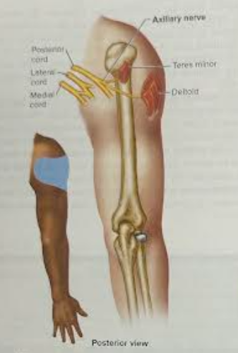
radial nerve
major nerve of the brachial plexus that innervates the extensions of the hand

Lumbosacral Plexus
sometimes the lumbar plexus and sacral plexus are combined and called ______
note: both the lumbar and sacral plexus’ take sensory information from and motor info to the lower limbs
(take sensory info from the lower limbs and send motor info to the lower limbs)
Lumbar plexus
L1-L4
contains the obturator nerve and the femoral nerve

Obturator Nerve
major nerve of the lumbar plexus that innervates the adductor muscles of the thigh

Femoral Nerve
major nerve of the lumbar plexus that innervates muscles of the anterior thigh
Sacral Plexus
L4-S4
the tibial nerve and the common fibular nerve unite to form the Sciatic nerve which innervates the leg and foot
Coccygeal Plexus
S5-Co
Sciatic Nerve
composed of the medial tibial nerve and the lateral common fibular nerve and can be seen at the distal part of the thigh

Tibial Nerve
found in the sacral plexus and helps make up the sciatic nerve

Common Fibular Nerve
found in the sacral plexus and is the second nerve that helps make up the sciatic nerve