N212B - Exam #2 Study Guide
 Call Kai
 Learn
Learn Practice Test
 Spaced Repetition
 Match
Match Flashcards
 Knowt Play
1/52

There's no tags or description
Looks like no tags are added yet.
Name | Mastery | Learn | Test | Matching | Spaced | Call with Kai | Chat |
|---|
No analytics yet
Send a link to your students to track their progress
53 Terms

Anemia
Reduction in the total number of erythrocytes in the circulating blood or in the quality/quantity of hemoglobin
Could be caused by:
Impaired erythrocyte production
Acute/chronic blood loss
Inc. erythrocyte destruction
Combination of the above
Classified by size and hemoglobin
Size → - cytic
Macrocytic, microcytic, and normocytic
Hemoglobin content → -chromic
Normochromic and hypochromic

Folate deficiency
Anemias of diminished erythropoiesis
deficiencies in vitamin b12 or folate → results in defective DNA synthesis
Folate deficiency anemia → a Macrocytic (megaloblastic) anemia
Humans are dependent on dietary intake of folate
Absorption of folate occurs in the upper small intestine
Not dependent on any other factor
Similar symptoms to pernicious anemia except neurologic manifestations are generally not seen
Treatment requires daily oral admin of folate until body levels are adequate
Anemia is a reduction in the number of red blood cells (RBCs), the amount of hemoglobin, or the hematocrit (percentage of packed RBCs per deciliter of blood). It can also be defined as a decrease in the quality or quantity of hemoglobin.
Important: Anemia is a clinical indicator, not a specific disease. It occurs with many health problems and is a pathophysiologic manifestation of various underlying disease states.
Diagnostic Criteria
For adults:
Men: Hemoglobin <14 g/dL (8.7 mmol/L)
Women: Hemoglobin <12.0 g/dL (7.4 mmol/L)
Anemia refers to a true decrease in erythrocyte number, not a relative decrease from increased plasma volume (hemodilution)
Definition and Pathophysiology
Folate (folic acid) is an essential water-soluble vitamin required for RNA and DNA synthesis in maturing red blood cells. The daily adult requirement is 400 mcg, obtained entirely through diet—fruits, green leafy vegetables, liver, and fortified grains.
Folate deficiency is more common than B12 deficiency, especially in individuals with alcoholism or chronic malnourishment. Deficient thymine production affects rapidly dividing cells in bone marrow, causing megaloblastic precursor cells and macrocytic anemia.
Unlike B12, folate is absorbed from the upper small intestine and does not require intrinsic factor. After absorption, it circulates through and is stored in the liver.
High-Risk Groups
Females of childbearing age, non-Black Hispanic females, and older adults
Pregnant and lactating individuals (increased requirements; deficiency causes neural tube defects)
Individuals with alcoholism or chronic malnourishment
Inflammatory bowel disease (Crohn disease, ulcerative colitis) causing malabsorption
Those taking sulfasalazine (decreases folate absorption)
Clinical Manifestations
Distinctive GI symptoms:
Cheilosis (scales/fissures of mouth)
Stomatitis and painful oral/tongue ulcerations (burning mouth syndrome)
Dysphagia, flatulence, watery diarrhea
GI histologic changes similar to celiac disease
Blood findings: Macrocytic RBCs (high MCV)
Key difference from B12 deficiency: Paresthesias do not occur because folate deficiency alone does not affect nerve function. If neurologic symptoms are present, consider thiamine deficiency, which often accompanies folate deficiency in malnourished individuals.
Folate deficiency may suppress intestinal mucosa proliferation, worsening GI damage.
Diagnosis and Treatment
Evaluation: Clinical manifestations, macrocytic RBCs (high MCV), and serum folate levels
Treatment:
Oral folate preparations until adequate blood levels are achieved
Dietary adjustments: folate-rich foods
Manifestations disappear within 1-2 weeks
Long-term therapy unnecessary if diet is corrected

Leukemia Med Admin
Nursing implications:
Assess baseline blood counts before administering antineoplastic drugs
Follow specific administration guidelines for each antineoplastic drug
Chemo-certified to give IV meds
Remember that all rapidly dividing cells are affected by antineoplastic drugs
GI/Mucous membranes → stomatitis, altered bowel function w/ high risk for poor appetite, nausea, vomiting, diarrhea, and inflammation/ulcerations of GI mucosa
Hair follicles → alopecia
Bone marrow components → dangerously low (life-threatening) blood cell counts
Monitor for effects/complications of these tissues
Implement measures to monitor for and prevent infection in patients w/ neutropenia/leukopenia → hand hygiene, precautions
Implement measures to monitor for and prevent bleeding in pts w/ thrombocytopenia and anemia
Anticipate N/V, implement measures to reduce
Antiemetics work best when given 30-60 minutes before chemo is given
Safe Handling of Chemotherapy
All chemotherapy agents are designated hazardous drugs (HDs) and require strict safety protocols.
Key safety principles:
For IV chemotherapy:
Nurses must undergo certification to administer IV chemotherapy
Administer through a central line indwelling catheter device to minimize extravasation risk
Check device patency before administration
Monitor IV site constantly during infusion
For oral chemotherapy:
Can be administered by any RN (no certification required)
Never physically touch chemotherapy tablets—highly toxic to normal cells
Same exposure risks as IV forms
Toxicity causes DNA damage leading to cell death, cancer, birth defects, or immune dysfunction
Extravasation Management
Extravasation (drug leakage into surrounding tissue) is a serious complication when vesicants (tissue-damaging chemicals) are involved.
If extravasation occurs:
Stop drug immediately but leave IV cannula in place (for possible antidote administration)
Follow institutional protocol (treatment is drug-specific)
Use of antidotes, hot/cold packs outlined in facility policy
Monitor hourly with documentation
May result in pain, infection, tissue loss, or need for reconstructive surgery
Prevention: Regular monitoring of blood return at access site during infusion
Patient and Family Safety
Bodily fluid precautions: Patients excrete chemotherapy chemicals through urine, stool, vomit, sweat, and saliva. Families must be warned of exposure dangers when patients self-administer at home.
Key nursing considerations:
Establish therapeutic relationship with trust and empathy
Provide individualized education appropriate to patient's level
Address fear and anxiety
Coordinate with interdisciplinary team
Monitor for toxicities affecting multiple body systems
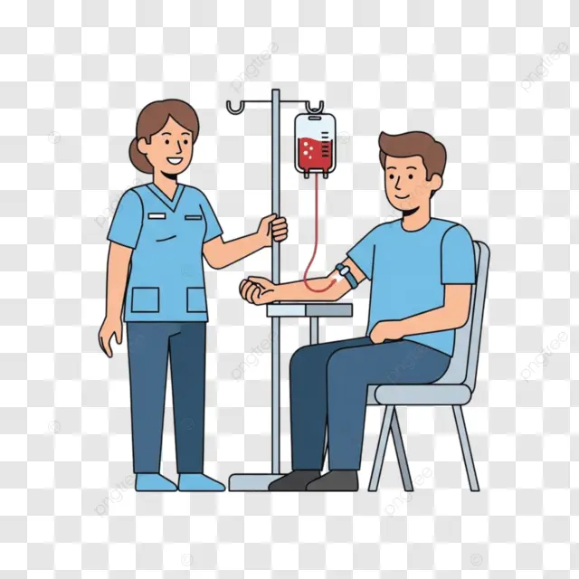
Blood administration
Pretransfusion Phase
Preparation is critical to prevent transfusion reactions.
Required steps:
Health care provider must discuss benefits/risks with patient
Obtain physician prescription specifying component type, volume, and special conditions
Obtain separate consent form per hospital policy
Collect blood specimen for type and crossmatch to test donor/recipient compatibility
Use blood filter (170 microns) to remove sediment
Prime tubing with normal saline only—never Ringer's lactate or dextrose (causes clotting/hemolysis)
Never add medications to blood (may cause clotting)
Critical safety check: Two registered nurses must verify:
Patient identification (name, health record number, date of birth)
ABO and Rh compatibility
Expiration date
Product appearance (no discoloration, gas bubbles, or cloudiness indicating bacterial growth)
During Transfusion
Initial phase (first 15-30 minutes):
Explain procedure to patient
Assess baseline vital signs and temperature
Start infusion slowly
Stay with patient—severe reactions typically occur within first 50 mL
Take vital signs at 15 minutes
Ask patient to report chills, shortness of breath, hives, itching, or back pain
Continuation:
If no reaction, increase rate to transfuse 1 unit over 2-4 hours (based on cardiac status)
Monitor vital signs hourly throughout transfusion
Blood products without large amounts of RBCs can infuse more quickly
Older patients may require slower infusion rates
Posttransfusion Phase
Completion steps:
Discontinue infusion
Dispose of bag/tubing per hazardous waste policy
Obtain vital signs and compare to baseline
Document: product type and number, volume infused, duration, vital signs, adverse reactions with interventions, patient tolerance
Special Considerations: Platelet Transfusions
Infuse immediately after bringing to room (platelets are fragile)
Infuse over 15-30 minutes
Use special transfusion set with smaller filter
May premedicate with diphenhydramine and acetaminophen if patient has history of reactions
Platelet administration
Indications for Platelet Transfusions
When to give platelets:
Platelet count <10,000/mm³ (10 × 10⁹/L)
Patient with thrombocytopenia who is actively bleeding
Patient scheduled for invasive procedure
Product Types
Pooled platelets:
Combined from up to 10 donors
Volume: ~300 mL
Do not need to match patient's blood type
Single-donor platelets:
From one donor only
Volume: ~200 mL
Preferred for patients receiving HSCT or requiring multiple transfusions
Reduces risk of allergic reactions1
Administration Procedure
Equipment:
Special transfusion set with smaller filter and shorter tubing
Platelet filters remove WBCs for patients with history of febrile reactions or needing multiple transfusions
Timing and rate:
Infuse immediately after bringing to patient's room (platelets are fragile)
Infusion duration: 15-30 minutes
Vital signs:
Before infusion starts
15 minutes after infusion starts
At completion
Preventing Reactions
Premedication for at-risk patients:
Diphenhydramine (antihistamine)
Acetaminophen (fever reducer)
Given to patients with history of transfusion reactions to reduce fever and severe chills (rigors)
Key Safety Points
Platelets are fragile—handle gently and infuse promptly
Never delay infusion once product arrives at bedside
Monitor closely during first 15 minutes when reactions most likely occur
Use appropriate filter to remove WBCs and prevent febrile reactions
Impaired hemostasis → Vitamin K deficiency
Hemostasis is the arrest of bleeding by formation of blood clots at sites of vascular injury.
As a result of hemostasis, damaged blood vessels may maintain a relatively steady state of blood volume, pressure, and flow. Three equally important interactive components of hemostasis are the vasculature (endothelial cells and subendothelial matrix), platelets, and clotting factors. The general sequence of events in hemostasis is (1) transient arteriolar vasoconstriction to limit blood flow to the affected site; (2) formation of a platelet plug (primary hemostasis); and (3) coagulation and clot formation with aggregation of proteins synthesized by the liver, endothelial cells, and platelets (secondary hemostasis). The relative importance of the hemostatic mechanisms clearly varies with vessel size. Hemostasis in capillaries and smaller veins and arteries occurs with sealing of the injury. Damage to large vessels cannot easily be controlled by hemostasis but requires vascular contraction to decrease blood flow from the damaged vessels.
Impaired hemostasis - inability of coagulation and stable fibrin clot
Vitamin K is necessary for the synthesis and regulation of prothrombin and the procoagulant factors (VII, XI, and X) and proteins C and S
Vitamin K's Role in Hemostasis
Vitamin K is essential for hepatic biosynthesis of clotting factors II, VII, IX, and X. It also helps regulate prothrombin and anticoagulant factors (proteins C and S) within the liver. Without adequate vitamin K, these clotting factors cannot be properly synthesized, leading to coagulation disorders caused by hypoprothrombinemia.
Causes of Vitamin K Deficiency
Primary sources of vitamin K:
Green leafy vegetables (dietary source)
Intestinal flora (synthesize sufficient amounts normally)
Most common cause:
Parenteral nutrition combined with antibiotics that destroy normal gut flora
Other causes:
Dietary deficiency (rare)
Bulimia (can suppress vitamin K–dependent activity)
Liver disease (cirrhosis, hepatocellular disease)—impairs production of vitamin K–dependent clotting factors
Clinical Manifestations
Vitamin K deficiency results in impaired hemostasis with bleeding tendencies due to decreased levels of multiple clotting factors. The liver produces most factors involved in hemostasis, so vitamin K deficiency or liver damage causes:
Factor VII decreases first (rapid turnover)
Factor IX levels decrease with advanced liver destruction
Deficient clotting factor synthesis leads to coagulation disorders
Treatment
Parenteral vitamin K administration:
Treatment of choice for vitamin K deficiency
Correction usually occurs within 8-12 hours
Fresh-frozen plasma (FFP):
Reserved for life-threatening hemorrhages
Used when emergency surgery is required
Provides immediate clotting factors while waiting for vitamin K to work
Key Point
Vitamin K deficiency is rare because intestinal flora normally synthesize adequate amounts. When deficiency develops, it can be corrected with supplementation. However, in patients on antibiotics with parenteral nutrition or those with severe liver disease, proactive monitoring and supplementation may be necessary to prevent bleeding complications

Chemotherapy
Care of pts receiving chemotherapy medications
Safe Handling and Administration
Protection requirements:
Never physically touch chemotherapy tablets—highly toxic to normal cells
Chemotherapy causes DNA damage leading to cell death, with potential injuries including cancer, birth defects, or immune dysfunction that may not appear for months or years
Nurses must undergo certification to administer IV chemotherapy; oral chemotherapy can be administered by any RN
Use proper protective techniques during unpacking, storage, handling, administration, and disposal
Patient excretion hazards:
Patients excrete chemotherapy through urine, stool, vomit, sweat, and saliva
Double-flush toilets during treatment and for 48 hours after discontinuing chemotherapy
Warn families about exposure dangers, especially for children and pets
Nursing Care During Treatment
Assessment priorities:
Complete physical assessment including vital signs, height/weight
Baseline hearing and vision testing
Assess bowel/bladder patterns, neurologic status, heart sounds, breath sounds
Evaluate skin and mucosa (turgor, hydration, color, temperature)
Pain assessment using 0-10 intensity scale
Monitor for chest pain, palpitations, dyspnea, numbness/tingling, bleeding signs
Critical monitoring during infusion:
Monitor for reactions within first few minutes of infusion
Watch for dyspnea, abdominal distention, lung crackles, dependent edema
Assess for cutaneous reactions (rash on hands/feet) requiring immediate attention
Ensure IV site patency—prevent extravasation of irritants/vesicants
Follow facility protocol immediately if extravasation occurs
Preventing complications:
Minimize tissue trauma (avoid IM injections, rectal temperatures if possible)
Monitor blood counts closely with every treatment
Address nausea/vomiting promptly to prevent dehydration and electrolyte disturbances
Be aware of numerous IV incompatibilities
Psychosocial Support
Reduce fear and anxiety:
Establish therapeutic relationship with trust and empathy
Provide individualized teaching appropriate to education level
Encourage relaxation techniques (music, meditation, guided imagery)
Connect with social services, spiritual guidance, financial assistance, and community resources

Chemotherapy and pregnancy
Strict contraindication during 1st trimester but not complete contraindication during 2nd and 3rd trimesters
Pregnancy Risk Classification
Chemotherapy drugs are classified as pregnancy category D or X, indicating significant risk to the fetus. The decision to use chemotherapy during pregnancy is based on risk versus benefit analysis.
Timing and Fetal Risk
First trimester—HIGHEST RISK:
Greatest danger of drug-caused developmental defects extends from fertilization through the first trimester
Both radiation and chemotherapy can cause significant permanent fetal harm or death
Many women may not yet realize they are pregnant during this critical period
Second and third trimesters:
Chemotherapy treatment is more likely to improve maternal outcome without significant fetal risk
Risk is reduced but not eliminated
Contraception Requirements
During treatment:
Sexually active women of childbearing age must protect themselves against pregnancy due to risk of embryonic death
Use contraceptive measures throughout chemotherapy
After treatment:
Continue contraception for up to 8 weeks after discontinuing therapy
Some antineoplastic drugs require contraception for up to 2 years after completion due to long-term risk of fetal genetic abnormalities
Reproductive Effects
Antineoplastic drugs negatively impact the reproductive tract by:
Destroying germinal epithelium of testes
Damaging ovaries
Causing teratogenesis (fetal malformations)
Male patients:
Risk of sterility, which may be irreversible
Discuss sperm banking before chemotherapy if appropriate
Female patients:
Sterility
Amenorrhea
Premature menopausal symptoms (hot flashes, decreased vaginal secretions, mood changes, irritability)
Decreased libido or sexual dysfunction
Special Consideration: Prepubertal Patients
Children who haven't reached puberty are more resilient and can have normal puberty and fertility after receiving chemotherapy

Diabetes Management
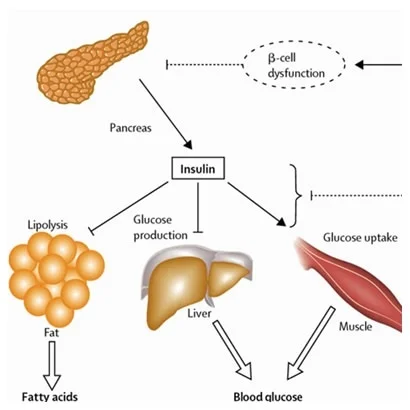
Insulin production
Pancreas → located behind the stomach; both an endocrine and exocrine gland
Produces two hormones that are important in the regulation of glucose homeostasis: Insulin and glucagon
Glycogen → excess glucose stored in the liver and skeletal tissue
Glycogenolysis → conversion of glycogen into glucose when needed
Insulin:
Direct effect on fat metabolism
Stimulates lipogenesis and inhibits lipolysis
Stimulates protein synthesis
Promotes intracellular shift of potassium and magnesium into the cells
Cortisol, epinephrine, and growth hormone work synergistically w/ glucagon to counter the effects of insulin
Synthesis Process
Precursor molecules: Insulin synthesis begins with preproinsulin, which is converted to proinsulin. Proinsulin is composed of:
A peptide
B peptide
C peptide (connecting the A and B peptides)
Two disulfide bonds
Final formation: Proteolytic enzymes cleave the C peptide, leaving the bonded A and B peptides as the active insulin molecule. The mature insulin protein consists of 51 amino acids.
Location
Insulin is synthesized by beta cells in the islets of Langerhans (pancreatic islets). The pancreas is located behind the stomach, between the spleen and duodenum.
Regulation of Secretion
Stimulating factors:
Primary stimulus: Increased blood glucose levels
Parasympathetic nervous system (usually before eating)
Certain amino acids (leucine, arginine, lysine)
Gastrointestinal hormones (glucagon, gastrin, cholecystokinin, secretin)
Inhibiting factors:
Low blood glucose (hypoglycemia)
High insulin levels (negative feedback to beta cells)
Sympathetic nervous system stimulation of beta cells
Circulation and Action
Transport: Insulin circulates freely in the plasma and is not bound to a carrier protein.
Target cells: Insulin binds to cell surface receptors found on cells throughout the body. This receptor activation initiates insulin signaling.
Function: Insulin prevents hyperglycemia by allowing body cells to take up, use, and store carbohydrates, fat, and protein. It's called the "hormone of plenty" because it's secreted when food intake is high and works to move glucose from blood into cells.
Clinical Application: C-Peptide Measurement
C-peptide level can be measured in the blood as an indirect measurement of serum insulin synthesis. This is useful clinically to evaluate β-cell destruction in diabetes, as C-peptide is released during insulin production
Diabetes management (oral and insulin)
Insulin + Oral hypoglycemic drugs → both aim to produce normal blood glucose levels
Non-insulin antidiabetic drugs:
Used for DM type 2
Effective treatment involves several elements:
Careful monitoring of blood sugar levels
Therapy w/ one or more drugs
Treatment of associated comorbid conditions like HTN and high cholesterol
Biguanides (metformin)
MOA:
Decreases glucose production in liver
Decreases intestinal absorption of glucose
Increase uptake of glucose by tissues
Does not increase insulin secretion from the pancreas (doesn’t cause hypoglycemia)
Primarily affects GI tract: abdominal pain, bloating, nausea, cramping, diarrhea, feeling of fullness
May have metallic taste, reduced vitamin B12 levels
Lactic acidosis rare but life-threatening if it occurs
Contraindicated in renal/hepatic diseases
Type 2 Diabetes Management First-Line Treatment
Metformin is the first-line medication for new-onset type 2 diabetes, combined with lifestyle interventions. If A1C goals aren't achieved after 3-6 months with metformin at maximum tolerated dose and lifestyle modifications, add a second agent.
Metformin mechanism:
Decreases glucose production by the liver
May decrease intestinal glucose absorption
Improves insulin receptor sensitivity
Increases peripheral glucose uptake
Decreases hepatic triglyceride and cholesterol production
Does NOT stimulate insulin secretion—no significant hypoglycemia or weight gain when used alone
Additional Oral/Injectable Agents
GLP-1 agonists:
Dulaglutide, exenatide, liraglutide, lixisenatide, semaglutide
Work like natural gut hormones released after food intake
Taken subcutaneously (daily or weekly); semaglutide available orally or subcutaneously
SGLT2 inhibitors:
Prevent kidney reabsorption of filtered glucose
Excess glucose is excreted in urine
When to Add Insulin
Indications for insulin in Type 2 DM:
Target glucose levels not met with 2-3 different antidiabetic agents (including GLP-1 agonists)
A1C above 10% with diabetes symptoms at presentation
Beta cell function declines over time—some patients eventually require insulin alone or with other drugs
Insulin Regimens
Basal coverage:
Intermediate-acting (NPH), long-acting (glargine, detemir), or ultra-long-acting (degludec)
Single daily injection for basal coverage, often combined with oral agents or GLP-1 for bolus stimulation
Effectiveness assessed by fasting blood glucose
Multiple daily injections (MDI):
Basal dose (intermediate/long/ultra-long-acting)
Mealtime bolus (short/rapid-acting) to bring glucose into target range
Correction doses for elevations above target
Combination therapy:
Two-thirds daily dose before breakfast, one-third before evening meal
Bedtime snack may prevent nocturnal hypoglycemia with NPH
Treatment Selection Factors
Consider:
Efficacy, cost, side effects
Comorbidities, weight effects, hypoglycemia risk
Patient preference and ability to manage multiple dosages
Once-daily or once-weekly dosing improves adherence
Always combine with balanced eating and regular physical activity
GLP-1s → Mimic naturally secreted incretin hormones
Incretin hormones increase insulin synthesis and lower glucagon secretions
Incretins increase insulin synthesis and lowers glucagon secretion
Non-insulin Antidiabetic drugs
Metformin (Glucophage)
1st-line drug and most commonly used oral med for treatment of DM type 2
Not used for DM type 1
Sulfonylureas
2nd gen → glimepiride (Amaryl), glipizide (Glucotrol), glyburide (DiaBeta)
Stimulate insulin secretion from the beta cells of the pancreas, increasing insulin levels
Beta cell function must be present
Improves sensitivity to insulin in tissues
Lowers blood glucose level
Adverse effects: hypoglycemia, hematologic effects, nausea, epigastric fullness, heartburn, and others
Glinides:
Repaglinide (Prandin), nateglinide (Starlix)
Used for type 2
MOA is similar to Sulfonylureas
Increases insulin secretion from the pancreas
Adverse effects: hypoglycemia and weight gain
Thiazolidinediones (glitazones)
Pioglitazone (Actos)
Rosiglitazone (Avandia) → only available through specialized manufacturer programs
“Insulin-sensitizing drugs”
Used for type 2
MOA: Decreases insulin resistance, increases glucose uptake and use in skeletal muscle, inhibits glucose and triglyceride production in the liver
Contraindications: HF; can exacerbate or cause HF→ peripheral edema, weight gain and decreased bone marrow density
Incretin Mimetics
Mimic naturally secreted incretin hormones
Incretin hormones increase insulin synthesis and lower glucagon secretions
Incretins increase insulin synthesis and lower glucagon secretion
Oral + injectable
Dipeptidyl peptidase IV inhibitors (DPP-IV)
“gliptins”
Sitagliptin (Januvia)
Saxagliptin (Onglyza)
Improve glycemic control in type 2
Used as an adjunct w/ diet and exercise
Adverse effects: upper respiratory tract infections, H/A, and diarrhea, possible pancreatitis
Glucagon-like peptide-1 (GLP-1) receptor agonists
Semaglutide (Ozempic)
Exenatide (Byetta, Bydureon)
Liraglutide (Victoza)
Enhance glucose-dependent insulin secretion
Slows gastric emptying
Inc. 1st and 2nd phase insulin secretion
Indicated for pts w/ type 2 who have not been able to achieve blood glucose control w/ metformin, a sulfonylurea and/or a glitazone
Adverse effects:
Black box warning → risk of developing thyroid C-cell tumors
N/V
Diarrhea
Rare - hemorrhagic or necrotizing pancreatitis
Pts may experience weight loss of 5-10 pounds
GLP-1s
Mechanism of Action
GLP-1 agonists mimic natural incretin hormones released by the GI tract in response to food. They work by:
Enhancing glucose-dependent insulin secretion
Suppressing elevated glucagon secretion
Slowing gastric emptying
Increasing satiety
Improving first- and second-phase insulin secretion
Natural incretins are rapidly broken down by the enzyme DPP-IV, but GLP-1 agonists resist this degradation.
Available Medications
Injectable GLP-1s:
Exenatide (Byetta, Bydureon)—twice daily or once weekly
Dulaglutide (Trulicity)—once weekly
Liraglutide (Victoza)—once daily
Albiglutide (Tanzeum)—once weekly
Lixisenatide (Adlyxin)
Semaglutide (Ozempic)—once weekly
Oral option:
Semaglutide (Rybelsus)—once daily
Combination products:
Soliqua (insulin glargine + lixisenatide)
Xultophy (insulin degludec + liraglutide)
Clinical Use
Indications:
Type 2 diabetes patients unable to achieve glucose control with metformin, sulfonylureas, and/or glitazones
Added when lifestyle modifications and maximum metformin dose don't achieve A1C goals after 3-6 months
Administration:
Best given 60 minutes before a meal
Subcutaneous injection (except oral semaglutide)
Cardiovascular benefits:
GLP-1s directly improve cardiovascular outcomes according to 2020 American College of Cardiology consensus
Reduce risk for renal and heart disease, even in obese individuals without diabetes
Additional Benefits
Weight management:
GLP-1 receptor agonists cause significant weight loss4
Helps control postprandial glucose by promoting glucose-dependent insulin secretion
Lower HgA1C effectively
Safety note:
All GLP-1s are pregnancy category C

Gestational diabetes
Hyperglycemia that develops during pregnancy
Insulin must be given to prevent birth defects
Usually subsides after delivery
30% of patients may develop DM type 2 within 10-15 years
Definition
Gestational Diabetes Mellitus (GDM) is carbohydrate intolerance that develops during pregnancy, regardless of whether management includes medication (insulin or oral hypoglycemic agents) or only dietary changes. The American Diabetes Association defines GDM as diabetes diagnosed during pregnancy that is clearly not preexisting (overt) diabetes.
Pathophysiology
GDM develops during the second or third trimester when:
Fetal nutrient demands increase
Maternal insulin resistance rises due to placental hormones, cortisol, and insulinase
Maternal insulin demands increase as much as threefold
The pancreas cannot produce sufficient insulin or insulin isn't used effectively
Most pregnant women compensate by increasing insulin production, but those who cannot develop GDM.
Screening
Timing: 24-28 weeks of gestation
Two-step method (ACOG recommended):
Step 1: 50-g oral glucose load (no fasting required), plasma glucose measured at 1 hour
Positive screen: 130-140 mg/dL or higher
Step 2: 3-hour (100-g) oral glucose tolerance test (OGTT) on another day if step 1 positive
Requires overnight fast and at least 3 days of unrestricted diet (≥150 g carbohydrates)
Avoid caffeine and smoking for 12 hours before test
Maternal and Fetal Risks
Pregnancy complications:
Infant macrosomia (extra-large baby)
Neonatal hypoglycemia
Preeclampsia
Cesarean delivery
Long-term maternal risks:
Up to one-third will have diabetes or impaired glucose metabolism postpartum
High risk for recurrent GDM in future pregnancies
Up to 70% chance of developing type 2 diabetes within 10-20 years
Child health risks:
Increased risk for obesity and type 2 diabetes later in life
Management
CDC recommendations:
Eat healthy foods
Exercise regularly
Monitor blood sugar often
Take insulin if needed
Get tested for diabetes after pregnancy
Postpartum Follow-up
Immediate screening:
75-g, 2-hour OGTT or fasting plasma glucose at 6-12 weeks postpartum
Lifelong monitoring:
Repeat screening at least every 3 years with normal postpartum results
Use hemoglobin A1c, fasting plasma glucose, or 75-g 2-hour OGTT
Diabetes medications and contraindications/contrast
Metformin will need to be discontinued if the patient is to undergo studies w/ contrast dye because of possible renal effects → check w/ prescriber
Diabetes medications and NPO status
If a pt is NPO for a test or procedure, consult the PCP to clarify orders for antidiabetic drug therapy
General Principles
When a patient is NPO, it's crucial to follow the prescriber's orders regarding diabetes medication administration. If there are no written orders about NPO status and diabetes medications, contact the prescriber for further instructions.
Oral Diabetes Medications
Timing is critical:
Ensure food will be or is being tolerated before the dose is given
If oral medication is taken without a meal or the meal is delayed, hypoglycemia may result and cause negative health consequences, even unconsciousness
Preoperative management:
Sulfonylureas: Discontinued 1 day before surgery
Metformin: Stopped at least 24 hours before surgery; restarted only after kidney function documented as normal
All other oral drugs: Stopped the day of surgery
SGLT2 inhibitors:
Usually given in the morning before the first meal
Follow prescriber's orders if patient is NPO
Insulin Management
Rapid-acting and short-acting insulin:
Will cause hypoglycemia if patient is not eating
Should NOT be given when NPO
Amylin and incretin mimetics:
Also cause hypoglycemia if patient is not eating
Should NOT be given when NPO
Basal insulin (intermediate or long-acting):
Should be given when patient is NPO (often at a reduced dose)
Controls baseline glucose levels independent of food intake
Special consideration: If patient is NPO but receiving IV dextrose solution, the prescriber may still order insulin—always clarify the order
Preoperative insulin adjustments:
Long-acting insulin may need to be switched to intermediate-acting forms 1-2 days before surgery
Target preoperative blood glucose: <200 mg/dL
Inpatient Insulin Management
Hospitalized patients:
Continuous IV insulin (insulin drip): Most effective for critically ill patients
Scheduled subcutaneous injections: Basal, meal, and correction doses for non-critically ill patients


Rapid-acting insulin (Peak and Duration)
Most rapid onset of action → 5-15 minutes
Peak: 1 to 2 hours
Duration: 3 to 5 hours
Pt must eat a meal after injection
Don’t LAG -
lispro (Humalog)
aspart (Novolog)
glulisine (apidra)
Given SUBQ or via continuous SUBQ infusion pump, NOT IV

Short-acting insulin (Peak and Duration)
Humulin R
Routes: IV bolus, IV infusion, IM, and SUBQ
SUBQ route -
Onset: 30 to 60 minutes
Peak: 2.5 hours
Duration: 6 - 10 hours
IV route -
Immediate onset
Duration: 2 to 6 hours

Intermediate acting insulin (Peak and Duration)
Insulin isophane suspension (NPH) *cloudy
Clear (regular) → cloudy (NPH) when mixing
Onset: 1 to 2 hours
Peak: 4 to 8 hours
Duration: 10 to 18 hours

Long-acting insulin (Peak and Duration)
Insulin glargine (Lantus) or “basal insulin”
Clear and colorless, constant level of insulin in the body, usually dosed once daily but can be dosed every 12 hours
Onset: 1 to 2 hours
Peak: None
Duration: 24 hours
Detemir (Levemir)
Duration of action is dose dependent
Degludec (Tresiba)
Ultra-long-acting
Once daily
GLP-1 Effects
Adverse effects:
Black box warning → risk of developing thyroid C-cell tumors
N/V
Diarrhea
Rare - hemorrhagic or necrotizing pancreatitis
Pts may experience weight loss of 5-10 pounds
Physiological Effects
Insulin and glucagon regulation:
Enhance glucose-dependent insulin secretion
Suppress elevated glucagon production
Improve first- and second-phase insulin secretion
Gastrointestinal effects:
Slow gastric emptying
Increase satiety (feeling of fullness)
Background: GLP-1 is an endogenous incretin hormone naturally released by the GI tract in response to food intake. In type 2 diabetes, β-cell responsiveness to GLP-1 is decreased, reducing insulin secretion after meals.
Clinical Benefits
Glucose control:
Help control postprandial glucose levels by promoting glucose-dependent insulin secretion
Lower HgA1C effectively
Weight management:
Cause significant weight loss
Improve weight management for individuals with type 2 diabetes
Cardiovascular and renal protection:
Reduce risk for renal and heart disease, even in obese individuals without type 2 diabetes
Directly improve cardiovascular outcomes
How They Work
Natural incretin hormones like GLP-1 are rapidly broken down by the enzyme DPP-IV. GLP-1 agonist medications resist this degradation, allowing prolonged therapeutic effects.
Effect is glucose-dependent: GLP-1s stimulate insulin secretion primarily when blood glucose is elevated, which reduces the risk of hypoglycemia compared to some other diabetes medications.
Summary of Key Effects
✓ Stimulate insulin release (when glucose is elevated)
✓ Reduce glucagon secretion
✓ Slow stomach emptying
✓ Increase fullness after eating
✓ Promote weight loss
✓ Protect heart and kidneys
✓ Lower A1C levels
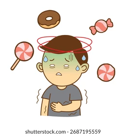
Treatment of Hypoglycemia
Hypoglycemia → abnormally low blood glucose level (below 70 mg/dL)
Mild cases can be treated w/ diet → higher protein intake and lower carbohydrate intake to prevent rebound hypoglycemia
Glucose-elevating drugs:
Oral forms of concentrated glucose → buccal tablets and semisolid gel
IV → 50% dextrose in water (D50W)
SUBQ → glucagon
Recognition of Hypoglycemia
Definition: Blood glucose below 70 mg/dL (3.9 mmol/L)
Early warning symptoms (autonomic):
Sweating, irritability, tremors
Anxiety, tachycardia, hunger
Advanced symptoms (neuroglycopenic):
Confusion, paralysis
Seizures, coma
The 15-15 Rule (Hospital Protocol)
Step 1: Give fast-acting carbohydrate
15 g of carbohydrate if blood glucose <70 mg/dL (3.9 mmol/L)
30 g of carbohydrate if blood glucose <50 mg/dL
Preferred: Liquid form (if patient can swallow)
Avoid: High-potassium options like orange juice; chocolate and concentrated sweets (fat slows absorption)
Step 2: Recheck in 15 minutes
If still low: Repeat same treatment
If above hypoglycemic range: Follow with complex carbohydrate (crackers with nut butter)
Why complex carbs matter: They help prevent subsequent hypoglycemia after initial treatment
When Patient Cannot Swallow
Drug options:
IV concentrated dextrose (hospital setting)
IM or nasal glucagon (first-line therapy for severe hypoglycemia)
Glucagon precautions:
Can induce vomiting
Position patient on side to prevent aspiration
IV dextrose precautions:
Give carefully to avoid extravasation (hyperosmolar solution can damage tissue)
Monitoring After Treatment
Short-term:
Monitor blood glucose every 15 minutes until hypoglycemia resolves
Continue monitoring for several hours (effects of glucagon and dextrose are temporary)
Target glucose level: 70-110 mg/dL (3.9-6.2 mmol/L)
Additional treatment may be needed if initial response is inadequate
Best Treatment Approach
Most effective: Glucose or glucose-containing foods, NOT complex carbohydrates initially
Treatment sequence:
Fast-acting simple carbohydrate (glucose tablets, gel, juice, non-diet soda)
Recheck blood glucose
Follow with complex carbohydrate once resolved
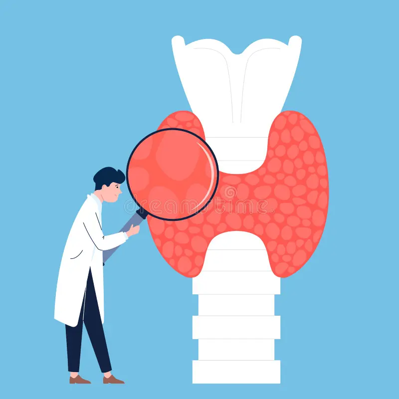
Endocrine and Thyroid Disease
Treatment of Diabetes Insipidus (DI)
Diabetes insipidus → insufficiency of ADH causing loss of too much water in the urine (3-5L/day)
Two forms:
Neurogenic (central) - insufficient secretion of ADH
doesn’t release enough vasopressin
Nephrogenic - inadequate response to ADH
vasopressin is present but doesn’t respond properly
Symptoms:
Polyuria
Nocturia
Continuous thirst
Treatment:
Desmopressin (DDAVP) → replaces the missing hormone vasopressin
Nephrogenic: also uses a thiazide diuretic (HCTZ)
Diabetes insipidus (DI) presents as excessive thirst (polydipsia), excessive urination (polyuria), and dehydration due to deficiency or resistance to antidiuretic hormone (ADH/vasopressin).
Two main types:
AVP-D (Arginine Vasopressin Deficiency): Insufficient ADH production
AVP-R (Arginine Vasopressin Resistance): Kidneys don't respond to ADH
Drug Therapy for AVP-D
Primary medication: Desmopressin (DDAVP)
Synthetic replacement for natural vasopressin (ADH)
Routes available: Oral, sublingual "melt," subcutaneous, intranasal metered spray
Dosing frequency: Varies with individual patient response
Alternative: Vasopressin (Pitressin)
Available as nasal spray or injection (IM/IV)
Used to prevent or control polydipsia, polyuria, and dehydration in DI
Route selection considerations:
Intranasal preparations may cause mucosal ulceration, allergy, chest tightness, or lung inhalation
Intranasal changes can lead to unpredictable drug absorption
If side effects occur or patient has upper respiratory infection, switch to oral or subcutaneous route
Treatment of AVP-R
Non-drug approaches:
Low-salt diet: <2.3 g sodium/day
Low-to-normal protein: <1.0 mg/kg daily
Thiazide diuretics: Induce mild volume depletion; even 1-1.5 kg weight loss significantly reduces urine output
Key Nursing Monitoring
Risk: Fluid overload/water toxicity
Drug therapy induces water retention
Daily weights: Same scale, time, clothing
Warning signs: Weight gain >2.2 lb (1 kg), persistent headache, confusion, nausea, vomiting
Action: Go to ED or call 911 immediately
Hospitalized patients:
Accurately measure I&O
Check urine specific gravity
Daily weights
Ensure fluid intake equals urine output
Missed doses: Can cause severe electrolyte abnormalities leading to significant harm or death
Patient Education
✓ Wear medical alert bracelet
✓ Recognize polyuria/polydipsia as signs needing another dose
✓ May require lifelong therapy
✓ Children need unrestricted water/bathroom access at school
Administration of thyroid medication
Hypothyroidism is treated w/ levothyroxine (Synthroid) (T4), which replaces the hormone the thyroid isn’t making enough of
Take on an empty stomach 30-60 minutes before food intake
Meds and supplements can interfere w/ absorption
Do not stop taking thyroid medication abruptly
TSH level checked 6-8 weeks after change of dose
Timing and Food Interactions
Take on an empty stomach
30 to 60 minutes before breakfast
Tube feedings can impair absorption
Why timing matters: Food interferes with medication absorption, reducing effectiveness
Consistency is Critical
Same brand throughout treatment
Switching between brands can destabilize treatment
If brand change occurs, monitor thyroid function tests more carefully
Daily routine:
Take consistently every morning
Helps maintain stable hormone levels
Dosing Precautions
Common medication error:
Levothyroxine is dosed in micrograms (mcg), NOT milligrams (mg)
Writing dose in milligrams results in 1000-fold overdose
Doses higher than 200 mcg need to be questioned
Starting therapy safely:
Start at lowest dose for:
Older adults
Patients with cardiac disease
Severe hypothyroidism
Unknown degree of hypothyroidism
Dose gradually increased over weeks if needed
Too high or too rapid increases can cause abnormal heart rhythms, heart failure, or myocardial infarction
Special Situations
Myxedema coma:
May require IV administration due to severely reduced GI motility and absorption
Before thyroid scans:
Usually discontinued 4 weeks before radioactive iodine studies (as prescribed)
Older adults (≥60 years):
May require 25% dose reduction
Drug Interactions to Avoid
Fiber supplements:
May interfere with thyroid hormone absorption
Over-the-counter medications:
Do not take without consulting provider—thyroid hormones interact with many drugs and supplements
Monitoring Effectiveness
Two easiest self-monitoring parameters:
Sleep requirements (need for more sleep may indicate low levels)
Bowel elimination (constipation may indicate low levels)
If these worsen: Contact provider for potential dose adjustment

Digestive Health
Treatment of Diarrhea
Diarrhea: Presence of loose, watery stools, acute or persistent
Large-volume diarrhea → Caused by excessive amounts of water or secretions, or both, in the intestines
Small-volume diarrhea → Volume of feces is not increased; usually results from excessive intestinal motility
Treatment:
Fluid restoration, antimotility, or water-absorbent medications, and treatment of causal factors
Oral rehydration if tolerated
BRAT diet
Imodium (loperamide) → if there is no blood in the stool, “don’t love” as overuse can cause constipation
Pepto-Bismol → avoid if allergic to ASA or taking blood thinners
Antidiarrhea Drug Classes:
Antimotility/anticholinergics → Imodium (loperamide)
Decrease intestinal muscle tone and peristalsis of the GI tract, slowing the movement of fecal matter; reduce peristalsis and gastric secretions
Absorbents → Pepto-Bismol
Coat the walls of the GI tract and bind to the causative bacteria or toxin, which is then eliminated through the stool; avoid if allergic to ASA or taking blood thinners
Probiotics → Lactobacillus acidophilus (Bacid)
AKA intestinal flora modifiers and bacterial replacement drugs; Lactobacillus supplies missing bacteria and suppresses growth of diarrhea-causing bacteria
Anticholinergics and opiates used for more serious cases
Primary objectives:
Stop stool frequency and alleviate abdominal cramps
Replenish fluids and electrolytes
Prevent weight loss and nutritional deficits from malabsorption
Often fluid replacement is the only treatment needed
Oral Rehydration Therapy (ORT)
First-line treatment for acute diarrhea with dehydration
Why ORT is preferred:
More effective, safer, less painful, and less costly than IV rehydration
Recommended by American Academy of Pediatrics, WHO, and CDC
Oral rehydration solutions (ORS):
Enhance and promote reabsorption of sodium and water
Greatly reduce vomiting, illness duration, and need for IV fluids
Available as commercially prepared solutions in the U.S.
Administration:
Give in small quantities at frequent intervals4
Vomiting is NOT a contraindication unless severe
After rehydration:
Alternate ORS with low-sodium fluids (breast milk, lactose-free formula, or half-strength lactose-containing formula) during maintenance therapy
Antidiarrheal Medications
Three main categories:
Adsorbents
Antimotility drugs (anticholinergics and opiates)
Probiotics (intestinal flora modifiers)
Bismuth subsalicylate (specific example):
Turns stool black or gray
Chewable tablets: chew thoroughly with ≥6 oz water
Non-chewable tablets: take with 6-8 oz water
Do NOT combine with other salicylates (risk of toxicity)
Avoid in children/teens with viral infections (chickenpox, influenza) due to Reye's syndrome risk
Dietary Management
Reintroduce normal diet after rehydration
Slightly higher stool output initially is expected
Benefits of continuing normal diet: Better nutritional outcomes, fewer complications, shorter illness duration
These benefits outweigh temporary stool frequency increase
When to Seek Medical Attention
Contact provider if:
Symptoms persist despite treatment
Signs of dehydration worsen
Hospitalization may require:
NPO status for 12-48 hours
Parenteral fluid therapy
Treatment of constipation – medication actions
Laxatives
Bulk forming
Hyperosmotic
Stimulant
Emollient (stool softeners, lubricant laxatives)
Saline
Laxatives: Bulk Forming
Examples:
Psyllium (Metamucil)
Methylcellulose (Citrucel)
MOA:
High fiber
Absorbs water to increase bulk
Distends the bowel to initiate reflex bowel activity
Indications:
Acute and chronic constipation
IBS
Diverticulosis
Bulk-forming laxatives
Absorb water into the intestine, increasing bulk and distending the bowel
Trigger reflex bowel activity promoting movement
Produce normal, formed stools
Safest for long-term use
Must take with liberal amounts of water to prevent esophageal obstruction or fecal impaction
Examples: psyllium (natural), methylcellulose (synthetic)
Laxatives: Emollient
Examples:
Docusate salts (Colace, Surfak) → stool softeners
Mineral oil → lubricant
MOA:
Stool softeners and lubricants
Promote more water and fat in the stools
Lubricate the fecal material and intestinal walls
Indications:
Acute and chronic constipation
Fecal impaction
Facilitation of bowel movements in anorectal conditions
Emollient laxatives (stool softeners and lubricants)
Docusate salts: Lower surface tension of GI fluids so more water and fat absorb into stool
Facilitate passage of water and lipids into fecal mass
Do NOT cause defecation—only soften stool to ease passage
Mineral oil: Lubricates fecal material and intestinal wall, preventing water absorption
Water remains in bowel to soften and expand stool
Promotes bowel distention and reflex peristalsis
Laxatives: Hyperosmotic
Examples:
Polyethylene glycol (PEG)
Sorbitol, lactitol, glycerin
Lactulose (also used to reduce serum ammonia levels)
MOA:
Increased fecal water content
Results in bowel distention, increased peristalsis, and evacuation
Indications:
Chronic constipation
Diagnostic and surgical preps
Hyperosmotic laxatives
Increase fecal water content, causing distention
Result: increased peristalsis and evacuation
Action limited to large intestine
Laxatives: Saline
Examples:
Magnesium hydroxide (Milk of Magnesia)
Magnesium citrate
MOA:
Increases osmotic pressure within the intestinal tract, causing more water to enter the intestines
Results in bowel distention, increased peristalsis, and evacuation
“works from above” → pushes down
Indications:
Constipation
Diagnostic and surgical preps
Saline laxatives
Increase osmotic pressure in small intestine
Inhibit water absorption
Increase water and electrolyte secretions from bowel wall into bowel lumen
Produce watery stool
Increased distention promotes peristalsis and evacuation
Rectal enemas: produce defecation in 2-5 minutes
Laxatives: Stimulant
Examples:
Senna (Senokot)
Bisacodyl (Dulcolax)
MOA:
Increases peristalsis via intestinal nerve stimulation
Common after surgery
Anesthesia slows smooth muscle activity
Indications:
Acute constipation
Diagnostic and surgical preps
Stimulant laxatives
Stimulate nerves that innervate the intestines
Result: increased peristalsis
Also increase fluid in the colon, increasing bulk and softening stool
Renal
Adrenergic Blockers
Drugs that stimulate the sympathetic nervous system (SNS)
Fight-or-flight
Run faster → Increases HR
Breathe better → widens airway
Pumps harder → more blood to muscles
Catecholamines: are the “emergency drugs” that activate the adrenergic and dopaminergic receptors, fight-or-flight chemicals
Norepinephrine (NE)
Epinephrine (EPI)
Dopamine
Located throughout the body, there are receptors for the sympathetic neurotransmitters
Alpha-adrenergic receptors
Beta-adrenergic receptors
Dopaminergic receptors → only respond only to dopamine
Receptors determine where these effects occur
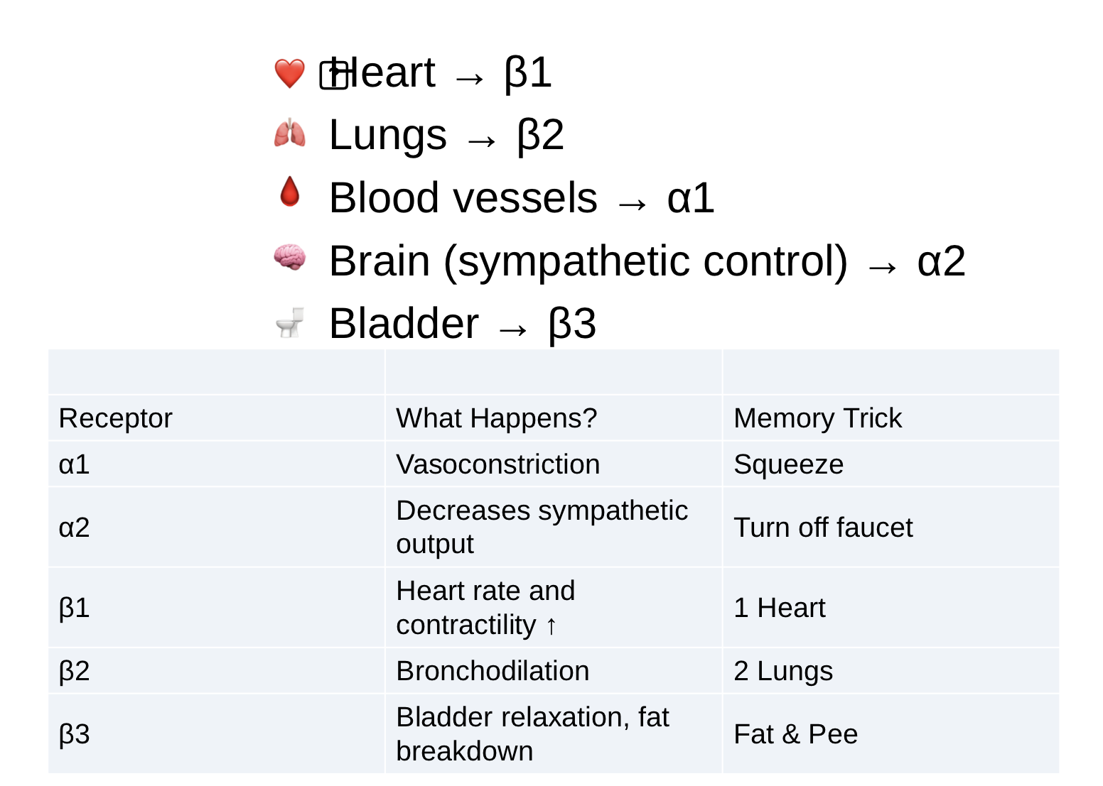
The Adrenergic Receptors
Alpha-1 adrenergic receptors are located on postsynaptic effector cells (the cell, muscle, or organ that the nerve stimulates)
α1 = constricts blood vessels
Vasoconstriction
Increased BP
Pupil dilation
Urinary retention
e.g. → phenylephrine
Alpha-2 adrenergic receptors are located in the brain (the nerve that stimulates the effector cells)
α2 stimulation = BP decreases
α2 = shuts down norepinephrine release
Decreases sympathetic activity
Lowers BP
Slows HR
e.g. → clonidine
Beta-Adrenergic Receptors → all located on postsynaptic effector cells
Beta-1 adrenergic receptors are located primarily in the heart
B1 = One heart
Effects:
Increased HR
Increased contractility
Increased cardiac output
e.g. → dobutamine
Beta- 2 adrenergic receptors are located in the cmooth muscle of the bronchioles, arterioles, and visceral organs
B2 = Two lungs
Effects:
Bronchodialation
Vasodialation
Relaxed uterus
e.g. → albuterol and terbutaline
Dopaminergic Receptors
D1 = “Dilate and Diurese”
Causes dilation of the following blood vessels, resulting in increased blood flow
Renal
Mesenteric
Coronary
Cerebral
D2 = Don’t release dopamine
Effects:
Inhibits dopamine release
Affects movement, mood, and nausea
Example → Tetrabenazine, lowers the amount of dopamine released to decrease involuntary movements
Clinical correlation:
Antipsychotics block D2 receptors
Metoclopramide blocks D2 receptors to reduce nausea
Parkinsons meds

Epinephrine
Epinephrine = The Superhero
“Works everywhere”
Stimulates:
a1
B1
B2
Effects:
Increases HR
Increases contractility
Increases bronchodilation
Increases BP
Uses:
Anaphylaxis
Cardiac arrest
Severe asthma reactions
If the pt can’t breathe and their BP is crashing, think → epinephrine

Norepinephrine
Norepinephrine = The Pressure Drug
“Squeeze the vessels”
Stimulates:
a1 strongly
B1 somewhat
Effects:
Vasoconstriction
Increased SVR
Increased BP
Uses:
Septic shock
Vasodilatory shock
“any kind of BP support”

Dopamine
Dopamine = The Dose Drug
Dopamine is a dopaminergic drug that becomes an adrenergic drug as the dose rises
Effects change w/ the dose
Low → kidneys
Medium → heart
High → blood vessels
Receptors
D1
B1
a1
Low-dose dopamine
Low-dose dopamine (0.5 - 2 mcg/kg/min)
Simulates D1 receptors → D for diuresis
Effects:
Increases kidney perfusion
Increases urine output
“Kick to the kidneys to work”
Medium-dose dopamine
Medium-dose dopamine (2 - 10 mcg/kg/min)
Stimulates B1 receptors → works more on the heart
Effects:
Increases HR
Increases contractility
Increases cardiac output
“Dopamine wakes up the heart”
Used as opposed to open heart surgery
High-dose dopamine
High-dose dopamine (>10 mcg/kg/min)
Activates a1 receptors
Effects:
Vasoconstriction
Increases BP
“Dopamine starts squeezing vessels”

Medication and administration of the patient with hypotension
Vasoactive Adrenergic Medications
Primary drugs for hypotension/shock:
Norepinephrine (Levophed)
Dopamine
Epinephrine
Phenylephrine (Neo-Synephrine)
Ephedrine
Dobutamine
Midodrine
Fenoldopam
Also available: Angiotensin II (Giapreza) - not an adrenergic but used as vasoactive agent for septic or other shock
Administration Guidelines
Route: IV infusion only (except midodrine)
Use continuous IV infusion
Only use clear solutions
Administer with IV infusion pump
Follow manufacturer's instructions and suggested infusion rates
Dosing:
Titrated to desired physiologic response
Very potent and quick-acting
Effects cease quickly when stopped
Critical Nursing Monitoring
IV site assessment:
Check every hour for infiltration
Infiltration causes tissue necrosis from excessive vasoconstriction
Phentolamine used to treat infiltration
Cardiac monitoring:
Continuous vital signs
Heart sounds
ECG monitoring
Dangers of improper administration:
Too rapid infusion causes precipitous, dangerously high blood pressure2
Elevated pulse rate with subsequent complications
Important Safety Alerts
Medication error risk:
Norepinephrine (Levophed) vs. Phenylephrine (Neo-Synephrine) sound alike
Both may be ordered simultaneously
Wrong drug administration possible—verify carefully
Epinephrine concentration:
Now labeled in mg/mL (no longer ratios)
Available in two strengths for IV use
Carefully check correct concentration against order
Specific Drug: Dopamine
Unique renal benefit:
Lacks renal vasculature vasoconstriction
Improves perfusion through kidneys
Salvages kidney function while increasing blood pressure
Oral Option for Chronic Hypotension
Droxidopa (Northera):
For neurogenic orthostatic hypotension
Converted to norepinephrine in the body
Black box warning: Supine hypertension risk
Sleep with head/upper body elevated
Last dose ≥3 hours before bedtime

Overactive Bladder - treatment

Overactive Bladder - treatment cont.
Lifestyle modifications:
Pelvic floor (Kegel) exercises—muscle contraction inhibits detrusor contraction, providing time to reach toilet
Bladder training with timed voiding
Managing fluid intake, caffeine, and alcohol
Managing constipation
Biofeedback techniques
Beta₃-Adrenergic Agonists
Medications:
Mirabegron (Myrbetriq)
Vibegron (Gemtesa)
Mechanism:
Stimulates beta₃ receptors in urothelium and detrusor muscles
Relaxes detrusor muscle during storage phase
Increases bladder capacity
Adverse effects (different from antimuscarinics):
Hypertension—monitor blood pressure
Urinary tract infection
Headache
Nasopharyngitis
Nausea and dizziness
Advanced Therapies (Neuromodulation)
When behavioral/pharmacologic therapy insufficient:
Intradetrusor botulinum toxin A (Botox) injections—inhibits acetylcholine release
Peripheral tibial nerve stimulation
Sacral neuromodulation
Nursing Monitoring
When starting antimuscarinics:
Use bladder diary to track effectiveness
Monitor for decreased urgency, frequency, and incontinence episodes
Regularly assess for constipation via bowel movement record
For mirabegron: Monitor blood pressure regularly