Unit 1A Psychology
 Call Kai
 Learn
Learn Practice Test
 Spaced Repetition
 Match
Match Flashcards
 Knowt Play
1/60

There's no tags or description
Looks like no tags are added yet.
Name | Mastery | Learn | Test | Matching | Spaced | Call with Kai | Chat |
|---|
No analytics yet
Send a link to your students to track their progress
61 Terms
Cerebral Cortex/Cerebrum
Outer or top layer, tissue is wrinkled and is referred to as fissures. Named after its color, its composed of grey matter which is densely packed with neurons. This portion is in control of housing long term memories and decision making.
White Matter
Acts as the brain wiring that transmits signals between different brain regions which is made of myelinated axons.
Hemispheres
The brain has two of these on each side, the left controls the right side of the body. Lateralization
Lobes
Areas of the cerebral cortex. There is four of these in each hemisphere. Any area dealing with our senses or muscle movement is called Association Area (AA).
Corpus Callosum
Bundle of nerve fibers that connect two hemispheres of the cerebral cortex. Messages move from side to side of the hemispheres.
Lateralization
There is some evidence that each brain hemispheres has its own distinct function. The left side is language.
Frontal Lobes
Located in the front and top of the brain. This is where abstract thought like reason, ability to plan, linguistic processing, emotional control and personality. Contains Motor cortex and Broca’s area.
Prefrontal Cortex
Located in the front portion of the Frontal Lobes. It enables judgement, social interactions, personality and emotional regulation.
Motor Cortex
Located in the frontal lobe (AA). This area receives information from various lobes and utilizes this information to carry out body and muscle movements.
Broca’s Area
this controls speech production and language comprehension (left side). This area has an aphasia that causes an inability to speak frequently, they use few word that are broken up like younger kids. Though they know what they want, it often makes the person frustrated.
Parietal Lobe
Contains the centers from processing tactile sensory signals like touch, pressure, and pain. Made up of (AA).
Somatosensory Cortex
Located in the Parietal lobe (AA). This cortex receives incoming sensations like pressure, touch, pain, and temperature from the rest of the body.
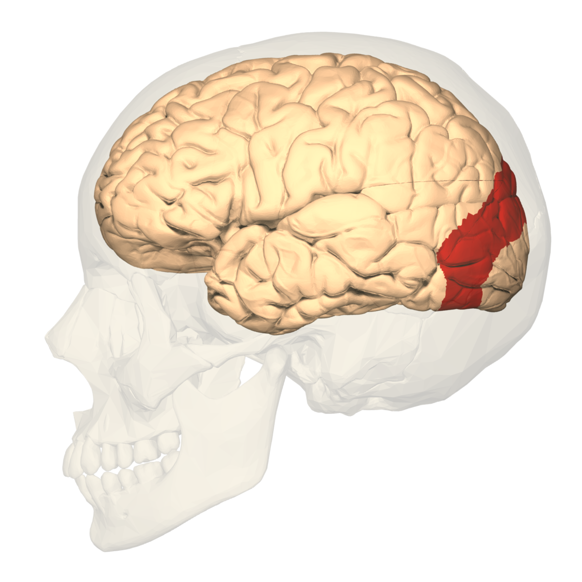
Occipital Lobe
Located in the back of the head is made up of (AA). It handles visual input from your eyes. Its lateralized by it gets information from the left part of each eye in the right visual field.
Visual Association Cortex/Area
The parts that recognize lines, angles, shapes, and shadows.
Temporal Lobe
Processes sound sensed by our ears and houses long term memory (not lateralized).
Wernicke’s Area
Located mostly in the temporal lobe. Interprets written and spoken speech (left hemisphere). Has a type of aphasia that causes a person to not understand spoken or written language but can still speak.
Brain Stem
The most primitive part of the brain, which is the base of the brain connected to the spinal cord. It contains the mid-brain, pons, medulla oblongata.
Medulla Oblongata
Located at the top of the spinal cord. It controls heart rate, breathing, blood pressure, and swallowing.
Pons
The larger swelling above the medulla. Controls things like breathing, facial expressions, and the sleep cycle. If there is damage to this area, it can cause locked in syndrome.
Midbrain
Located at the top of the brain stem. It controls digestion, eye movement, and swallowing.
Reticular Activating System
A network of nerves (NOT tissue) that carry messages between parts of the brain stem. It regulates the intensity of pain, arousal, alertness, and eye movement. Helps people focus on useful sensory input but filters out unneeded things. Damage to this area causes comas.
Thalamas
Located on top of the brain stem. Sensory “relay station” of the brain, passing information to the cerebral cortex. Receives information from all senses except smell.
Cerebellum “Little Brain”
Helps control posture, balance, fluid movement, and coordination of fine voluntary movements.
Limbic System
Located beneath the cerebral cortex. Its the emotional control center of the brain. It houses the reward system (dopamine). It includes thalamus, hypothalamus, pituitary gland, hippocampus, and amygdala.
Hypothalamus
Controls hunger, thirst, body temperature, circadian rhythm. The four F’s ( Fighting, Fleeing, Feeding, Fucking). If this area is damaged it impacts signals for hunger.
Amygdala “Fear Center”
Vital for basic emotions and survival responses. Is the reason for fight or flight response and fear or aggression.
Glial Cells
The brain is made up of these cells; support and nourish neurons; create myelin sheath. Einstein had more than normal of these in his brain.
Neuroscience
Conduct research on the brain and nervous system. They study brains through case studies like Phineas Gage.
Phineas Gage Accident
Tamping iron went through the left frontal lobe and he stayed alive. This case study help to reveal that personality is held in the prefrontal cortex.
Prefrontal Lobotomy
Operation that removes parts of the brain to control behavior like bipolar .
Brain Autopsy
Examination of brain tissue after death, is currently the only definitive way to diagnose certain neurodegenerative disorder.
Hemispherectomy
Brain surgery that removes one of the halves of the brain to control seizures.
Lesioning Studies
Humans with brain lesions (damage) are often the subjects of research with a goal of understanding the function in the part of the brain, like Phineas Gage.
Brain Scanning
Is not used to treat disorders rather they provide information about the structure of the brain.
Electroencephalogram (EEG)
A measurement of electrical activity of the brain by recording from electrodes placed on the scalp. Its used to diagnoses damage and disorders.
Computer Tomography (CT or CAT Scan)
X-ray photos from different angles and used to create a 3D representation of living tissue without surgery. It can identify abnormalities like a muscle or bone disorder, tumor, or blood clot. Reveals vivid horizontal slices of the brain and is the least expensive option.
Magnetic Resonance Imaging (MRI Scan)
Using radio waves and magnetic fields of the body to produce 3D detailed images of the brain’s damage. Is it more precise than other scans but more expensive. The machine uses energy to construct and image.
Functional MRI (fMRI)
Measures the brain activity, detects the changes in blood oxygenation and flow that occurs in response to neural activity. More activity = more oxygen. This Scan shows brain structure and function.
Positron Emission Tomography (PET scan)
A visual display of activity that detects where a radioactive form of glucose goes while the brain preforms a given tasks on living cells.
Brain Plasticity
Constantly change both structure and function of many cells in the brain in response to experience and trauma.
Split Brain
Hemispheres can’t communicate due to a lack of corpus callosum, each side of the brain acts by itself. Important Neuroscientists include Roger Sperry and Michael Gazzaniga. This procedure is done for epilepsy.
Nervous System
The body’s fast communication system between the brain and nerve cells or neurons.
Nerve Cell or Neurons
Specialized cells that send a neuron impulse.
Spinal Cord
A two way information processing system connecting the CNS and PNS.
Central Nervous System (CNS)
Coordinates action of the brain and spinal cord.
Sensory Neurons
Carry’s information from nerves and tissue inward to the CNS.
Motor Neurons
Carry’s information outward to body’s muscles and glands (PNS)
Interneurons
Neurons within the brain and spinal cord that communicates internally. Middleman neurons between sensory and neurons.
Peripheral Nervous System (PNS)
Relays messages from the CNS to the rest of the body.This includes the somatic and autonomic systems.
Somatic Nervous System
Controls voluntary movements of muscles and is a nervous system.
Autonomic Nervous System
Controls automatic, involuntary functions of the body. Auto pilot. Divided into sympathetic and parasympathetic.
Spinal Reflex Arc
A signal is sent from a sensory organ to the spinal cord, which processes the information instead of passing it all the way to the brain. Shows how CNS and PNS work together. Sensory, motor, and interneurons all work together in this reflex.
Sympathetic Nervous System
Emergency response system; fight, flight, or freeze. Scared or excited. Automatically accelerates your heartbeat, blood pressure, sweat, sugar levels, slows your digestion, and dilates purples.
Parasympathetic
Function to calm a person after a stressful event. Everything slows and returns to normal.
Endocrine System
A collection of glands that slowly secrete hormones into your blood. Slow to take effect, long lasting.
Hormones
Produced in the Endocrine system and are secreted into blood. Slow to take effect but is long lasting.
Pituitary Gland
Master gland; located in the brain, controlled by hypothalamus. Regulates stress, growth, and reproduction. It sends stress messages to adrenal glands.
Thyroid Gland
Located in the lower neck, regulates metabolism, growth, and appetite.
Pineal Gland
Lacated in the back of the brain, secretes a hormone called melatonin which regulates sleep.
Adrenal Gland
Two glands on the top of each kidney involved in stress response. Releases epinephrine and norepinephrine (adrenaline) and cortisol (primary stress hormone). Increases heart rate, blood flow, and blood sugar, creates energy. Dilates airways, used with allergic reactions.
Oxytocin
Powerful hormone that regulates social interaction, sexual reproduction, and bonding.