Orbit and Nasal Cavity
 Call Kai
 Learn
 Practice Test
 Spaced Repetition
 Match
 Flashcards
 Knowt Play
1/27

There's no tags or description
Looks like no tags are added yet.
28 Terms
What is the FUNCTION of the Nose?
1) Nose is a projection from the face.
Function is to filter dust entering the respiratory system.
What is the STRUCTURE of the Nose?
1) Bones, comprised of paired nasal bones, maxillae, frontal bone and septum.
2) Cartilages, comprised of 3 main: lateral, major alar and septal

What is the STRUCTURE of the Nasal Cavity?
1) Septum divides the chamber into two cavities
Internal surface, lateral wall has three projections; superior, middle and inferior conchae
2) Nasal cavity opens into nasopharynx
3) Supplied arterial branches from ophthalmic and maxillary.
4) Nerve supply from olfactory nerve, ophthalmic (V1 and maxillary V2)

Cranial Nerve 1 - OLFACTORY

What is OLFACTION?
1) Olfaction is aided by the air flow through the nasal cavity
2) Conchae help warm and moisten the air
3) Olfaction receptors are in the epithelium lining the roof and walls, the epithelium secretes a mucus fluid, odoriferous gases are dissolved into the fluid and then detected by the olfactory nerves

What is STRUCTURE and FUNCTION of the Paranasal Sinuses
1) Paranasal sinuses are air filled extensions of the nasal cavity
2) Paired frontal sinuses in frontal bone that drain through frontonasal duct.
3) Ethmoidal cells are invaginations in the ethmoid bone and drain into middle meatus (if anterior, or middle) or superior meatus (if posterior).
4) Sphenoidal sinuses in sphenoid bone that drain into spheno-ethmoidal recess.
5) Paired maxillary sinuses in maxilla drains into middle meatus.

The ORBIT - Bones
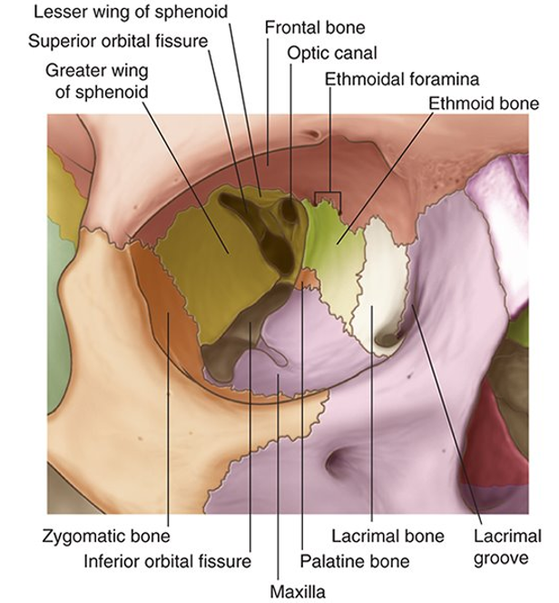
Made up 7 bones:
1) Frontal
2) Maxilla
3) Ethmoid
4) Lacrimal
5) Sphenoid
6) Zygomatic
7)Palatine

The ORBIT - Openings
Several openings into the orbit:
1) Optic canal
2) CN II and ophthalmic artery
3) Superior orbital fissure
4) CN III, IV, V1 & VI
5) Inferior orbital fissure
6) CN V2
What is the FUNCTION of the Eye?
The eyeball contains the optical apparatus for the visual system
What is the STRUCTURE of the Eye?
1) Located in orbit, suspended by muscles
2) 3 layers
Outer fibrous- sclera and cornea
Vascular- iris
Inner- retina
3) Divided into anterior, posterior, vitreous chamber

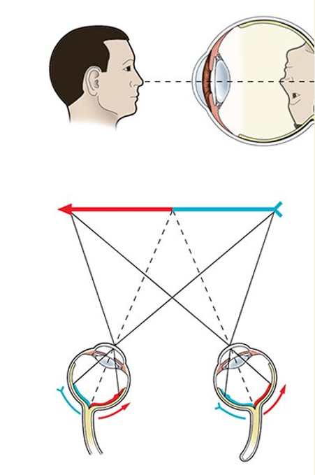
How does the VISUAL FIELD function?
1) Visual information received by the retina
2) Impulses carried through CN II (Optic nerve)
3) Lateral visual field is processed by contralateral portion of the occipital lobe

Why is the Visual Fields important?
1) The crossing of visual fields between eyes is important for binocular vision
3D vision
Depth perception
2) Things are seen backwards and inverted
3) Visual cortex processes the information to “flip” the image

Cranial Nerve 2 - OPTIC

Clinical Consideration of Optic Nerve
Swelling of the gland can impact on the optic nerve causing disturbances in vision.

What does the MOVEMENT OF THE EYE involve?
7 extraocular muscles:
1) Levator Palpebrae
2) Superior Oblique
3) Superior Rectus
4) Medial Rectus
5) Lateral Rectus
6) Inferior Rectus
7) Inferior Oblique

Movement of the Eye

Anatomical Organization of the Eye

Extra Ocular Muscles responsible for Eye Movement

Overview of Eye Movement

Cranial Nerve 3 - OCULOMOTOR

Cranial Nerve 4 - TROCHLEAR


Cranial Nerve 6 - ABDUCENT

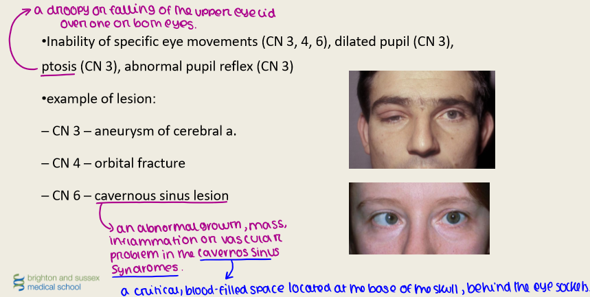
Clinical Considerations of Cranial Nerve 3,4,6
What is the Mechanism of CRYING?

Parasympathetic Supply to the Lacrimal Gland
1) Sensory innervation
Lacrimal nerve (branch of V1)
2) Parasympathetic innervation
Secretomotor function via facial nerve (CN VII)
3) Sympathetic innervation
Vasoconstrictive (sup. cervical ganglion, via internal carotid)

Cranial Nerve 5 - TRIGEMINAL

V1 - OPTHALMIC

Question 1
