OMS1- CRS I
 Call Kai
 Learn
 Practice Test
 Spaced Repetition
 Match
 Flashcards
 Knowt Play
1/239

There's no tags or description
Looks like no tags are added yet.
240 Terms
thoracic vertebra
have costal facets where ribs will articulate with vertebral bodies
superior and inferior demifascets on vertebral bodies that make up the articulation with the ribs when put together. Except rib one, where T1 has a complete fascet
transverse costal facets on transverse processes where rib tubercles articulate while running under the transverse process (although ribs 11-12 do not have costotransverse joint as they do not have tubercles)

parts of sternum
jugular notch
manubrium
body
xiphoid process
on sides are articular facets for clavicle and costal cartilage
sternal angle
formed from manubrium articulation with the body, major landmarks or orientation
corresponds to second costal cartilage (easier to find than rib one)
T4/T5 intervertebral level, forms transverse thoracic plane (separted superior and inferior mediastinum)
types of ribs
True Ribs (1-7): have own costal cartilage that articulates to sternum
False ribs (8-10): costal cartilage joints cartilage above instead of going to sternum
floating ribs (11-12): no anterior attachment. more mobile.
what is in the costal groove?
on interior and inferior aspect of rib, contains intercostal vein, artery, and nerve.
important for invasive procedures
a thoracentesis needle should be immidiately superior to rib

atypical rib features: rib 1
scalene muscles attachments
subclavian artery/vein attachment
brachial plexus
atypical rib features: rib 2
attachments for serratus anterior
whis ribs articulate with 1 vertebral body?
ribs 1, 10, 11, 12
sternocostal joints
1st rib joint: syncondrosis (hyaline cartilage => immobile)
ribs 2-7 are synovial plane joints
costochondrial joint
between ribs and costal artilage
syncondroses joints
little to no movement
stuctures entering thoracic aperture
aka throacic inlet, opening boardered by rib one
trachea
esophagus
apex of lungs
great vessels
vagus nerves
phrenic nerves
sympathetic trunks
inferior thoracic aperture boundaries
T12
Ribs 11-12
costal margin
xiphisternal joint
closed by diaphram
how are intercostal spaces named?
named for the rib above it
phrenic nerve
arises from C3-C5
C3, 4, 5 keep the diaphragm alive
enters thorax anterior to subclavian artery
passes anterior to lung root then a long pericardium to diaphram

diaphragmic openings
I 8 10 Eggs At 12
T8: caval opening (inferior vena cava)
T10: esophagus
T12: aorta

rib movement- pump handle
for ribs 2-6
anterior aspect and sternum of ribs move upwards and forward
increases chest depth (AP diameter)
rib movment- bucket handle
in ribs 7-10
ribs elevate, lateral aspect move outward/upward
increases thorax transverse diameter
overall, increasing thoracic volume creates a negative pressure gradient so air naturally flows into lungs
rib movement- caliper motion
in floating ribs (11-12)
has greater mobility, anterior aspect moves laterally during inhalation
increases expansion of lower thoracic cavity
what ribs are most commonly fractured
ribs 5-9 since they have little protection over the lateral thoracic wall.
most dangerous ribs to get fractures are 1-2 due to neurovasculatiue structures
ribs 9-12 are more mobiles and fracture less, but pose an organ damage risk
mediastinum boundaries
superior: superior thoracic aperture
lateral: parietal pleura
inferior: diaphram
anterior: ribs
posterior: thoracic vertebrae bodies
inferior mediateinum divisions
anterior: in front of heart
middle: heart
posterior: behind heart
superior mediastinum boundaries
supeiror thoracic aperture
transverse thoracic plane
manubrium
T1-T7 vertebral bodies
superior mediastinum contents
superficial to deep
thymus: anterior to fiberous pericardium, aorta, and great veins. posterior to manubrium
brachiocephalic veins
superior vena cava
aortic arch plus branches
phrenic nerve
vagus nerve
trachea
esophagus
brachiocephalic veins
fusion of internal jugular and subclavian veins
anterior to aortic arch branches, trachea, vagus nerve
left is longer than the right since it crosses over to right side of heart to drain into superior vena cava
superior vena cava
drops into median mediastinum to heart
anterior to trachea and right vegus nerve
right of the ascending aorta and brachiocephalic trunk.
aortic arch
starts to curve at TV4/TV5. once it drops, it becomes descending aorta at that level
arches towards the back over left lung root
3 banches: brachicephlic trunk, left carotid artery, left subclavian artery
attaches to pulmanary trunk through ligamentum arteriosum
vagus nerve
enters superior mediastinum lateral to common carotid arteries, posterior to lung root (phrenic is anterior)
parasympathetic and visceral sensory to thoracic and abdominal viscera
anterior vegal trunk travels with esophagus into abdomen
posterior vagal trunk travels with trachea into abdomen
left recurrent laryngeal nerve
loops under aortic arch to ascend to larynx
aortic aneurysm can cause voice issues
trachea
starts an inferior boarder of cricoid cartilage (C6)
bifurcates at TV4/TV5
posterior to great vessles
esophogus
continuation of pharynx starting at C6
posterior to trachea, at T10 exits thorax at the esophageal hiatus of diaphram
narrowing points: Pharyngoesophageal junction, aortic arch and tracheal bifurcation, diaphragm.
anterior mediastinum contents
part of thymus
fat and connective tissue
lymph nodes
sternopericardial ligaments
middle mediastinum contents
heart (left atrium is anterior to esophagus)
great vessels
phrenic nerves => anterior to lung roots
main bronchi lateral to heart
esophagus
posterior mediastinum contents
descending thoracic aorta: passes through diaphragm at aortic hiatus (T12)
Azygous system of veins: ascends vertebral column, arches over right lung to superior vena cava
thoracic lymphatic ducts
thoracic splanchnic nerves
azygous system of veins
azygous vein is supplied by:
hemiazygous vein: on left side, ascends vertebral column and drains around T9
Accesory hemiazygous vein: descends on left side, crossed to azygous at T8
thoracic duct
major lymphatic drainage of body, driven by diaphragm motion
starts at cisternae chyli (large lyphatic duct that collect lymph from lower body) at L2
ascends behind esophagus on right
crosses to the left around T5, drains to left venous angle (where subclavian and internal jugular veins fuse)
thoracic splanchnic nerves
pregaglionic sympathetic fibers off the sympathetic chain (most of thoracic wall), will synapse at prevertebral ganglia in abdomen.
Greater splanchnic nerves (T5-T9)
Lesser splanchnic nerves (T10-T11)
Least splanchnic nerves (T12)
diaphram development
starts in week 4, after embryo folding. starts in cervical region and body will elongate around that. phrenic nerve will elongate as well
steptum transversum: a mesnchyme partition between embryonic thorax and abdomen
pleuroperitoneal membranes: grows in between thoracic and abdominal cavity. Two, directly inferior to where lungs will grow and continuous with septum transversium
mesentery of the esophagus: surround esophagus
muscles cells will arise from cervical somite mytotomes and fuse with membranous complex.
diaphramatic hernia
caused by failure of pleuroperitoneal membranes to grow across intramembranous body cavity. typically on the left
abdominal organs can extend into the thorax, shifting the mediastinum to the right. underdevelopment of lungs
with surgery, 70% survival
respirtory development
starts in 4th week, 5 stages. “Early pulms can’t suck air”
embryonic
pseudoglandular
canalicular
saccular (past this point, respiration is possible)
alveolar
embronic stage of respiratory development
lung bud appears, starts at gut tube. aka Laryngotracheal diverticulum. Will start to bifurcate into Bronchi, Bronchi bifurcate into lobar bronchi, lobar into segmental.
pulmonary veins and artery start to develop
lined by endoderm
cartilage, muscles, and CT (visceral pleura) arises from splanchnic mesenchyme surrounding lung bud during development
somatic mesoderm forms parietal pleura
separation of primordial pharynx
in 4th week
lung bud separates
tracheoesophageal folds must develop to separate GI from resp.
fold will fuse to form septum, forms a distinct esophagus dorsally and larngeotrachial tube ventrally
trachesophageal fistulas
congenital defect in the foregut structure separation
most common (85% of cases) is when the proximal end of esophagus is a dead end and distal end of esophagus is used to trachea. can’t eat, air in abdomen
esogagus can be disconinuous
communication between trachea and esophagus
esophagus can connect to trachea to food goes into lungs
esophagus can pass through trachea
visceral pleura
from splanchnic mesoderm
line parenchyma of lungs, is integrated into lungs
parietal pleura
from somatic mesoderm
lines thoracic body wall
pseudoglandular phase of respiratory development
6-16 weeks
conducting airways and terminal bronchioles develop
immature neuronal network
pre-acinar blood vessels
canalicular phase of embryonic development
16-26 weeks
primative alveoli, terminal bronchioles elarge.
terminal bronchioles will give rise to 2+ respiratory bronchioli, which will divide into 3-6 primative alveolar ducts
type 1 (wall of alveolus) and 2 (produces surfactants) cells
surfactant synthesis
lung tissue become highly vascularized
respiration is possible by the end of this stage
saccular phase of respiratory development
alveoli saccules develop
extracellular matrix and neural network mature
surfactant forms a film in alveolar sacs (prevents collapse)
acinus
terminal bronchial + respiratory bronchile + alveoli
respiratory development nmeuonics
Can’t breath till end of canalicular phase
capillaries invade during canalicular phase
surfactant is secreted in saccular stage
alveolar stage
32 weeks to 8 years
gas exchange area continues to expand
nerves and capillaries grow
continued proliferation in general
how does aminiotic fluid affect lung development
breathing it in opens lungs and kidneys to development. Low amniotic fluid will impair development.
respiratory distress syndrome
usually caused by surfactant deficiency in newbonrs
congenital lung hypoplasia
often associated with low amniotic fluid
decreased amounts of lung
can also be caused by a diaphamic hernia
picornaviridae family characteristics
small size
+RNA, icosahedral hapsid
non-enveloped
Three genera: enterovirus, hepatovirus, parechovirus
enterovirus genera
most are acid stable, transmitted fecal-oral
poliovirus
coxsackie A and B
Echovirus
rhinovirus (although it’s very acid sensitive compared to other enteroviruses, respiratory transmission)
numbered and lettered enteroviruses
parechovirus genus of picornaviridae
human parechoviruses replicate in respiratory and GI tract
diseases are asymptomatic or mild
Type 3: sever neonatal sepsis and central nervous system damage
coxsakivirus A24
upper respiratory infection
may cause acute hemorrhagic conjuctivitis
coxsakivirus A16
Hand-foot-and-mouth disease
coxsakivirus B1-B6
myocarditis
pericarditis
pleurodynia
respiratory infections
enterovirus A71
hand foot and mouth
neurologic complications- acute flaccid paralysis
echovirus
meningitis
febrile illness
rash
respiratory infection
enterovial disease
replicates in upper respiratory tract. wide variety of incubation times.
summer/fall disease
can spread into blood in primary viremia, can be stopped by memory antibodies
migrated to target tissues: skin, muscle, brain, meningies, liver, heart
replication in target tissues causes secondary viremia, leads to a more serious infection (such as polio => paralysis)
enteroviruses and rashes
most create some kind of rash with cold-like symptoms, especially coxsackie (A21 and A24) and echovirus (11 and 20).
macular rashes: flat red spots
macularpapilar rashes: flat red spots with one bump in the center
petechial rashes: small blood blisters
vesicular rashes: large boils
coxsaki A vs B
A: common cold and rashes, hand foot and mouth disease, and herpangina
B: can cause cardiac disease, responsible for half of viral cardiomyopathy. chest pain. rashes and cold-like symptoms
herpangina
typically caused by coxsackie A (8,10,16)
lesions appear on roof of mouth, not the tounge. Causes pain and difficulty swallowing
abrupt fever
no skin rash
will resolve on it’s own in a few days
typically infects 3-10yo
hand foot and mouth
typically caused by coxsackie A16 and enterovirus 71
lesion in mouth (including tounge), hands, and feet
slow, progressing fever
exanthem: macules or papules on skin
enanthem: oral lesion in the tounge and palet
coxsackievirus A viral tagert
ICAM-1; epithelial cells, fibroblasts, endothelial. Also rhinovirus target
a few bind to CD55-deacy accelerating factor. mainly in cells associated with blood stream
coxsackievirus B viral tagert
CAR (or CXADR), transmembrane adhesion molecule on cardiac muscle, brain, epithelial and endothelial cells
enterovirus 71 viral target
human scavenger receptor class B member 2 (SCARB2). on several tissue types
poliovirus viral target
CD155
hepatovirus viral target
HAVCR1 (TIM-1). allows it to enter hepatocytes
coxsackie B and pleurodynia
chest pain affecting pleura with sudden onset and worsens when breathing
abdominal pain and vomiting
lasts 4 days before full recovery
typically affects children under 15
coxsackie B and myocardial/pericardial infection
causes about half of the viral myocarditis infections
inflammation of myocardium, sudden worsening of heart failure. diagnosed with inflamatory markers.
inflamation of the pericardium surrounding heart, increased fluid within puts pressure on the heart (cardiac tamponade).
especially coxsackie B3,4,5
how is pericarditis diagnosed
imaging: heart has a flask-like shape
sharp chest pain, worsens when inhaling and laying back
pertinent positive: pain is reduced by sitting up and leaning forward
picornavairus genome
not large
looks like messenger RNA with a poly A tail but no 5’ cap.
at 5’ end is a IRES (internal ribosome entry site), an RNA stem loop. will be linked to VpG protein, has more to do with transcription than tanslation
one open reading frame creates a large polyprotein that is cleaved by 3Cpro. produces structural protein from P1, and nonstuctual protein from P2 and P3
picornovirus poly protein cleavage
by 3Cpro
primary cleavage: cleaves structural proteins P1,2,3
secondary cleavage: splits proteins up into individual, mature proteins
creates an RNA dependent RNA polymerase for amplification
picornivirus life cycle
virus binds to cells surface receptor through Vp1.
can be endocytosed or just have RNA injected into cell. VP4 is released to weaken capsid
RNA will undergo translation (Vpg is cleaved beforehand). protein processing.
at same time, genome is amplified. first transcribed into -RNA by RNA dependent RNA polymerase. then will be switched back to cyclic +RNA VpG will be added on
genetic material is packaged with help of VpG protein. creates provirion
another cleavage event produced mature virion.
picornovirus viral protein functions
3a: inhibits host cell protein secretion so more can be put towards viral assembly
3B: encodes VpG for transcription
3C: cysteine protease, cleaved polyprotein. has an RNA binding domain. also shuts off host cell transcription
3D: RNA dependent RNA polymerase
3CD: zymogen of 3Cpro. also recognizes cloverleaf sutucture at 5’ end of RNA to trigger replication
IRES
recruits ribosomal subunits for initiation. some host genes have these, mainly those associated with stress, mitosis, apoptosis
Vpg in transcription
viral protein
required for both + and - RNA sequence
primer to start transcription, causes it to be covalently attached to genomic RNA
needs to uridylated by 3Dpol before it can work as a primer, attached to Cre.
once uridylated, it will cause RNA to loop around and bind to 3’ end on poly A tail. 3Dpol them elongated
IDK, come back to this later. Schizo shit
structures in the pleural cavities
endothoracic fascia, outside parietal pleura and lines thoracic wall.
pleura: membrane surrounding lung. outer layer is parietal pleura, inner is visceral pleura. meet and fold back into the root of the lung. forms serous sack
costodiaphragmatic recess: the space between 2 layers of pleura along the inferior aspect as lung expands/contracts
costomediastinal recess: space between ribs as lungs change volume
pleural margins
places you want to insert a needle
midclavicular line: anterior, in the middle of clavical. Lung margin is rib 6, pleural margin is rib 8.
midaxillary lines: coronal, down the center of armpit. lung margin is rib 8, pleural 10
midscapular line: posterior, through the center of the inferior angle. lung, rib 10, pleural, rib 12
costophrenic angle
a radiography landmark. angle formed between diapragm and thoracic wall
can be blunted by fluid in pleural cavity
pleural effusion (hydrothorax)
fluid in the pleural cavity. can be blood (hemothorax) or air (pneumothorax)
removed through thoracentesis
structures unique to left lung
cardiac notch along superior lobe where heart protudes
lingula at the inferior aspect of the cardiac notch
2 lobes
root of lung
where two layers of pleura fold over eachother
contains the neurovasculatiue and trachea that enters lungs
pulmanary artery and veins
primary bronchus
bronchial arteries and veins
on right lung bronchus is anterior to pulmanary artery
on left lung bronchus is inferior to pulmanary artery
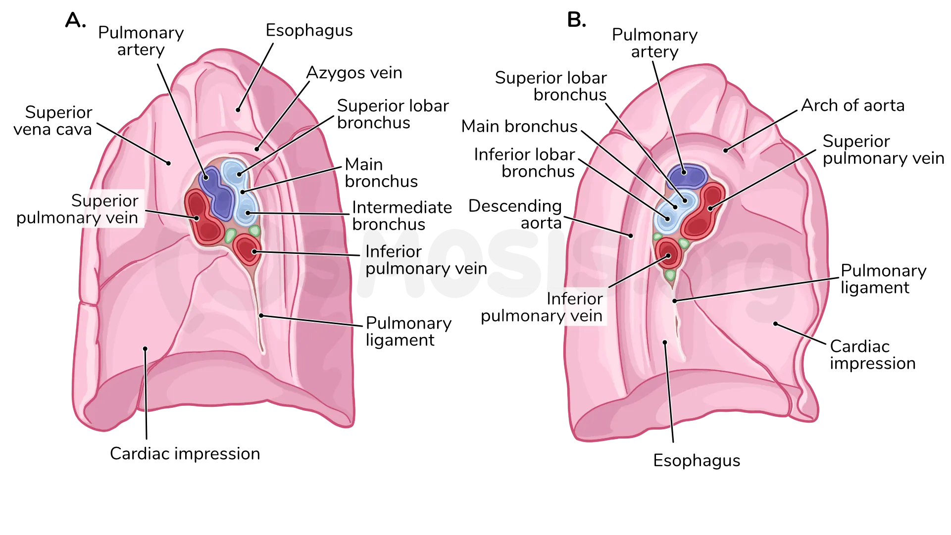
trachea
composed of C shaped hylan cartilage
where it sits against esophagus is just smooth muscle/connective tissue to allow for some movmenet
bifurcates at the TV4/TV5. midline strip at the junction is called the Carina.
Primary Bronchi
sits outside the elungs. Also C shaped hyaline cartilage
right opening is larger, so a forgine object will probably go there
left bronchus curves up more as it enters lung
lobar bronchi
branches off main bronchi
right lung has 3, left has 2
segmental/tertiary bronchi
segments off lobar branchi, 10 per lung. as they are functionally independent, one can be operated on and the rest of the lung is okay.
pulmanary vs bronchial circulated
pulmanary: blood that needs to be oxygenated
bronchial: much smaller, supplies lung tissues with blood. mainly branches off aorta
what nerves innervate pleural cavity
parietal pleura: somatic sensory, from phrenic (mediastina aspectl) and intercostal (anterior aspect) nerves
visceral pleura: visceral sensory innervation and autonomic pathway. localized somatic sensory pain.
lungs: both sympathetic (cardiopulmonary splanchnic nerves) and parasympathetic affect bronchodilation.
lymph flow of lungs
flows medially then superiorly, following the bronchi
bronchopulmonary nodes: at hilum
tracheobonchial nodes on primary bronchi
bronchomediastinal trunks: along trachea
right lyphatic duct/thoracic duct
normal echocardiogram checks
parasternal long axis (PLAX)
parasternal short axis(PSAX)
4 chamber
subcostal
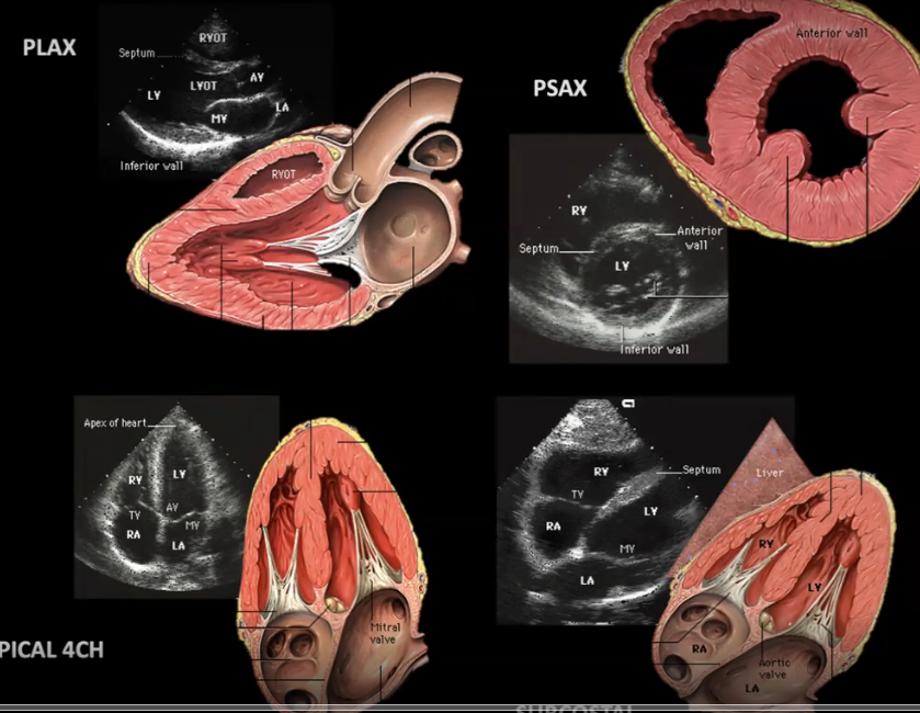
procedure for an echo
low frequency, phased array transducer. have pt roll to left side.
PLAX: 10 oclock or pt right shoulder
PSAX: 2 o clock or pt left shoulder
Apical: 3 o clock
SUBC: straight to pt left

PLAX
scan left of the sternum, to pt 10 o clock

PSAX
want to scan
(A) Base- asess anatomy and regurgitation
(B) mitral valve level- asseses mitral valve anatomy
(C)papillary muscle level- assess segmental wall motion and left ventricular systolic function
(D)apex- rule out apicalthrombosis

apical ultrasound
shows 4 chambers

SUBC
subcostal view. asses left ventrical function and right heart sive

layers of heart
parietal pericardium- on outside of heart, made of areolar tissue and mesothelium.
epicardium- separated from pericardium via pericardial cavity. areolar tissue and simple squamous mesothelium (epithelium of cavities). also called viseral pericardium.
myocardium- cardiac cells with connective tissues
endocardium- made of areolar tissue and simple squamous endothelia