A&P1 Nervous System
 Call Kai
 Learn
 Practice Test
 Spaced Repetition
 Match
 Flashcards
 Knowt Play
1/103

There's no tags or description
Looks like no tags are added yet.
104 Terms
Neurology
The branch of medicine that deals with the diagnosis and treatment of nervous system disorders (brain, spinal cord, and neurons)
Central Nervous System (CNS)
Makes up the brain and the spinal cord
Peripheral Nervous System (PNS)
Makes up the nervous tissue outside of the brain and the spinal cord
Somatic Nervous System (subdivision of the PNS)
Part of the PNS that includes neurons in the skin, limbs and sense organs (involuntary control), and motor neurons to skeletal muscles only (voluntary control)
Autonomic Nervous System
Part of the PNS that includes involuntary control of organs, cardiac & smooth muscles, and glands
Autonomic Nervous System (Sympathetic Division)
Controls flight or fight response under stress (increased heart rate & breathing, dilation of pupils, muscle tension, and decreased digestion)
Autonomic Nervous System (Parasympathetic Division)
Controls rest & digest response (decreased heart rate & breathing, increased digestion & salvation, normal bladder and bowel)
Enteric Nervous System
Involuntary, controls GI track
Sensory Function
Detects external and internal stimuli (see, hear, taste, touch, smell) (headache, hungry, stomach ache)
Integrative Function
Analyzes and stores sensory information and makes decisions about how to respond (cold = shiver, hot = sweat, hungry = eat)
Motor Function
Responds to information via muscle contractions or glands (hot = body sends signal to sweat glands to secrete sweat)
Two types of nervous tissue
Neurons & neuroglia
Neurons (nerve cells)
Produce nerve impulses (action potential)
Cell Body of Nerve Cells
Main part of the neuron, contains the nucleus and organelles
Dendrites of Nerve Cells
Branches that receive information and carry nerve impulses to the cell body
Axon
Usually a single branch which carries a nerve impulse away from the cell body to other neurons, muscles, or glands
Axoplasm
Cytoplasm of an axon
Axolemma
Plasma membrane of an axon
Sensory Neurons (afferent neurons)
Carry sensory information to the central nervous system (CNS)
Motor Neurons (efferent neurons)
Carries information from the CNS to muscles or glands
Interneuron (association neuron)
Connect sensory neurons to motor neurons, responsible for integration
Neuroglia
Makes up roughly ½ the volume of the (CNS), small and more numerous than neurons, provides support, nourishment, and protection to neurons.
How many types of Neuroglia are there?
There are six main types of neuroglia in the central nervous system: astrocytes, oligodendrocytes, microglia, ependymal cells, and in the peripheral nervous system, Schwann cells and satellite cells.
Astrocytes (neuroglia found in the CNS)
Support neurons - contains microfilaments that give them strength to support neurons
Make up the blood brain barrier (BBB)
Maintain appropriate chemical balances
May be involved in learning & memory by influencing formation of neural synapses
Oligodendrocytes (found in the CNS)
Form myelin sheaths in the central nervous system
Microglia (found in the CNS)
Phagocytize bacteria, dead cells
Ependymal Cells (found in CNS)
Forms cerebrospinal fluid
Schwann cells (found in the PNS)
Forms myelin sheathes around axons in the PNS
Satellite cells (found in the PNS)
Supports neurons and regulates exchanges
Myelin Sheath
Composed of lipids & proteins, produced by Schwann cells & Oligodendrocytes insulates an axon and increases speed or nerve impulse
Nodes of Ranvier
Gapes in myelin sheath
Nerve
A bundle of axons located in the PNS
Cranial Nerves
Connect brain to PNS (12 in body)
Spinal Nerves
Connect spinal cord to the PNS
Endonerium
Inner layer of nerve that surrounds individual axons and their myelin sheaths.
Perineum
Middle layer of nerve that surrounds nerve fascicles, providing support and protecting the axons within.
Epineurium
Outer layer of nerve that surrounds entire nerve, providing protection and support.
Ganglion
Group of nerve cells bodies in the PNS
Nucleus
A collection of nerve cell bodies in the CNS
Nerve Coverings
The three layers of tissue that surround and protect nerves, including the epineurium, perineurium, and endoneurium

Tracts
A bundle of axons in the PNS
Ascending Tracts
tracs that go up the spinal cord and carry sensry information
Descending Tracts
tracts that go down the spinal cord and carry motor information.
White Matter
Myelinated matter
Grey Matter
Unmyelinated matter
Graded Potentials
Used for short distance ocmmunication
Action Potentials
Used for long distance communication (muscles)
Nerve Action Potentials (nerve impulse)
The potential in a neuron (nerve cell)
Resting Membrane Potential (RMP)
Electrical potential difference (voltage) across the membrane of an excitable cell (-40 to -90mv) (millivolts)
Presence of Specific Types of Ion Channels
Allow specific ions to go across the plasma membrane
Types of Ion Channels
Leakage, mechanical, ligand, voltage-gated
Leakage Channel
Non-gated, always / mostly open
Mechanical Channel
Mechanical stimulation to open (vibration, pressure, stretching tissue)
Ligand Channel
Chemical stimulation to open (neurotransmitter, hormone, ions)
Voltage-Gated Channel
Require a change of voltage to open
Action Potential (impulse)
A sequence of rapidly occurring events that decrease and reverse the membrane potential and then eventually restore it to the resting state.
Generation of Action Potential
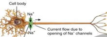
Voltage gated Na+ channels allow Na+ into the cell causing depolarization phase.
Voltage gated K+ channels allow K+ to flow out of the cell, producing repolarization phase and restoring the resting membrane potential.
The after-hyperpolarization phase occurs when the K+ channels remain open after the repolarization phase.
Action Potential Threshold
An action potential occurs when depolarization reaches the level of threshold, about -55mV in many neurons, that triggers the opening of voltage-gated Na+ channels, leading to a rapid influx of Na+ ions.
All or None Principle
Once the threshold for depolarization is reached, an action potential will fire at full strength, or not at all, regardless of the strength of the stimulus.
Depolarization phase
The initial phase of an action potential where the membrane potential becomes more positive than the resting membrane potential, due to the influx of Na+ ions through voltage-gated sodium channels.
Repolarization Phase
The phase of an action potential during which the membrane potential returns to a more negative value, due to the efflux of K+ ions through voltage-gated potassium channels.
After-Hyperpolarization Phase
The phase following repolarization where the membrane potential becomes even more negative (-90mV) than the resting potential, due to the continued efflux of K+ ions.
Refractory Period
The period during which a neuron is incapable of firing a second action potential, occurring after depolarization and repolarization phases due to inactivation of sodium channels.
Continuous Conduction (propagation of action potentials)
A type of nerve impulse transmission along an UNMYLINATED axon where the action potential is continuously regenerated at adjacent points along the membrane. (slower)
Saltatory Conduction (propagation of action potentials)
A type of nerve impulse transmission along a MYLINATED axon where action potentials jump between the nodes of Ranvier, increasing the speed of conduction. (faster)

Conduction Velocity
The speed at which an action potential travels along an axon, influenced by the presence of myelination, the diameter of the axon and temperature
Amount of Myelination
Action potentials propagate more quickly along myelinated axons
Axon Diameter
Larger diameter axons propagate action potentials faster than smaller ones
Temperature of Axons Effect
Axons propagate action potentials at lower speeds when cooled
Stimulus Intensity
Light touch - low frequency of action potentials
Firm pressure - high frequency of action potentials
(number of sensory neurons activated by the stimulus)
Synapse
Places where 2 neurons or a neuron and a muscle cell or gland meet, but don’t touch
Presynaptic Neuron
Neuron sending the impulse
Postsynaptic Neuron
Neuron receiving the impulse
Crossing the Synapse
Nerve impulses cannot cross the synapse without the release of neurotransmitters

Excitatory
Makes permeability less negative by increasing Na+ permeability
Inhibitory
Makes membrane more negative by increasing K+ permeability, inhibits or makes it less likely to transmit an impulse
Removal of Neurotransmitter
Diffusion
Enzymatic degradation (destruction) certain neurotransmitters are inactivate by enzymatic breakdown in the synaptic cleft
Re-uptake by cells, many neurotransmitters are actively transported back into the neuron that released them
Neurotransmitters
Both inhibitory & excitatory are in the CNS and PNS
Some neuron transmitters can be inhibitory & excitatory
Important neurotransmitters (acetylcholine, glutamate, aspartate, gamma aminobutyric acid, glycine, norepinephrine, epinephrine, and dopamine)
Neurotransmitter Effects
Agonist - anything that enhances a transmitters effects
Antagonist - anything that blocks the action of a neurotransmitter
Neuron Regeneration & Repair
plasticity maintained throughout life (sprouting new dendrites, synthesis of new proteins, changes in synaptic contacts with other neurons)
Limited repair for PNS, no repair for CNS
Neurogenesis
Formation of new neurons, can occur from stem cells in the PNS
Depression (Clinical Connections)
Linked to an imbalance of the neurotransmitters, serotonin, norepinephrine, and dopamine in the brain.
SSRI’s (selective serotonin reuptake inhibiters)
Seasonal Effective Disorder (Clinical Connections)
Depression symptoms experienced during the winter months, when days are short, overproduction of melatonin
Multiple Sclerosis (Clinical Connections)
Progressive destruction of myelin sheath surrounding neurons in the CNS
Unsure of cause (genetics, environment)
Feeling weak, abnormal sensations, double vision
Local Anesthetics (Clinical Connections)
Lidocaine (Novocain) - blocks nerve transmitters to pain centers in the CNS by binding to and inhibiting the function of Na+ channels
Epilepsy (Clinical Connections)
Periodic episodes of seizures caused by abnormal electrical firing of neurons in the brain
causes thought to be (brain damage, brain tumor, brain infections, drug use, unknown causes)
Treatment - Antiepileptic drugs, Keto diet can help