Stomach & Duodenum
 Call Kai
 Learn
 Practice Test
 Spaced Repetition
 Match
 Flashcards
 Knowt Play
1/81

There's no tags or description
Looks like no tags are added yet.
82 Terms
Where does the celiac trunk arise
Just underneath the diaphragm at T12
From the abdominal aorta is the celiac trunk the 1st, 2nd, 3rd,… single branch
1st single branch
3 main arteries in the celiac trunk
Common hepatic artery
Splenic artery
Left gastric artery
Common hepatic artery goes in what direction
Right
Common hepatic artery splits into what
Gastroduodenal artery
Hepatic artery proper (proper hepatic artery)
Gastroduodenal artery splits into what
Right gastro-omental artery
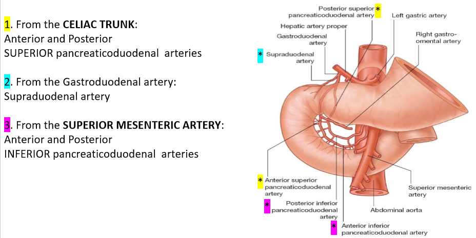
Supraduodenal artery
Superior pancreaticoduodenal arteries (Anterior and Posterior)
Hepatic artery proper splits into what
Right gastric artery
Left hepatic artery
Right hepatic artery → Cystic artery
The splenic artery splits into
Short gastric artery
Left gastro-omental artery
(In a spot test if there is a wiggly vessel superior to the spleen, it is the splenic artery. If there is a vessel on the back of the spleen it is the splenic vein)
The left gastric artery splits into
Oesophageal branches
Parasympathetic innervation of the celiac trunk
Vagus nerves (CN X)
sympathetic innervation of the celiac trunk
Thoracic splanchnic nerves (T5-T9)
Celiac ganglion
What arteries supply the midgut
Superior Mesenteric Artery
What arteries does the Superior Mesenteric Artery give rise to
anterior & posterior inferior pancreaticoduodenal arteries
How does the oesophagus travel through the diaphragm
The oesophagus travels through the oesophageal hiatus of the diaphragm (T10)
How is the oesophagus attached to the diaphragm
phrenico-oesophageal ligament
Where & with what does the oesophagus terminate
Terminates at the cardial orifice of the stomach with the inferior oesophageal sphincter (physiological) (Clinically significant!)
How many atomical & functional sphincters does the stomach have
1 anatomical
2 functional
What types of muscle are found in the oesophagus
1st 3rd = skeletal muscle
2nd 3rd = mix of 2
3rd 3rd = smooth muscle
What type of epithelium is the oesophagus lined with
non-keratinized stratified squamous epithelium
At what point does the epithelium change in the oesophagus when coming down to the stomach? What does it change to?
Changes at the Z line from non-keratinized stratified squamous epithelium to simple columnar epithelium
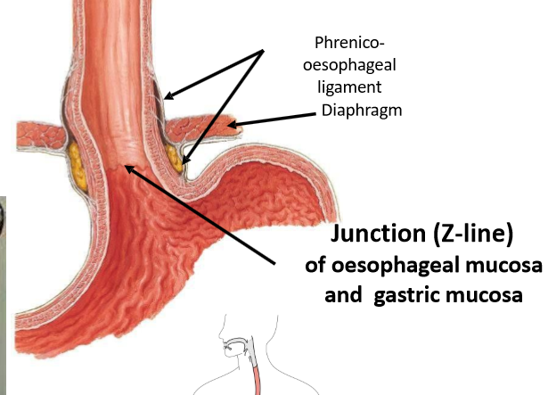
Arterial supply of oesophagus
oesophageal branches from the left gastric artery
Venous drainage of oesophagus
Left gastric vein → Portal venous system
Oesophageal veins → Systemic venous system (via azygos v.)
Lymphatics of oesophagus
Left gastric lymph nodes → Celiac lymph nodes
Innervation of oesophagus
Vagus
Greater splanchnic nerve
Why do babies spit up food a lot
They don’t have a fully developed diaphragm or sphincter
4 parts of the stomach
Cardia
Fundus
Body
Pylorus
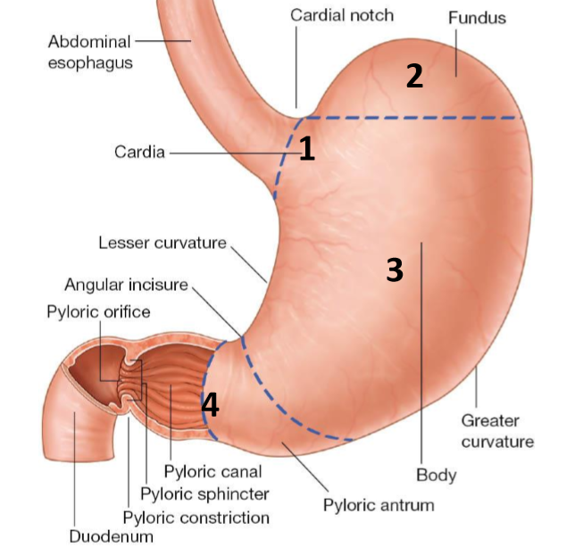
What vertebral level is the cardia stomach at
T11
Which part of the stomach is the dilated part
Fundus (contains gas / fluids)
Name the curvatures of the body of the stomach
Greater curvature
Lesser curvature
Role of pylorus
“Gate Keeper” that controls the outflow of chyme
Name the parts of the pylorus
Pyloric antrum → Chamber
Pyloric canal → Outflow region
What are gastric rugae made up of
Longitudinal folds of mucosa
Are gastric rugae temporary/permanent
Both:
Can be temporary - diminish as stomach distends
Can be permanent - form the gastric canal
What is the gastric canal
Groove that appears along the lesser curvature during swallowing
What is the Pyloric sphincter
A circular band of smooth muscle that forms the junction between stomach and small intestine - it is anatomical as it is circular
Meaning of rugose
Wrinkled
Arterial supply of the lesser curvature of the stomach
Right & Left gastric arteries
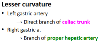
Arterial supply of the greater curvature of the stomach
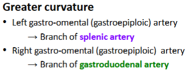
Arterial supply of the fundus of the stomach
All under umbrella of celiac trunk
Blood vessels to the stomach are anastomotic - what does this mean
If 1 blood vessel gets blocked there are many other vessels that can take over . Important because we don't want contents of stomach to enter abdomen
2 venous systems in the GI system
Portal & Systemic
Venous drainage of the stomach
hepatic portal vein
superior mesenteric vein
splenic vein
right gastric vein
Summary of stomach
Arterial supply:
Venous drainage:
Lymphatics:
Innervation:
Summary of stomach
Arterial supply: Celiac trunk
Venous drainage: Ultimately to the Hepatic portal vein → Portal venous system
Lymphatics: Ultimately drain by following the arteries towards → Celiac lymph nodes
Innervation: Vagus & Greater Splanchnic nerves
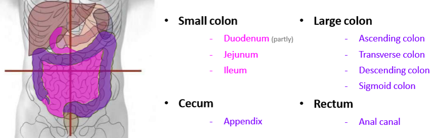
What are infracolic abdominal organs
The visceral structures related to the gastrointestinal tract that are located below the transverse colon
Name the infracolic abdominal organs
What are the parts of the duodenum
Superior part
Descending part
Transverse/Inferior/Horizontal part
Ascending/Fourth
Which part of the duodenum is the longest
Transverse part
Is the superior part of the duodenum Intraperitoneal / Secondarily Retroperitoneal / Mostly Retroperitoneal
Intraperitoneal
What is the part of the duodenum that is continuous with the pylorus of the stomach called
Duodenal cap or ampulla (ampulla = collecting area for stuff)
At what vertebral level does the stomach meet the duodenum
L1
Attachments to the superior part of the duodenum
Hepatoduodenal ligament connects superiorly
Greater omentum connects inferiorly
Blood supply to the superior part of the duodenum
Celiac trunk
Is the descending part of the duodenum Intraperitoneal / Secondarily Retroperitoneal / Mostly Retroperitoneal
Secondarily Retroperitoneal
What organs does the descending part of the duodenum have important relationships with
Pancreas & gallbladder
Position of the descending part of the duodenum
Lies right of the midline (approx. L2) and is anterior to the hilum of the right kidney
Blood supply to the descending part of the duodenum
celiac trunk AND superior mesenteric artery
Internal features of the descending part of the duodenum
Major duodenal papilla (aka greater duodenal papilla)
Minor duodenal papilla (aka lesser duodenal papilla)
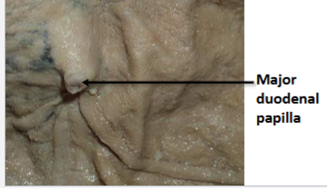
What is the major duodenal papilla (what is it made up of? what are its functions)
Elevation of mucosa protruding into the duodenum and is larger due to the hepatopancreatic ampulla and sphincter internally within the head of the pancreas
Opening of the common bile duct and the main pancreatic duct
Releases pancreatic juice and bile
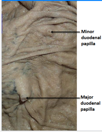
What is the minor duodenal papilla (location? what are its functions)
Releases pancreatic juices only
Where the accessory pancreatic duct opens
Located approximately 2cm superior to the major duodenal papilla
Is the transverse part of the duodenum Intraperitoneal / Secondarily Retroperitoneal / Mostly Retroperitoneal
Secondarily Retroperitoneal
The path of the transverse part of the duodenum runs over/under some blood vessels. What vessels? Over/Under?
Crosses posteriorly under the superior mesenteric artery (and vein)
Passes anteriorly over the inferior vena cava
What part of the pancreas & mesentery does the transverse part of the duodenum have a relationship
Closely related to the uncinate process of the pancreas and the root of the mesentery
The transverse part of the duodenum lies at what vertebral level
L3
In what direction does the transverse part of the duodenum go
Crosses from right to left
Blood supply to the transverse part of the duodenum
Superior mesenteric artery
Is the ascending part of the duodenum Intraperitoneal / Secondarily Retroperitoneal / Mostly Retroperitoneal
Mostly Retroperitoneal
Where does the ascending part of the duodenum terminate
Duodenojejunal flexure
What blood vessel is the ascending part of the duodenum closely related to
abdominal aorta
What vertebral level is ascending part of the duodenum at
Ascends up to approx. L2
Blood supply to the ascending part of the duodenum
superior mesenteric artery
The ascending part of the duodenum is supported by a structure. What structure
A suspensory ligament: The Ligament of Treitz
What does the Ligament of Treitz connect the duodenum to
to the diaphragm and the posterior abdominal wall
Function of the Ligament of Treitz
Supports the ascending part of the duodenum
Facilitates with the movement of the intestinal content
Why is the Ligament of Treitz clinically significant
Ligament clinically divides the gastrointestinal tract:
An upper GI tract bleed is a bleed that is proximal to the Ligament of Treitz:
Blood appears in vomit (Haematemesis)(M for mouth - haemateMesis)
Stool is dark & “tarry” (Melena)
A lower GI tract bleed is a bleed that is distal to the Ligament of Treitz:
Red blood appears in the stool (Haematochezia)
Blood supply to the duodenum
Summary of the Duodenum
Arterial Supply:
Venous Drainage:
Lymphatics:
Innervation:
Summary of the Duodenum
Arterial Supply: Celiac trunk & Superior mesenteric artery
Venous Drainage: Ultimately to the Hepatic portal vein → Portal venous system
Lymphatics: Ultimately drain by following the arteries. Anterior surface towards → Celiac lymph nodes. Posterior surface towards → Superior Mesenteric lymph nodes
Innervation: Vagus nerve, Greater splanchnic & lesser splanchnic nerves
What marks the duodenojejunal flexure – junction between the duodenum and jejunum
Ligament of Treitz
What is a bezoar
an impacted mass of material in the GIT
What can cause a bezoar
Rapunzel syndrome - Psychiatric condition that generally affects young girls - get addicted to eating hair - Hair forms a trichobezoar - can't be digested
What is a phytobezoar made up of
Vegetable fibres
How is a phytobezoar cleared
Coca cola
What supplies the midgut
Superior mesenteric artery