bacterial and parasitic persisters TUESDAY 11/3
 Call Kai
 Learn
 Practice Test
 Spaced Repetition
 Match
 Flashcards
 Knowt Play
1/74

There's no tags or description
Looks like no tags are added yet.
75 Terms
bacterial persisters to know:
brucella abortus
chlamydia abortus
coxiella burnetii (q fever)
mycobacterium avium subsp. paratuberculosis (johne’s)
salmonella enterica subsp. enterica serotype dublin
which bacterial persistors are G-?
all of them (b. abortus; c. abortus, c. burnetii, salmonella enterica…) except mycobacterium, which is acid-fast
bacterial persisters that shed through birthing material:
b. abortus (also milk), c. abortus, c. burnetii (also feces, urine)
bacterial persistors that shed through feces
mycobacterium (also mammary secretions) and salmonella
bacterial persistors that are facult. intracellular:
b. abortus, mycobacterium and salmonella
bac. persisters that are obligate intracellular
c. abortus and c. burnetii
b. abortus and chlamydia abortus cause what dz in the reservoir?
primiparous abortion
c. burnetii causes what dz in reservoir?
abortion - any parity
mycobacterium causes what dz. in reservoir?
chronic diarrhea w malabsorption / wasting
salmonella causes
septicemia (sepsis) especially w embolic spread to lung (interstitial pnuemonia)
these bac have a bovine reservoir
b. abortus, mycobacterium, and salmonella (adults shed, calf gets dz)
ovine reservoir
c. abortus and c. burnetii (also ovine reservoir)
short environmental bacterial persisters
b. abortus and c. abortus
which bac persister lasts months to years on surfaces (bacterial persister)
salmonella
(4-7 months in soil)
long lasting bacterial persisters
c. burnetii and mycobacterium - both depending on surface
effective vx for these bacterial persisters?
b. abortus (vx F cattle); c abortus (vx F sheep - marginal shed reduction)
no vx for these persisters:
c. burnetii (in US); mycobacterium; salmonella has vx but ineffective!
bac persistors that remove birthin material for biosecurity prevention/control
c. abortus and c. burnetii
bac persistors that use culling to improve biosecurity
b abortus, mycobacteriu, and salmonella
protozoan examples:
giardia (a diplomonad); tritrichomonas (a trichomonad); cystoisospora and eimeria (apicomplexans / coccidia)
taxonomy of giardia
a flagellated protozoan
worldwide dist.
in small intestine
direct life cycle
giardia morphology
trophozoites in small intestine
cysts are closer to external environment, giardia form cysts to protect themselves, very HARDY
no replication in cysts
giardia diagnostic tests
immunofluorescent assays - (giardia cysts & cryptosporidium cysts go GREEN)
done regularly in lab
wet mount - mix stool + saline, look for motile trophozoites
centrifugal flotation - finds cysts (need sugar solution - salt too high SpG)
immunodiagnostics like ELISA
swab STOOL
challenges with diagnosing giardia:
intermittent shedding - repeat testing good
often confused w/ other stuff (bc small and refractile)
low sensitivity in wet mounts
persistent detection of cyst wall antigen in chronically infected asymp adult dogs
giardia infective stages
cysts! are infective
giardia diagnostic stages
both trophozoites and cysts can be diagnosed, but only cysts are infectious bc trophozoites don’t survive in the enviro
how is giardia transmitted?
through ingestion of cysts
very prevalent in small animals
what is a prepatent period?
the time from ingestion of cysts to shedding cysts in feces (for giardia)
clinical signs of giardia caused by (which stage)
trophozoites caused clin signs
attach → blunt intestinal villi → maldigestion, malabsorption, diarrhea
clinical signs of giardia are highly variable, and can be asymptomatic or can include
chronic/pasty diarrhea (could also be normal school), wt loss
lethargy, farting
failure to thrive, impacts on growth
+/- dry skin, poor haircoat
protocols for prevention and control of giardia
avoid contact w contaminated water/food sources
utilize disinfectants where possible
bathe them once treated (cysts sometimes on fur, can reingest when grooming)
giardia has different __ which means when both animals and humans are infected in the same house, it does not necessarily imply zoonotic transmission
_ assemblages _ (diff assemblages in diff species probably)
tritrichomonas blagburni: what to know
“trich”
casuses GI dz in small animals, devastating dz and repro dz in herd animals
no hardy enviro form, ONLY trophozoites
cystoisospora and eimeria are
apicomplexans (“coccidia”)
pretty hardy in environment (pick up poop before oocysts sporulate and become infectious!)
apicomplexan life cycles always include a phase called “__”
“sporogony”
oocysts produced → sporogony → sporulate
sporogny happens in environment (unsporulated oocyst exits host)
becomes infectious after sporulation (needs time and conditions)
a platyhelminth is a type of helminth/worm characterized by
dorsoventrally flattened body
a trematode is a type of platyhelminth, known as a __
_ fluke _
fasciola hepatica lives WHERE in the host?
in the liver/bile ducts
(“common liver fluke”)
hosts for f. hepatica:
common in herbivores (cattle, sheep, goats, rums, horses, pigs)
diagnosis of flukes/trematodes uses
sedimentation (can use the “fluke finder”)
can find fluke eggs bc heavy stuff sinks
can +/- use ELISA, slaughter data, seasonal occurrence, previous history, knowledge of snail habitat, and liver enzyme tests
important intermediate host for f. hepatica:
snail (important intermediate host)
morphology of f. hepatica
~1 cm long and leaf-like appearance
microscopic spines
ORAL SUCKER
f. hepatica form that penetrates snail host:
miracidium (ciliated motile form comes out of egg, moves through water looks for snail)
(egg leave liver of definitive host through bile ducts, travel thru SI, leave in stool)
in the snail, f hepatica goes from miracidium to…
…sporocyst, rediae, then cercariae
motile cercariae of f. hepatica leave snail and then…
…encyst on grass and form infective metacercariae (6 wks to months)
infective stage of f. hepatica is
metacercariae stage
diagnostic stage of f. hepatica
diagnostic stage is the eggs that leave in stool
sedimentate stool to find eggs
what intermediate host does the f. hepatica trematode use?
lymnae spp. snails are the IH
is f. hepatica zoonotic?
yes, it is zoonotic
also, has indirect life cycle
is a platyhelminth
f. hepatica and f. gigantica have ___ periods as well
_ prepatent _
(weeks in fasciola)
clinical signs of fasciola
hepatomegaly (enlarged liver)
ANEMIA!!!
sudden death (depends on host)
biliary obstruction and inflammation, fibrotic changes over time
ascities, bottle jaw (submandib edema), emaciation
fasciola causes ___ signs in cattle and __ signs in sheep
_ chronic_ and _acute_
chronic signs of fasciola in cattle
gradual wt loss, poor feed conversion, constipation
+/- anemia in calves
sometimes aberrant migration to lungs (cattle)
acute signs of fasciola in sheep
bloody nasal discharge, anemia, sudden death
associated w black’s disease (sheep)
treatment of fasciola
parziquantel (a common platyhelminth drug, used for tapeworms too)
albendazole (another drug)
avoid eating veg w metacercariae
kill snails in habitate
rotate pastures (reservoir hosts maintain pasture contamination)
comparing impact of environment (giardia vs trichomonads vs coccidia vs fasciola)
giardia: persistance of cysts in direct life cycle
trich: fragility of trophozoite (only stage)
coccidia: oocyst development - sporulate in enviro
fasciola: prevalence related to IH population (snails)
coccidian life cycles steps:
sporulated oocyst enters gut (has motile organisms in oocyst)
motile organisms released and start replicating
complete asexual reproduction (2nd round)
cells filled with asexual stages burst → find new cells to inhabit → more rounds of asex rep before making M and F
then you have M and F sexual stages that make oocysts
oocysts leave gut and reinfect next host
important details of coccidian life cycle as they relate to healthy animals:
coccidia is self-limiting, so in healthy animals eventually the coccidian life cycle will stop and the oocysts will be shed out
cytoisospora overview
an apicomplexan, commonly called “coccidia”
SMALL INTESTINE
life cycle of coccidia
merogony/schizogony = asexual phase
form invasive, motile forms
gamogeny/gametogony = sex phase & gamete production
form oocyst
sporogony = sporulation
dev of oocyst to infectivity
merogony/schizogony of a coccidian life cycle is…
…asexual, and forms invasive, motile forms
gamogony/gametogony in the life cycle of a coccidian is the…
…sexual phase with gamete production, and includes formation of the oocyst
sporogony in the life cycle of a coccidia is…
…sporulation and development of the oocyst to infectivity
cystoisospora and eimeria need to sporulate…
…in order to be infectious (as opposed to cryptosporidium, an apicomplexan that is shed already infectious bc sporulates endogenously)
a different depiction of the coccidian life cycle
consider: how time since infection change what is found on fecal examination
pre patent period might lead to false negatives in a sick animal
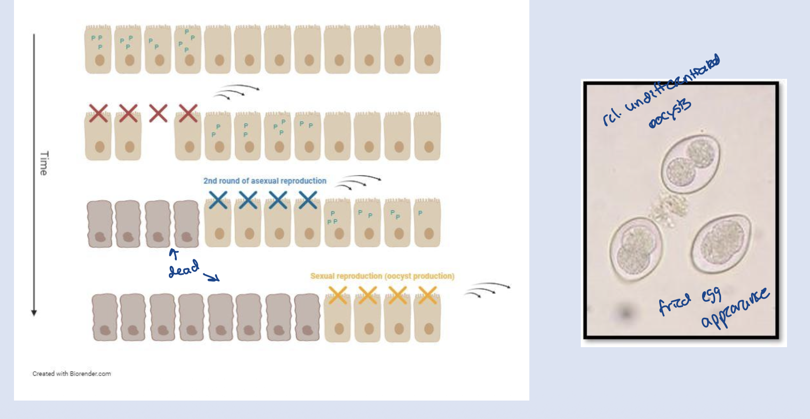
in the asexual phase of coccidia…
…the cell fills with high numbers of the organism and then burst (oh no!)
what is used to identify the various species of coccidia (crystoisospora spp.)?
sporulated oocyts are usually used to ID the various spp.
coccidia infections are more common in (what age group)?
more common in young animals!
cystoisospora are host specific or not host specific?
cystoisospora spp. are highly host specific!
prevalence of cystoisospora in dogs in north america:
prevalence ranges from 3% to 38%
clinical signs of coccidiosis include
diarrhea w/ wt loss
dehydration
+- hemorrhage (and/or bloody diarrhea)
severely affected animals may present w anorexia, vomiting, depression
treatment for coccidiosis
sulfadimethoxine is ONLY drug label approved for enteritis associated w/ coccidiosis
among newer drugs, ponazuril appears to be effective
control considerations for coccidia
once infective, oocysts are resistant to most disinfectants
pressure wash kennels, seal floors
pick up poop before sporulates!
flukes (trematodes) we have discussed
fasciola sp.
paragonimus sp.
tapeworms (cestodes) we have discussed:
diphyllobothrium sp. → marine life cycle, pi
echinococcus spp. and taenia spp. → scavenging/predation