Ch 7: Liver
 Call Kai
 Learn
Learn Practice Test
 Spaced Repetition
 Match
Match Flashcards
 Knowt Play
1/162

There's no tags or description
Looks like no tags are added yet.
Name | Mastery | Learn | Test | Matching | Spaced | Call with Kai | Chat |
|---|
No analytics yet
Send a link to your students to track their progress
163 Terms
Normal liver parenchyma sonographic appearance
homogeneous with medium level echoes. portal and hepatic veins appear as tubular fluid-filled structures. slightly more echogenic/isoechoic than renal cortex but less echogenic than pancreas and spleen
Liver Embryology: 4th week of development
Liver, GB, and bile duct system form from hepatic diverticulum
Liver Embryology: 6th week of development
right lobe increases in size, caudate forms from right lobe, and left lobe degenerates (becomes smaller). hemopoiesis occurs (formation of blood)
When does hemopoiesis occur, peak, and cease
occurs at week 6, peaks at 12-24 weeks, and ceases at birth
Liver Embryology: 10th week of development
Lymphocyte (WBC) formation and coagulation factors (10-12 weeks)
Liver Embryology: 13-16th week of development
Bile production begins
When does liver function in digestion
not until birth
What does the umbilical vein do
provides oxygenated blood and nutrients to fetus, and divides into left and right branches
Left branch of umbilical vein
Joins portal vein and enters the liver. after birth it becomes ligamentum teres (right ligament)
Right branch of umbilical vein
ductus venous, bypasses liver and flows directly into IVC. after birth it becomes ligamentum venosum
What ligaments can become recanalized and by what
ligamentum teres and venosum from portal hypertension
Why is color Doppler useful when evaluating a possible recanalized umbilical vessel
A recanalized vessel can resemble a contracted gallbladder, and color Doppler can confirm blood flow
Liver Physiology
It is an essential organ for life by performing >500 activities. some activities are:
factory for many chemical compounds
warehouse with short and long term storage capabilities
power plant producing heat
waste disposal plant excreting waste
chemistry lab regenerating tissue not too severely damaged
The types of cells that carry out liver functions
Hepatocyte in parenchyma, biliary epithelial cells, and Kupffer cells

Function of Hepatocytes
most abundant cell carries out most metabolic functions
Function of Biliary epithelial cells
line biliary system, bile ducts, canaliculi, and gallbladder
Function of Kupffer cells
Phagocytic (breakdown/eat structures/toxins that are not good for liver), belong to the reticuloendothelial system, and line the sinusoids
What does hepatic disease do to cell function
It alters the functions and produce identifiable clinical manifestations
What is Hepatomegaly
Enlarged liver beyond normal size
Hepatomegaly causes/presentation
Has nonspecific findings (does not mean pathological process occurring), could be caused from infection, hepatic tumors, congenital, or metabolic disorders. Will often present as abdominal mass
What is fatty infiltration (steatosis)
An acquired and reversible (if cause is eliminated) condition of extra fat build up inside your liver cells
Causes of fatty infiltration (steatosis)
2 main causes are alcohol abuse or obesity but also comes from accumulation of triglycerides in blood, or diabetes, steroid use, pregnancy, metabolic disorders, or high cholesterol
fatty infiltration (steatosis) clinical findings
may be asymptomatic (unless there’s significant amount of damage that changes physiology), hepatomegaly, or elevated LFTs
fatty infiltration (steatosis) Sonographic appearance/features
Diffuse increased echogenicity and results with decreased penetration, poor delineation of vessel walls (blends with hyperechoic liver tissue), and may be focal.
can decrease frequency or turn off harmonics to improve penetration

What is focal fatty sparing
When the liver is all fatty with some spots not affected, specifically near porta hepatis
Image: 2 areas of focal fatty sparing are seen in classic location as hypoechoic, irregularly shaped areas anterior to GB and portal veins. b/c the liver is now hyperechoic from fatty infiltration, normal tissue appears hypoechoic

What is focal fatty infiltration
When only specific areas of liver are fatty but otherwise it is normal
Image: A local irregular increased echogenic area in right lobe of liver compatible with focal fatty infiltration. does not displace vessels, suggesting that it is not a mass

What are two important clues that favor focal fatty change over a true liver mass
Irregular shape and lack of vessel displacement/internal vascularity
What is Glycogen Storage Disease
An autosomal recessive disorder (genetic) that allows excessive deposits of glycogen to be stored in the liver, intestines, and kidneys. Glycogen is stored in tissues but the body cannot synthesize it into glucose
Glycogen storage disease symptoms
Hypoglycemia, abdominal distension, fatigue, and irritability
What is vonGierke disease and sonographic appearance
Type I glycogen storage disease and is the most common type.
Appears with diffuse increased echogenicity and hepatomegaly
What liver mass can occur in von Gierke disease
Liver cell adenomas (40% of cases)
Types 3 and 4 glycogen storage disease complications
More often associated with cirrhosis and hepatocellular carcinoma (HCC)
How can viral hepatitis range in severity
mild or extensive
Types of hepatitis and their transmission
Hep A – parenteral or ingestion of contaminated food
Hep B, C, and D – sexual contact, contaminated blood, needles, body fluid
Hep E – contaminated water
Acute hepatitis clinical findings
Elevated LFTs, jaundice, RUQ pain, hepatomegaly, nausea/vomiting, and fever
Acute hepatitis Sonographic appearance
Normal to hypoechoic liver secondary to liver swelling, and “starry night” sign (portal vein walls seem more hyperechoic against liver parenchyma). Unlike fatty liver, this isn’t a penetration issue, we just need to adjust the gain.

Chronic Hepatitis sonographic appearance
Fibrosis causes course/hyperechoic parenchyma, and portal vein walls are less discrete
Complications from chronic hepatitis
Portal HTN and sequelae, or could progress to cirrhosis or liver failure
What machine adjustment can improve visualization of hepatitis
increase overall gain
What imaging-setting problem is more typical of fatty liver than hepatitis
Increased attenuation with poor sound penetration
What is Cirrhosis
Chronic and severe damage that destroys normal liver lobule architecture and leads to inflammation and subsequent necrosis. Is one of 3 types of alcoholic liver disease (other 2 being alcoholic fatty liver and alcoholic hepatitis)
What changes follow chronic inflammation in cirrhosis
Dense fibrous septa form, parenchymal cells degenerate, and regenerative nodules form.
Cirrhosis Clinical Presentation
RUQ pain, ascites, hepatomegaly, LFT abnormalities (depending on stage/extent of disease), or biliary/vascular channel blockage (which can lead to jaundice or portal HTN)
Cirrhosis Sonographic appearance
Varies during disease progression
early:hepatomegaly and possible textural changes indicative of diffuse disease (course/granular appearance)
Late: liver atrophy with caudate hypertrophy, surface nodularity (irregular border/edges, and can be seen bc of ascites outlining the liver), and loss of delineation of intrahepatic vasculature

Secondary findings to Cirrhosis
Portal HTN and splenomegaly, varies (abnormally enlarged/twisted blood vessels), or ascites
What are collateral shunts in cirrhosis
Secondary/accessory blood vessels that allow portal venous blood flow to bypass/route around blocked/damaged main vessel/damaged liver
Common collateral or varices found with cirrhosis
Recanalized umbilical vein, esophageal varices, and splenic varices

End-stage cirrhosis sonographic appearance
A small, lobulated or nodular liver with heterogeneous parenchyma, often outlined by ascites

Where may esophageal varices be seen sonographically
As anechoic vascular spaces posterior to the liver and anterior to the aorta. suspected esophageal varix should be confirmed with color doppler to demonstrate blood flow.

What can a recanalized umbilical vein be mistaken for and how to confirm it
the GB bc is can appear as a fluid-filled tubular structure near the liver if sonographer isn’t oriented to anatomy. it can be confirmed with color doppler and spectral doppler to show venous flow
Vascular changes with cirrhosis
Portal veins: reduced velocities and/or hepatofugal flow
Hepatic artery: enlarged with elevated velocities and turbulent flow (may have a corkscrew appearance with increased flow and aliasing)
Hepatic veins: become polarized (continuous flow and loss of multi-phasic appearance)

What is Hepatopetal flow
blood flow towards the liver. shows above the baseline on spectral doppler waveform

What is hepatofugal flow
blood flow away the liver. shows below the baseline on the spectral doppler waveform

What is assessed during a full abdominal doppler scan
3 hepatic veins, MPV, RPV, LPV, and hepatic arteries
Normal liver vascular flow
Arterial: hepatopetal, low resistance, continuous forward flow (above baseline)
Portal vein: hepatopetal, continuous monophasic flow
Hepatic vein: hepatofugal, triphasic and sensitive to respiration (below baseline)

What is Budd-Chiari syndrome and when is it clinically significant
Obstruction of hepatic veins and is clinically significant when at least 2 hepatic veins are occluded
Budd-Chiari syndrome symptoms
RUQ pain, jaundice, ascites, or hepatomegaly
Budd-Chiari syndrome acute clinical triad
ascites, hepatomegaly, and abdominal pain
Budd-Chiari syndrome etiology
Birth-control pills, tumor invasion from HCC (hepatocellular carcinoma), renal or adrenal carcinoma, severe metastatic liver disease, and Wilms tumor in pediatric patients
Can hepatic vein obstruction in Budd-Chiari be caused by external compression as well as intraluminal material
Yes. The lecture emphasizes that obstruction can result from material within the vein or compression from outside it
Budd-Chiari syndrome Sonographic Findings
varies depending on degree of obstruction/cause, hepatic veins are either not visible, narrow, or reversed flow (hepatopetal), or hypertrophy of caudate lobe. could also see portal HTN, intrahepatic collaterals, ascites, or splenomegaly.
color doppler may demonstrate enlarged caudate veins draining into IVC
What is passive liver congestion
Liver edema secondary to vascular congestion. commonly related to heart failure, especially right side
clinical findings of acute passive liver congestion
hepatomegaly or RUQ discomfort
Passive Liver congestion sonographic appearance
dilated veins, hepatic veins flow is reversed during systole and have a highly pulsatile W-type pattern, and portal veins become more pulsatile.
image shows “Canadian Moose sign” which is a dilated IVC and hepatic veins from right-sided heart failure

How can chronic passive congestion affect liver and veins
liver may shrink and become fibrotic while hepatic and systemic veins remain dilated/pulsatile
What are Congenital Hepatic Cyst
True hepatic cysts resulting from developmental anomalies. incidence increases with age

Congenital hepatic cyst symptoms
typically asymptomatic and discovered incidentally
Congenital hepatic cyst appearance
usually solitary lesions with varying size
Imaging modality of choice for congenital hepatic cysts
sonography
Classic sonographic features of a simple hepatic cyst
Thin walls, anechoic content, and posterior acoustic enhancement. May see reverberation artifact

What can give a hepatic cyst a complex appearance and how can CEUS help
hemorrhage or infection.
It can help distinguish a complicated cyst from malignant process
What is isolated polycystic liver disease (isolated PLD)
Multiple cysts are only in the liver

When does PLD cysts become detectable and who does it commonly affect
often detectable it 3-4th decade and women are more affected than men

What is autosomal dominant polycystic kidney disease (ADPKD)
It is 80-90% of PLD where cysts are present in both the liver and kidney
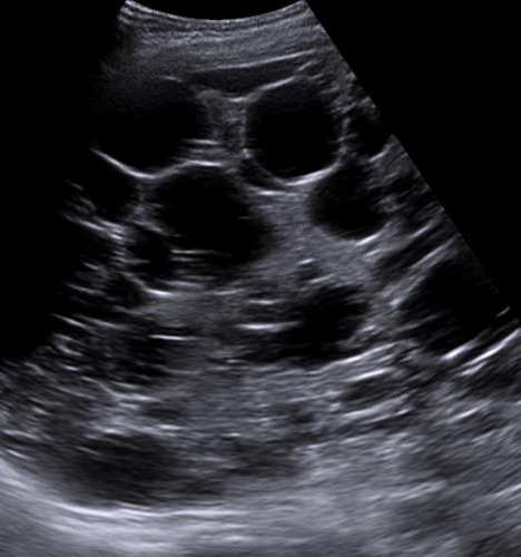
severe polycystic liver disease sonographic appearance
A “Swiss cheese” appearance with numerous cysts that may blend together.
Document “multiple cysts” and measure a clearly defined or largest representative cyst rather than every cyst
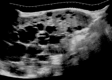
Acquired hepatic cyst causes/categories
trauma, parasites, or inflammatory reactions

would someone experience symptoms from Acquired hepatic cyst
yes
Sonographic appearance of acquired hepatic cysts
range from classic cystic to complex masses, and should always correlate your sonographic findings with clinical findings
Hepatic hematoma and how they are categorized
Collection of blood trapped inside the liver tissue or beneath its outer covering. they are categorized based on severity and location
Besides trauma, what causes hepatic hematoma
biopsy/interventional procedures, rupture of a vascular neoplasm or aneurysm, and pregnancy-induced hypertension
Hepatic Hematoma symptoms
a tender liver/abdominal pain and decreased hematocrit levels
What can occur if hepatic hematoma causes significant bleeding
patient can collapse with signs of shock
What clinical findings can you appear with significant hepatic hemorrhage
tender liver, collapse, shock with falling BP pulse and hematocrit
Hepatic hematoma sonographic findings
depends on age of the bleed (starts anechoic, then complex as its clotting then more solid mass appearance as it heals until it goes away).
can appear crescent shaped that displaces the liver medially.
left image is clotting stage and right is acute stage

hydatid cysts (echinococcal) cause
Most common cause is Taenia echinococcus or Echinococcus granulosus, which is a parasitic tapeworm. the larvae are ingested and travel to the liver through the SMV
Why is hydatid cyst rupture dangerous
it can cause severe anaphylactic shock and may be fatal
How can hydatid disease cause jaundice or portal HTN
cyst can obstruct bile ducts and larvae can invade or obstruct portal venous structures
hydatid cysts (echinococcal) sonographic appearance
varies depending on larval maturation but could be simple cyst or cyst with worms in it.
Active cyst can look like a large cyst containing daughter cysts (honeycomb appearance)
Inactive cyst can have more solid-appearing lesion with rim calcifications

What is a pyogenic hepatic abscess
A localized collection of pus
What organism is most commonly isolated from a pyogenic hepatic abscess
E. Coli
Most common source of pyogenic hepatic abscess and how
biliary tract. bile flow obstruction allows bacteria to grow and proliferate which can produce multiple abscesses
How does pyogenic bacteria enter liver
via portal vein or hepatic artery or direct extension from adjacent organs
What conditions are listed as sources of portal-vein seeding for a pyogenic abscess
Appendicitis, diverticulitis, or inflammatory bowel disease
What conditions are listed as sources of hepatic artery seeding for a pyogenic abscess
bacterial endocarditis and urinary sepsis
Pyogenic hepatic abscess symptoms/lab findings
Fever, elevated LFTs (AST, ALT, ALP, Bilirubin), RUQ pain, pleuritic pain, leukocytosis, and hepatomegaly
Pyogenic Hepatic abscess sonographic appearance
usually less echogenic than hepatic parenchyma, single or multiple masses usually >1cm, variable is shape (round, ovoid, poorly defined), and internal echo depends on debris, adhesions, or gas microbubbles. posterior acoustic enhancement differentiates debris filled pyogenic abscess from solid mass. irregular walls, peripheral hyperemia and heterogeneous

How severe is an untreated pyogenic liver abscess
it can reach 100% mortality rate
What is an Amebic abscess and where is it mostly located
a pus-filled mass in the liver caused by the parasite Entamoeba histolytica. most commonly in right lobe
Amebic abscess symptoms
usually asymptomatic when parasite is confined to GI tract.
Once it has developed (parasite is in the liver), you could experience RUQ pain and reactive hepatomegaly, diarrhea or black tarry stool, fever, jaundice, or moderate leukocytosis and elevated LFTs
Amebic abscess Sonographic appearance
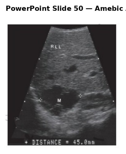
newly formed abscess appears echogenic and becomes hypoechoic as it matures.
color doppler usually shows no flow
image shows 4.5cm complex cystic mass in right lobe