lecture 27 - skeletal system: appendicular skeleton
 Call Kai
 Learn
 Practice Test
 Spaced Repetition
 Match
 Flashcards
 Knowt Play
1/92

There's no tags or description
Looks like no tags are added yet.
93 Terms
pectoral girdle
attachment of upper limb onto axial skeleton
made of 2 parts
2 parts of pectoral girdle
scapula
clavicle
sternoclavicular joint
where the clavicle articulates w/ the sternum
the ______________ joint is the only joint where the upper limb is connected to the axial skeleton
sternoclavicular
acromioclavicular joint
where clavicle articulates w/ scapula
glenohumeral joint
where upper limb attaches to pectoral girdle
scapula
shoulder blade

glenoid cavity
cavity of scapula that forms joint w/ head of humerus

scapular spine
process on scapula
attachment pt for muscles

acromion process
process on scapula
articulates w/ clavicle

supraspinous fossa
area above scapular spine on scapula

infraspinous fossa
area below scapular spine on scapula

subscapular fossa
basically the face of the scapula (from anterior view)

coracoid process
process on scapula
sticks out anteriorly + laterally
attachment pt for muscles that extend into upper limb + muscles for breathing

body of clavicle
middle region of clavicle

acromial (lateral) end
lateral end of clavicle
forms acromioclavicular joint
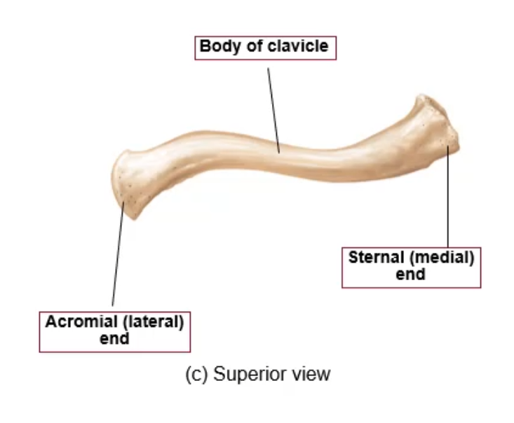
sternal (medial) end
medial end of clavicle
forms sternoclavicular joint
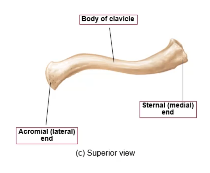
where is the weakest point (point that tends to break) of the clavicle?
right where we switch from the lateral → medial curve

superior border of scapula
top of scapula

humerus
upper arm

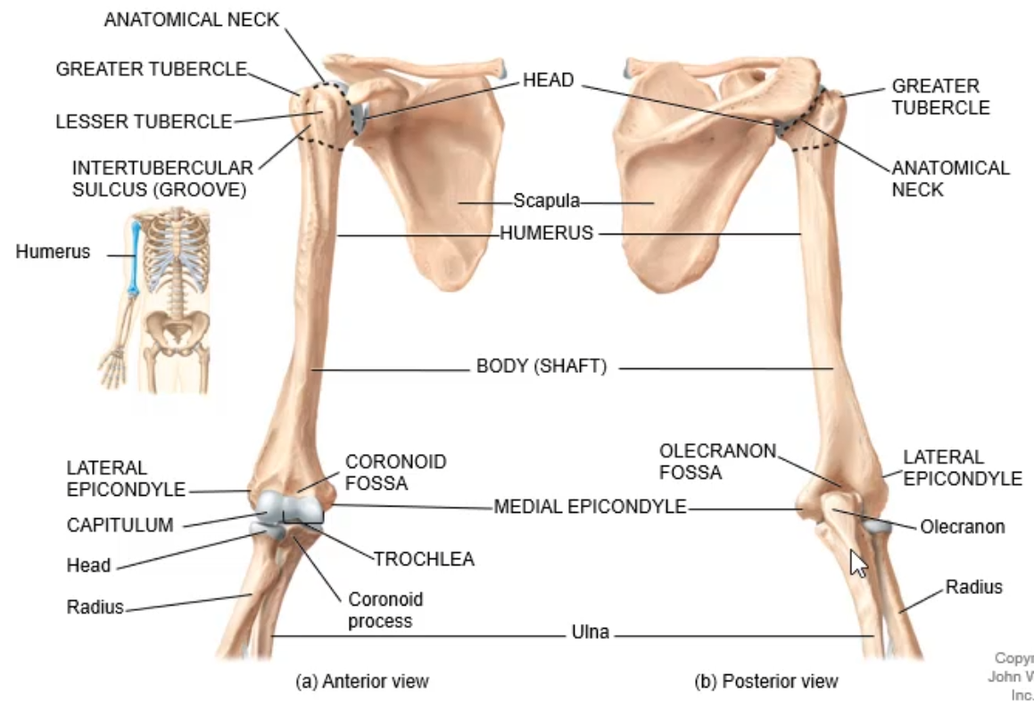
anatomical neck
just distal to head of humerus
surgical neck
more distal to the anatomical neck (narrowed region)

greater tubercle
feature of humerus on lateral side

lesser tubercle
feature of humerus on anterior side
attachment pt for muscles that form shoulder joint + arm muscles

intertubercular sulcus (groove)
in b/w greater + lesser tubercle
provides space for a tendon of biceps brachii

lateral epicondyle
projection on humerus (lateral)
pt of attachment for upper limb muscles

medial epicondyle
projection on humerus (medial)
pt of attachment for upper limb muscles

capitulum
feature on humerus (lateral side)
forms joint w/ radius
seen on anterior side

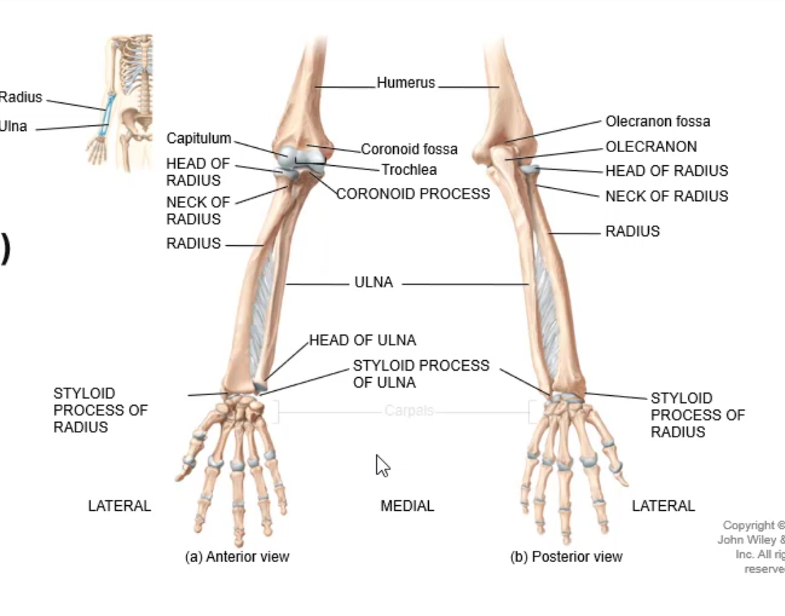
head of radius
flattened top region

trochlea
feature on humerus (medial side)
forms hinge joint w/ ulna
seen on anterior side

coronoid process
feature of ulna (proximal end)
seen on anterior side

coronoid fossa
indentation in humerus
seen on anterior side

olecranon fossa
indentation in humerus
seen on posterior side

olecranon
process on ulna
elbow

radius
thumb side

ulna
pinky side

the elbow joint is mostly made of what bone?
ulna

on the distal end of the forearm, is the ulna or radius larger?
radius
the radius forms an ______ joint w/ the wrist bones
ellipsoid
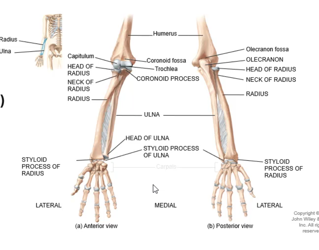
styloid process of radius
feature of radius
attachment pt for wrist ligaments

the head of the radius is at the _______ end while the head of the ulna is at the ______ end
proximal, distal

styloid process of ulna
feature of ulna
attachment pt for wrist ligaments

carpals
wrist bones

the wrist consists of _ carpal bones
8

how are the carpals arranged?
2 rows of 4 bones each

name the carpal bones in the proximal row
(lateral → medial): scaphoid bone, lunate bone, triquetrum, pisiform bone
these bones articulate w/ radius

name the carpal bones in the distal row
(lateral → medial): trapezium, trapezoid, capitate bone, hamate
articulate w/ metacarpal bones of palm
have gliding joints that allow small degrees of mvmt

mnemonic to remember carpal bones
Some Lovers Try Positions That They Can’t Handle
(Scaphoid, Lunate, Triquetrum, Pisiform, Trapezium, Trapezoid, Capitate, Hamate)

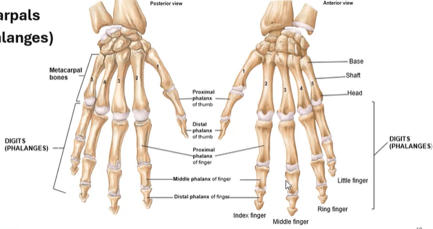
how many metacarpals are there
5

how are the metacarpal bones + phalanges numbered
1 to 5 as you move from lateral → medial

structure of metacarpals
base
shaft
head

phalanges
fingers

the head of each metacarpal forms a knuckle joint w/ the _______
phalanges

there are __ phalanges in each hand
14

phalanges structure (OTHER THAN THUMB)
proximal phalanx of finger
middle phalanx of finger
distal phalanx of finger

thumb phalanges structure
proximal phalanx of thumb
distal phalanx of thumb
what does the pelvic girdle consist of
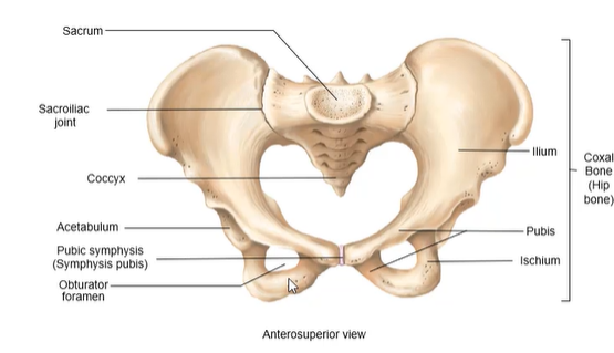
sacrum
hip bone

pelvic/hip girdle
attachment pt for lower limbs to axial skeleton
made of 2 coxal/hip bones

pubic symphysis/symphysis pubis
made of fibrocartilage

sacroiliac joint
sacrum + ilium joint

coccyx
protrusion at very base of spine

bony pelvis
pelvic girdle + coccyx

3 bones that join to form the coxal bone
ilium
pubis
ischium
ilium
most superior portion of coxal bone

pubis
middle (anterior + inferior) portion of coxal bone

ischium
inferior + posterior portion of coxal bone

acetabulum
fossa where the ilium, pubis and ischium meet
where the femur articulates

obturator foramen
large opening on inferior part of coxal bones
space for obturator nerve to travel to lower limbs

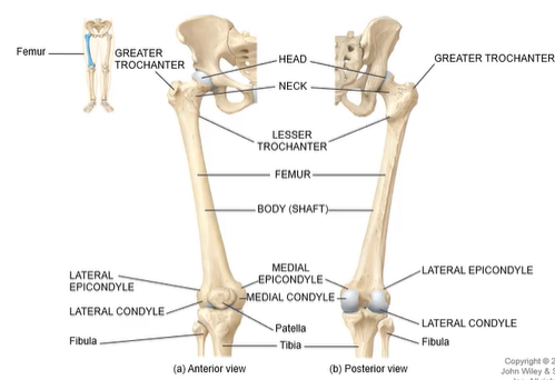
what is the longest + strongest bone in the body?
femur

where is a common site for fractures in the femur?
neck (bc its narrower + longer than it is in the humerus)

greater trochanter
protrusion on femur
attachment pt for hip + thigh muscles
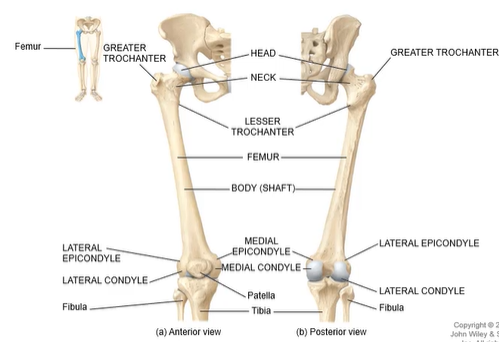
lesser trochanter
protrusion on femur
attachment pt for hip + thigh muscles

medial + lateral condyles (femur)
rounded surfaces on distal end of femur
articulate w/ tibia

medial + lateral epicondyles
features of femur
attachment pts for ligaments of knee joint

patellar groove
seen on anterior side of femur
b/w the lateral + medial condyles
what kind of bone is the patella? what does this mean?
sesamoid; its embedded within a tendon

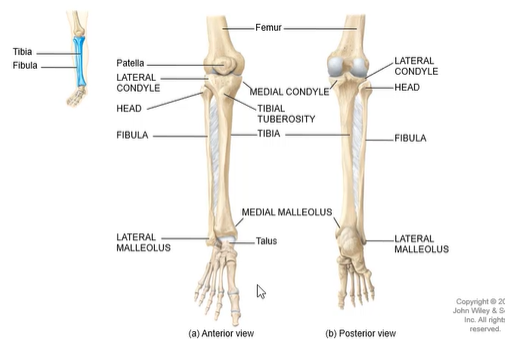
tibia
larger bone in lower leg
medial
weight-bearing bone

fibula
smaller bone in lower leg
lateral

medial + lateral condyles (tibia)
articulate w/ condyles of femur
pts of attachment for muscles

tibial tuberosity
seen on anterior side of tibia
attachment pt for tendon of quadriceps femoris muscle group

medial malleolus
distal end of tibia
forms medial side of ankle joint
can be felt (commonly thought of as ankle bone)

lateral malleolus
distal end of fibula
forms lateral side of ankle joint

is the head of the fibula part of the knee joint?
no; it is used for muscle attachment

talus
ankle
articulates w/ tibia + fibula
tarsals
foot bones

how many tarsals are there in each foot?
7

calcaneus
heel bone
largest + strongest bone in foot

mnemonic to remember tarsals
Tiger Cubs Need M I L C
Talus, Calcaneus, Navicular, Medial cuneiform, Intermediate cuneiform, Lateral cuneiform, Cuboid

metatarsals
literally metacarpals but for ur feet
but numbered 1-5 as you move medial → lateral

great toe
big toe
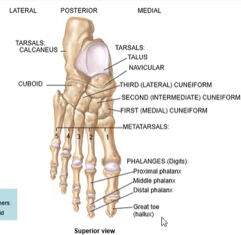
phalanges (foot) structure (EXCEPT GREAT TOE)
proximal phalanx
middle phalanx
distal phalanx
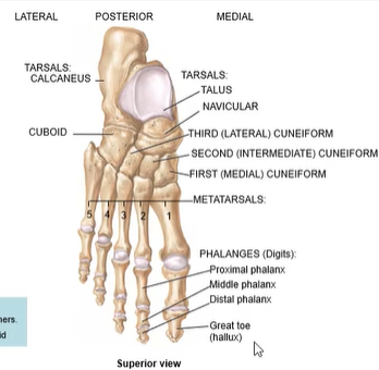
hallux
great/big toe

great toe phalanges structure
proximal phalanx
distal phalanx
